Abstract
Immunoelectron microscopy is a powerful tool to study biological molecules at the subcellular level. Antibodies coupled to electron-dense markers such as colloidal gold can reveal the localization and distribution of specific antigens in various tissues1. The two most widely used techniques are pre-embedding and post-embedding techniques. In pre-embedding immunogold-electron microscopy (EM) techniques, the tissue must be permeabilized to allow antibody penetration before it is embedded. These techniques are ideal for preserving structures but poor penetration of the antibody (often only the first few micrometers) is a considerable drawback2. The post-embedding labeling methods can avoid this problem because labeling takes place on sections of fixed tissues where antigens are more easily accessible. Over the years, a number of modifications have improved the post-embedding methods to enhance immunoreactivity and to preserve ultrastructure3-5.
Tissue fixation is a crucial part of EM studies. Fixatives chemically crosslink the macromolecules to lock the tissue structures in place. The choice of fixative affects not only structural preservation but also antigenicity and contrast. Osmium tetroxide (OsO4), formaldehyde, and glutaraldehyde have been the standard fixatives for decades, including for central nervous system (CNS) tissues that are especially prone to structural damage during chemical and physical processing. Unfortunately, OsO4 is highly reactive and has been shown to mask antigens6, resulting in poor and insufficient labeling. Alternative approaches to avoid chemical fixation include freezing the tissues. But these techniques are difficult to perform and require expensive instrumentation. To address some of these problems and to improve CNS tissue labeling, Phend et al. replaced OsO4 with uranyl acetate (UA) and tannic acid (TA), and successfully introduced additional modifications to improve the sensitivity of antigen detection and structural preservation in brain and spinal cord tissues7. We have adopted this osmium-free post-embedding method to rat brain tissue and optimized the immunogold labeling technique to detect and study synaptic proteins.
We present here a method to determine the ultrastructural localization of synaptic proteins in rat hippocampal CA1 pyramidal neurons. We use organotypic hippocampal cultured slices. These slices maintain the trisynaptic circuitry of the hippocampus, and thus are especially useful for studying synaptic plasticity, a mechanism widely thought to underlie learning and memory. Organotypic hippocampal slices from postnatal day 5 and 6 mouse/rat pups can be prepared as described previously8, and are especially useful to acutely knockdown or overexpress exogenous proteins. We have previously used this protocol to characterize neurogranin (Ng), a neuron-specific protein with a critical role in regulating synaptic function8,9 . We have also used it to characterize the ultrastructural localization of calmodulin (CaM) and Ca2+/CaM-dependent protein kinase II (CaMKII)10. As illustrated in the results, this protocol allows good ultrastructural preservation of dendritic spines and efficient labeling of Ng to help characterize its distribution in the spine8. Furthermore, the procedure described here can have wide applicability in studying many other proteins involved in neuronal functions.
Keywords: Neuroscience, Issue 74, Immunology, Neurobiology, Biochemistry, Molecular Biology, Cellular Biology, Genetics, Proteins, Immunohistochemistry, Immunological Synapses, Synapses, Hippocampus, Microscopy, Electron, Neuronal Plasticity, plasticity, Nervous System, Organotypic cultures, hippocampus, electron microscopy, post-embedding, immunogold labeling, fixation, cell culture, imaging
Protocol
1. Fixation
Fixatives are carcinogenic; wear gloves and handle the fixatives in a fume hood. Unless otherwise noted, all incubations are done on ice and all solutions should be filtered before use. Use electron microscopy-grade reagents.
Day 1
After experimental conditions (e.g. viral injection, drug treatment), place the membrane with organotypic hippocampal slices in a 60 x 15 mm polystyrene Petri dish containing ice-cold 0.1 M phosphate buffer (Sorensen's phosphate buffer), pH 7.3. Add 1 - 2 ml of 0.1 M phosphate buffer directly on top of the slices to keep them cold.
CRITICAL STEP: Always use freshly prepared buffers and fixatives. Make sure the pH of buffer is within desired range. A failure to do so could damage cellular structures.
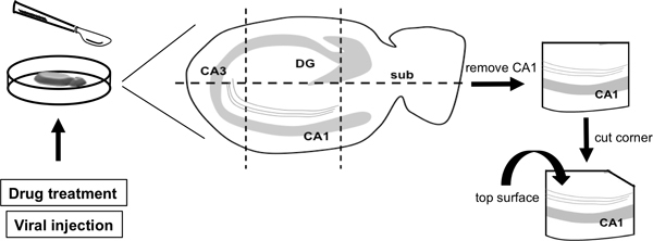
To isolate the CA1 subfield of the slice, use a disposable scalpel to gently cut across the slice next to the DG, parallel to the CA1 cell layer (Figure 1). Then cut the remaining slice vertically to remove the CA3 subfield and the subiculum. Cut a corner of the slice to help identify the top surface of the tissue.
NOTE: In case no viral delivery of protein of interest is required, the entire tissue slice can be fixed after the media is removed with a gentle rinse of ice-cold buffer. This is then followed by cutting out the CA1.
CRITICAL STEP: Keep track of the topside of the tissue in order for proper sectioning of the grids later on.
Carefully remove the tissue from the membrane using the backside of the scalpel. Use a Pasteur pipette to gently transfer the tissue to a 12-well plate containing 0.1 M phosphate buffer.
Remove the buffer in the well from the plate and add 500 μl - 1 ml of ice-cold fixative (pH 7.3) comprised of the following: 0.1% picric acid, 1% paraformaldehyde, and 2.5% glutaraldehyde in 0.1 M phosphate buffer. Incubate for 2 hr at 4 °C.
NOTE: Picric acid may be explosive when dry. Keep it wetted with water in a container tightly closed. Store in a dry and well-ventilated place.
Remove the fixative from the well and wash samples 3 times (20 min each) in 0.1 M phosphate buffer.
Incubate for 40 min in 1% tannic acid (w/v) in 0.1 M maleate buffer, pH 6.0.
Rinse twice (20 min each) in maleate buffer.
Incubate for 40 min in 1% uranyl acetate (w/v) in maleate buffer in the dark. Uranyl acetate is sensitive to light and is radioactive. Cover the beaker with Parafilm when dissolving and store any unused buffer in dark at 4 °C.
Rinse twice (20 min each) in maleate buffer.
Incubate for 20 min in 0.5% platinum chloride (w/v) in maleate buffer.
Rinse twice (20 min each) in maleate buffer. Store the samples at 4 °C until they are ready for further processing.
2. Dehydration and Embedding
Propylene oxide is carcinogenic. Avoid vapors by working with it in a fume hood. Use absolute, 100% pure ethanol that contains no trace of water. Dilute this absolute ethanol to produce different concentrations for dehydration.
Day 2
Incubate for 5 min in 50% ethanol and then for 5 min in 70% ethanol.
Incubate for 15 min in freshly prepared 1% p-phenylenediamine (PPD) in 70% ethanol.
Rinse three times in 70% ethanol.
Incubate for 5 min in 80% ethanol and then for 5 min in 95% ethanol.
Incubate in 100% ethanol twice 5 min each.
Prepare glass vials with screw caps and make sure they are clean and completely dry. Transfer slices to glass vials and label each vial simply and clearly.
Incubate for 5 min in 1:1 ethanol:propylene oxide.
Incubate in 100% propylene oxide twice 5 min each.
Add resin (Epon) to each vial to make a 1:1 mixture with propylene oxide. Gently mix for 2 hr. Do not shake the vials too rigorously to prevent bubbles that could interfere with embedding.
Add resin to make a 3:1 mixture with propylene oxide, and mix for 2 hr.
Transfer to 100% resin and incubate O/N.
Day 3
Sandwich samples between strips of ACLAR plastic and cure for 24 hr at 60 °C.
3. Sectioning and Mounting on Grids
Day 4
Cut semi-thin (0.5 μm) sections and stain with 1% toluidine blue + 1% borax to determine the correct orientation of samples.
Cut ultrathin sections (60 nm) and mount on nickel grids, one section per grid.
4. Immunohistochemistry
All buffers and water should be filtered before use.
Day 5
Place a drop (~50 μl) of 1% Tween-20/phosphate buffer (T/PB), pH 7.5, on a piece of clean Parafilm on a flat surface. Gently pick up the grid with clean forceps by the edge and float it on the buffer, section down, and incubate for 10 min at RT.
Drain excess liquid from the grid by placing section side up on to filter paper and then float on 50 mM glycine in T/PB for 15 min at RT.
CRITICAL STEP: To avoid excessive 'carry-over' of solutions from one solution droplet to the next during the incubations, drain excess liquid by gently touching the edge of the grid onto #1 filter paper between each solution change. Also remove any solution trapped between the arms of the EM forceps holding the grid by gently wicking with a sliver of filter paper between the forceps arms while ensuring the sections do not dry out completely.
Dry the grid with filter paper and then float on blocking solution containing 2.5% BSA and 2.5% serum from the animal of the secondary antibody in T/PB for 30 min at RT.
Incubate with primary antibody (in T/PB) at RT or O/N at 4 °C. The optimal time and temperature of incubation, as well as antibody concentration, need to be experimentally determined.
Wash the grid three times (2 min each) with T/PB.
Incubate with secondary antibody (1:20 anti-rabbit or anti-mouse coupled to 10 nm gold) for 1 hr at RT.
Wash three times (2 min each) with T/PB.
Postfix with 2% glutaraldehyde in T/PB for 5 min.
Wash three times (2 min each) with T/PB and then three times (2 min each) with filtered water.
Incubate with 2% uranyl acetate in water for 10 min.
While the grid is staining, prepare a CO2-free chamber by placing a piece of Parafilm in the center of a glass Petri dish. Then place 4-6 pellets of NaOH in the dish around the Parafilm to absorb CO2 in the air. Keep the top closed for a few minutes. Use a Pasteur pipette and quickly transfer a small volume of Reynold's lead citrate solution to the Parafilm in the glass Petri dish. Open the top just enough to insert the Pasteur pipette to minimize the re-introduction of CO2 into the chamber.
NOTE: To make Reynold's lead citrate solution, boil 100 ml of deionized water in a microwave oven and let cool in an airtight container. In a 50 ml volumetric flask with stopper, mix 1.33 g lead nitrate, 1.76 g sodium citrate and 30 ml of the boiled water by shaking vigorously for 1 min. Then shake intermittently for 30 min. To this cloudy solution, add 8 ml of 1 M sodium hydroxide and slowly invert the flask a few times. Solution will become clear. Bring the solution to 50 ml with the boiled water. Store the solution tightly sealed. If precipitate appears, discard and make a new one.
In three smaller beakers prepare warm, freshly boiled deionized water. Wash off the uranyl acetate by dipping the grid in the first beaker and gently swirl it around for 30 sec. Repeat this step in the other two beakers.
Open the top of the glass Petri dish just enough to place the grid on the drop of Reynold's lead citrate solution. If possible, wear a mask while doing this to avoid breathing on lead citrate and to prevent formation of precipitate. Incubate for 10 min.
Open the top just enough to remove the grid. Avoid breathing on the lead citrate. Wash the grid three times by dipping it sequentially in three beakers with warm, freshly boiled distilled water. Let the grid dry on a piece of filter paper, section up. The sample is now ready for the electron microscope.
Representative Results
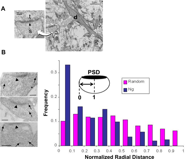
Figure 2B shows an example of the distribution of endogenous Ng molecules in dendritic spines of CA1 hippocampal pyramidal neurons. Nickel grids with ultrathin (60 nm) tissues containing CA1 region of the hippocampus (as seen in Figure 2A) were covered in 1% T/PB, 50 mM glycine, then blocked with 2.5% BSA and 2.5% serum prior to incubation with anti-Ng antibody. After washing with T/PB, grids were then covered in anti-rabbit secondary antibody coupled to 10 nm gold. Finally, grids were washed with T/PB and postfixed with 2% glutaraldehyde followed by 2% uranyl acetate and Reynold's lead citrate staining to enhance contrast. Left, representative electron micrographs showing asymmetrical synapses. Identification of the postsynaptic compartment is facilitated by the electron-dense PSD (arrow head) and well-defined plasma membrane. Right, the shortest distance between each Ng (arrows) and the plasma membrane was normalized to the radius of the spine. Ng molecules within the spine (but not at the PSD) exhibit preferential localization to the plasma membrane. This histogram was originally published in the EMBO Journal8.
 Figure 1. Schematic of CA1 area dissection from the hippocampal slice cultures. After experimental treatment to the slice, e.g. drug treatment or viral infection, the membrane containing the slice was placed in ice-cold phosphate buffer. The slice was first cut horizontally beneath the dentate gyrus (DG), parallel to the CA1 cell layer. The CA1 subfield was then isolated by removing the CA3 subfield and the subiculum (sub). To help identify the top surface of the CA1, a corner was cut to orient the tissue (in this case the upper right hand corner).
 Figure 2. Immunogold labeling of neurogranin at CA1 synapses in organotypic hippocampal slice cultures. (A) Transmission electron micrographs showing rat hippocampal CA1 area and synapses. Dendrites (d) of CA1 pyramidal neurons are easily distinguishable. Identification of asymmetrical synapses between axonal terminal 9t) and dendritic spines (s) is facilitated by the presence of post-synaptic density (PSD, arrowhead) and well-defined plasma membrane. (B) Distribution of neurogranin (Ng) in dendritic spines. Left, transmission electron micrographs showing immunogold-EM labeling of nG (arrows). For quantification, the distance of each gold particle (excluding the ones at PSD, arrow head) from the plasma membrane is normalize (x axis) to the radius of the spine. Hence, 0 corresponds to a particle lying on the membrane and 1 to a particle lying at the center of the spine. Right, a random distribution (pink) is generated by using a random number generator macro (Microsoft Excel) in a spine-shaped surface. Ng (blue) shows the highest peak distribution close to the plasma membrane. Scale bar, 200 nm. This histogram was originally published in the EMBO Journal8.
Discussion
In this protocol, we have adopted the Phend and Weinberg method for brain and spinal cord tissues to study dendritic spines in rat hippocampal slice cultures. Dendritic spines in the hippocampal CA3-CA1 area are delicate structures containing a vast variety of proteins that play important roles in regulating neuronal functions. The presented method provides a balanced approach for achieving enhanced antigenicity while maintaining good ultrastructural preservation (Figure 2A), permitting reasonable labeling efficiency and good reproducibility useful for characterizing the distribution of specific antigens within the spine. Here, we labeled Ng with 10 nm colloidal gold. We then quantified the gold to generate labeling pattern of Ng that helped to understand its role in synaptic function (Figure 2B)8.
Quantitative analysis of immunogold can be done in a variety of ways11-13. However, interpretation of such quantification is not always straightforward. Labeling efficiency, gold particle size, variability in processing as well as variability of antigen number across samples can all influence quantification of gold particles. These concerns are beyond the scope of this protocol but some of these issues have been addressed in a number of reviews14-17.
The replacement of osmium by TA, UA, and p-phenylenediamine (PPD) in this approach greatly enhanced the sensitivity of antigen detection in neuronal tissues compared to previous methods7. The use of formaldehyde and glutaraldehyde ensures structural preservation and retention of small soluble antigens such as amino acids. Epoxy-based resins, rather than acrylic plastic resins, provide better stability for embedding under the electron beam without compromising antigenicity7. Compared to standard osmium protocol, Phend et al. had shown that TA, UA, and PPD also improved structural preservation (see Figure 1 of the original Phend paper)7, and that efficient immunostaining was found for multiple proteins including substance P, calcitonin gene-related peptide (CGRP), AMPA receptor subunit 1 (GluA1), and gamma-aminobutyric acid (GABA)7. These findings demonstrate the capability of this procedure to study different proteins in neuronal tissues. Another advantage of this method is that TA, UA and PPD are easy to prepare and handle, and pose much smaller hazardous risks than osmium, which is extremely volatile and very toxic by inhalation and skin contact.
One limitation of this method is that although it preserves and retains small antigens such as amino acids, it does not show improved immunolabeling for these molecules compared to conventional osmium treatment. The advantage of the heavy metal platinum chloride to preserve structure and to enhance immunoreactivity also seems to be limited to the spinal cord, for reasons that are not completely understood7. Although the osmium-free method improves antigenicity and tissue contrast, it does not seem to work well for the preservation of other structures such as presynaptic vesicles (see Figure 2). In addition, Phend et al. had observed that the absence of osmium also resulted in reduced myelin preservation, as TA has limited penetrability into the intracellular components to preserve membranes7. For these reasons, freezing methods for specimen preparation may be better alternatives if ultrastructural preservation is the most important concern. Ultrarapid freezing of specimen allows the preservation of molecules in a more natural way18, without potential artifacts that could be introduced by the use of chemical fixatives.
Prior to performing the EM, the presence of the antigen in the sample should be confirmed through ways such as Western blot analysis. As with all immunolabeling studies, correct interpretation of data relies on the use of appropriate, specific antibodies. Highly purified monoclonal antibodies should always be used if available. Control experiments are essential to ensure that the labeling pattern generated is due to the specific interaction between the primary antibody and the immunogen. The specificity of the labeling can be tested by carrying out negative and positive controls. For example, in a negative control, the primary antibody can be replaced by an unrelated control serum of non-immune IgG, without changing any other steps in the staining protocol. Second, a protein that is present in one cell compartment but not in others can be used as a positive control. For example in glutamatergic synapses, PSD-95 is a postsynaptic marker while GAP-43 is primarily present in the presynaptic terminal19-22. If labeling is seen when primary antibody is omitted or in compartments where the protein is known not to be present, then the labeling is not specific.
Once the specificity of primary antibody is rigorously tested, one should be aware that samples with good labeling should not exhibit many clumping or have excessive gold particles in the background where no tissue is present. If the amount of gold is too high or too low, adjust the level or duration of blocking and the concentration of antibody accordingly. Maunsbach and Afzelius provide some excellent examples and tips for immunolabeling23.
Good structural preservation of samples can eliminate artifacts that may interfere with morphology and confound the interpretation of data. Hence, the superior preservation of structures illustrated by this method makes this technique applicable to a number of biological tissues especially where delicate structures can be easily distorted or destroyed during sample processing. Hence, in addition to organotypic hippocampal slices, this method can also be tremendously valuable for other neuronal preparations including acute brain slices and primary neuronal cultures. Organotypic hippocampal slices are easily amendable to drug treatment and expression of proteins through viral infection, which can be applicable for studying hippocampal synaptic plasticity. This method is also useful to examine the effects of different experimental conditions on antigen distribution or localization in the cell body, dendrites, or dendritic spines. Furthermore, colloidal gold particles with sizes of 6, 10, 15 or 25 nm are available, making it possible to carry out multiple labeling with no overlap. We believe that the advantage of enhanced antigenicity, flexibility of probes and reproducibility of immunogold labeling demonstrate the power of this technique to study a variety of antigens. It is potentially useful to characterize the localization and distribution of receptors, receptor-interacting proteins, transporter, ion channels and many others in different brain tissues including the cortex, thalamus, cerebellum and olfactory bulb.
In summary, the present protocol of an osmium-free, post-embedding immunogold labeling of rat hippocampal slice cultures is a valuable tool to study various antigens within dendritic spines. The potential applicability of this method in other delicate central nervous tissues will be very useful to examine the ultrastructural characteristics of different key molecules and to help understand their role in biological functions.
Disclosures
We have nothing to disclose.
Acknowledgments
The authors would like to thank Matthew Florence for preparation of the hippocampal slice cultures. This work was supported by grants from US National Institute on Aging and Alzheimer's Association to NZG.
References
- Faulk WP, Taylor GM. An immunocolloid method for the electron microscope. Immunochemistry. 1971;8:1081–1083. doi: 10.1016/0019-2791(71)90496-4. [DOI] [PubMed] [Google Scholar]
- Stirling JW. Immuno- and affinity probes for electron microscopy: a review of labeling and preparation techniques. J. Histochem. Cytochem. 1990;38:145–157. doi: 10.1177/38.2.2405054. [DOI] [PubMed] [Google Scholar]
- Phend KD, Weinberg RJ, Rustioni A. Techniques to optimize post-embedding single and double staining for amino acid neurotransmitters. Journal of Histochemistry & Cytochemistry. 1992;40:1011–1020. doi: 10.1177/40.7.1376741. [DOI] [PubMed] [Google Scholar]
- Berryman MA. Effects of tannic acid on antigenicity and membrane contrast in ultrastructural immunocytochemistry. J. Histochem. Cytochem. 1992;40:845–857. doi: 10.1177/40.6.1350287. [DOI] [PubMed] [Google Scholar]
- Akagi T, et al. Improved methods for ultracryotomy of CNS tissue for ultrastructural and immunogold analyses. J. Neurosci. Methods. 2006;153:276–282. doi: 10.1016/j.jneumeth.2005.11.007. [DOI] [PubMed] [Google Scholar]
- Stirling JW. Ultrastructual localization of lysozyme in human colon eosinophils using the protein A-gold technique: effects of processing on probe distribution. J. Histochem. Cytochem. 1989;37:709–714. doi: 10.1177/37.5.2467929. [DOI] [PubMed] [Google Scholar]
- Phend KD, Rustioni A, Weinberg RJ. An osmium-free method of epon embedment that preserves both ultrastructure and antigenicity for post-embedding immunocytochemistry. Journal of Histochemistry & Cytochemistry. 1995;43:283–292. doi: 10.1177/43.3.7532656. [DOI] [PubMed] [Google Scholar]
- Zhong L, Cherry T, Bies CE, Florence MA, Gerges NZ. Neurogranin enhances synaptic strength through its interaction with calmodulin. Embo J. 2009;28:3027–3039. doi: 10.1038/emboj.2009.236. [DOI] [PMC free article] [PubMed] [Google Scholar]
- Zhong L, Kaleka KS, Gerges NZ. Neurogranin phosphorylation fine-tunes long-term potentiation. Eur. J. Neurosci. 2011;33:244–250. doi: 10.1111/j.1460-9568.2010.07506.x. [DOI] [PMC free article] [PubMed] [Google Scholar]
- Zhong L, Gerges NZ. Neurogranin targets calmodulin and lowers the threshold for the induction of long-term potentiation. PloS one. 2012;7:e41275. doi: 10.1371/journal.pone.0041275. [DOI] [PMC free article] [PubMed] [Google Scholar]
- Horisberger M, Vauthey M. Labelling of colloidal gold with protein. A quantitative study using beta-lactoglobulin. Histochemistry. 1984;80:13–18. doi: 10.1007/BF00492765. [DOI] [PubMed] [Google Scholar]
- Bendayan M. A review of the potential and versatility of colloidal gold cytochemical labeling for molecular morphology. Biotech. Histochem. 2000;75:203–242. [PubMed] [Google Scholar]
- Racz B, Weinberg RJ. Spatial organization of coflin in dendritic spines. J. Neuroscience. 2006;138(2):447–456. doi: 10.1016/j.neuroscience.2005.11.025. [DOI] [PubMed] [Google Scholar]
- Posthuma G, Slot JW, Geuze HJ. Usefulness of the immunogold technique in quantitation of a soluble protein in ultra-thin sections. J. Histochem. Cytochem. 1987;35:405–410. doi: 10.1177/35.4.2434559. [DOI] [PubMed] [Google Scholar]
- Howell KE, Reuter-Carlson U, Devaney E, Luzio JP, Fuller SD. One antigen, one gold? A quantitative analysis of immunogold labeling of plasma membrane 5'-nucleotidase in frozen thin sections. Eur. J. Cell Biol. 44:318–327. [PubMed] [Google Scholar]
- Yokota E, Kawaguchi T, Kusaba T, Niho Y. [Immunologic analysis of families with complement receptor deficiency] Nihon. Rinsho. 1988;46:2040–2045. [PubMed] [Google Scholar]
- Kehle T, Herzog V. Interactions between protein-gold complexes and cell surfaces: a method for precise quantitation. Eur. J. Cell Biol. 1987;45:80–87. [PubMed] [Google Scholar]
- Bozzola JR, Lonnie . Electron Microscopy. Second Edition. Vol. 30. Jones and Bartlett Publishers; 1992. Ch. 2. [Google Scholar]
- Cho KO, Hunt CA, Kennedy MB. The rat brain postsynaptic density fraction contains a homolog of the Drosophila discs-large tumor suppressor protein. Neuron. 1992;9:929–942. doi: 10.1016/0896-6273(92)90245-9. [DOI] [PubMed] [Google Scholar]
- Hunt CA, Schenker LJ, Kennedy MB. PSD-95 is associated with the postsynaptic density and not with the presynaptic membrane at forebrain synapses. J. Neurosci. 1996;16:1380–1388. doi: 10.1523/JNEUROSCI.16-04-01380.1996. [DOI] [PMC free article] [PubMed] [Google Scholar]
- Gispen WH, Leunissen JL, Oestreicher AB, Verkleij AJ, Zwiers H. Presynaptic localization of B-50 phosphoprotein: the (ACTH)-sensitive protein kinase substrate involved in rat brain polyphosphoinositide metabolism. Brain Research. 1985;328:381–385. doi: 10.1016/0006-8993(85)91054-6. [DOI] [PubMed] [Google Scholar]
- Biewenga JE, Schrama LH, Gispen WH. Presynaptic phosphoprotein B-50/GAP-43 in neuronal and synaptic plasticity. Acta Biochim. Pol. 1996;43:327–338. [PubMed] [Google Scholar]
- Maunsbach AA, Bjorn . Biomedical Electron Microscopy Illustrated Methods and Interpretations. Academic Press; 1999. Ch. 16; pp. 383–426. [Google Scholar]


