Abstract
It is believed that sigmoid volvulus (SV) in Brazil is a frequent complication of megacolon caused by Chagas’ disease (CD), differing in some characteristics from volvulus found in other countries. Bowel obstruction in patients with CD, principally when the cause is SV, may be sometimes difficult to diagnosis exclusively with plain abdominal radiograph. Fecaloma impacted in retossigmoidal area is one of the differential diagnoses. In addition, the huge amount of gas and feces, and distension of the colon normally increase the difficulty to make the correct diagnostic. The use of computer tomography (CT) scan can easy elucidate the picture of SV, and can be a great tool in cases of patients with CD and suspicion of this entity. A 62-year-old man showed bowel distention and stop disposal of gas for 5 days. He had previous diagnosis of CD. He also had been suffering from chronic constipation for several years, including impacted fecaloma, with the necessity of manual extraction. Plain abdominal radiographs showed an important colon dilatation and gross amount of feces in the sigmoid colon. Abdominal computer tomography sacan revealed dilated colon filled with feces, as well, the “whirl sign” composed of mesentery and twisted colon. When abdominal radiograph films reveal gross colonic dilatation of unknown etiology in patients with CD, a whirl sign on CT scans raises the possibility of colonic volvulus.
Keywords: Sigmoid volvulos, Chaga’s disease, Whirl sign, Computer tomography scan, Bowel obstruction
Introduction
It is believed that sigmoid volvulus (SV) in Brazil is a frequent complication of megacolon caused by Chagas’ disease (CD), differing in some characteristics from volvulus found in other countries [1]. Until today, according with World Health Organization, CD is an endemic in some regions of Brazil and other parts of Latin America [2]. Because this, is not uncommon, principally in emergency hospital, the surgeon faced with patients with diagnosis of CD, and bowel obstruction.
Bowel obstruction in patients with CD, principally when the cause is SV, may be sometimes difficult to diagnosis exclusively with plain abdominal radiograph. Fecaloma impacted in retossigmoidal area is one of the differential diagnoses. In addition, the huge amount of gas and feces, and distension of the colon normally increase the difficulty to make the correct diagnostic.
The use of computer tomography (CT) scan can easy elucidate the picture of SV, and can be a great tool in cases of patients with CD and suspicion of this entity. The aim of this report is describe a CT whirl sing of SV in a patient with CD.
Case Report
A 62-year-old man, from a rural area, was admitted in our emergency hospital, with bowel distention and stop disposal of gas for 5 days. He had previous diagnosis of CD. He also had been suffering from chronic constipation for several years, including impacted fecaloma, with the necessity of manual extraction. Plain abdominal radiographs showed an important colon dilatation and gross amount of feces in the sigmoid colon.
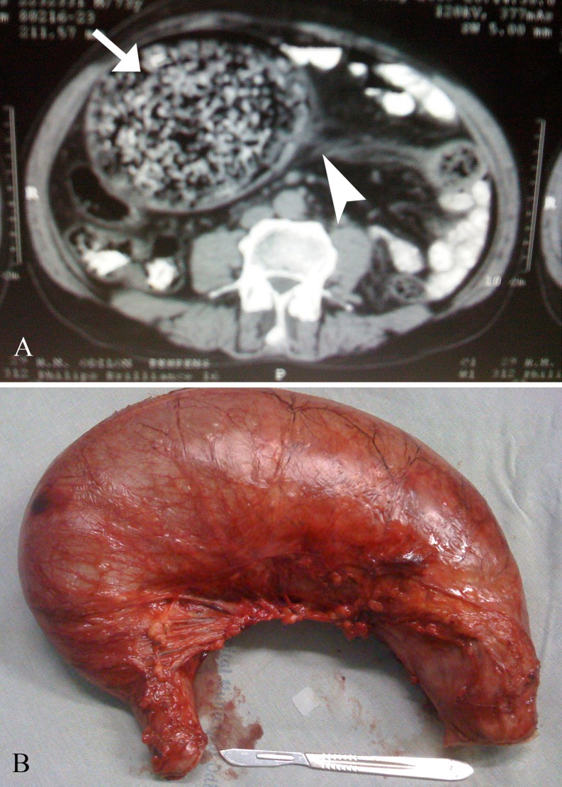
Because of doubt among fecaloma and SV, we decided to perform an abdominal CT to clarify the diagnosis. The exam revealed dilated colon filled with feces, as well, the “whirl sign” composed of mesentery and twisted colon (Fig. 1a).
Fig. 1.
a Abdominal computer tomography scan showed the “whirl sign” composed of mesentery (arrow head) and the twisted dilated colon, full with air and feces (arrow). b Surgical specimen (dilated sigmoid colon)
The patient underwent a laparotomy and sigmoidectomy with Hartmann procedure (Fig. 1b). Later he was discharge home with out any intercurrent.
Discussion
The correct diagnosis in patients with CD, and signs of bowel obstruction is very important. The treatment can vary from endoscopic emptying in cases of impacted fecaloma, to laparotomy, and Hartmann operation, in cases of complicated SV. To aggravate the situation, in most cases, beyond the emergency set, the patient with CD showed high operatory risk. They frequently are malnourished and have some heart disease, in consequence of megaesophagus and cardiomyophaty caused by CD.
The “whirl sign” was first described by Fisher in 1981, as suggesting volvulus of the small bowel, and it was considered that this sign represents the superior mesenteric artery at the center surrounded by bowel loops [3]. Subsequently, it has occasionally been recognized in patients with various forms of volvulus, such as sigmoid or cecal volvulus [4]. In a patient with SV, the whirl sign is caused by tightly twisted bowel and mesentery [5].
When abdominal radiograph films reveal gross colonic dilatation of unknown etiology in patients with CD, a whirl sign on CT scans raises the possibility of colonic volvulus.
References
- 1.Cutait DE, Cutait R. Surgery of chagasic megacolon. World J Surg. 1991;15:188–197. doi: 10.1007/BF01659052. [DOI] [PubMed] [Google Scholar]
- 2.Chagas disease, Brazil (2000) Wkly Epidemiol Rec 75:153-5 [PubMed]
- 3.Fisher JK. Computed tomographic diagnosis of volvulus in intestinal malrotation. Radiology. 1981;140:145–146. doi: 10.1148/radiology.140.1.7244217. [DOI] [PubMed] [Google Scholar]
- 4.Shaff MI, Himmelfarb E, Sacks GA, Burks DD, Kulkarni MV. The whirl sign: a CT finding in volvulus of the large bowel. J Comput Assist Tomogr. 1985;9:410. [PubMed] [Google Scholar]
- 5.Matsumoto S, Mori H, Okino Y, Tomonari K, Yamada Y, Kiyosue H. Computed tomographic imaging of abdominal volvulus: pictorial essay. Can Assoc Radiol J. 2004;55:297–303. [PubMed] [Google Scholar]


