Abstract
Splenic abscess due to acute brucellosis is a rare event. We report a case of splenic abscess caused by Brucella spp. in a 21-year-old man. The MRI revealed sharply demarcated lesions measuring 20 and 30 mm in diameter at the superior pole of spleen. Positive Wright agglutination test and positive blood culture confirmed the diagnosis. Antibiotic therapy, without surgical intervention, was successful.
Background
Brucellosis is one of the most widespread zoonoses worldwide.1 Brucellosis melitensis (small ruminants), Brucellosis abortus (cattle), Brucellosis suis (swine) and Brucellosis canis (dogs) are known to cause human disease. B melitensis is the most common casual agent of human cases.2 The disease is most prevalent in Mediterranean countries.3 Brucellosis is a systemic infection, ranging from asymptomatic disease to severe and/or fatal illness.4 Splenic abscess due to brucellosis is extremely rare. Diagnosis and localisation of the abscesses are relatively easy with the use of ultrasonography, CT and MR. Here we report a case of splenic abscess due to brucellosis in an adult patient with acute brucellosis.
Case presentation
A 21-year-old man was referred to our hospital with 2 weeks’ history of weakness, abdominal pain, arthralgia and night sweats. He was a farmer by occupation. On physical examination his axillary temparature was 38.9°C. The liver and spleen was palpable. No lymphadenopathy was present.
Investigations
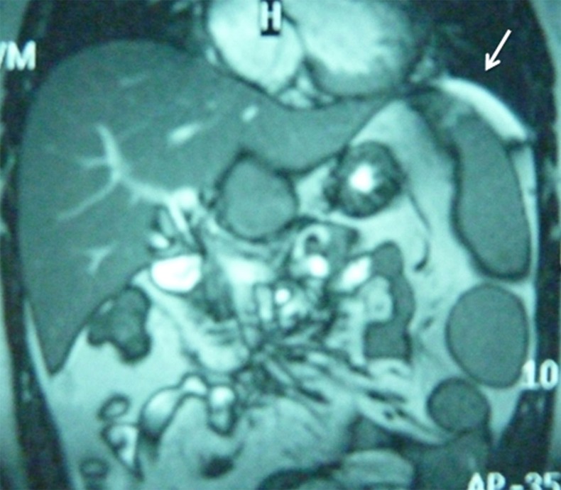
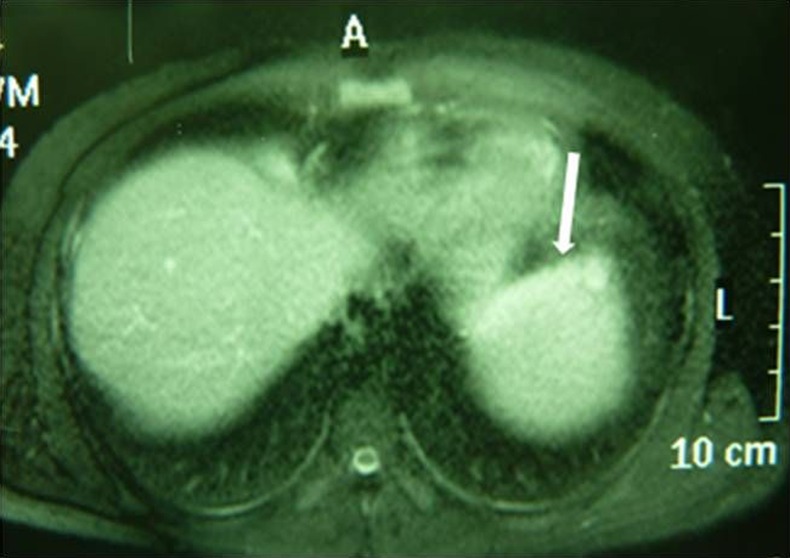
Laboratory tests revealed the following results: Haemoglobin 14.3 g/dl, leukocyte count 6300/mm3, (polymorphonuclear leukocyte %41, Lenfocyt %47.8), aspartate aminotransferase 261 IU/l(N:0–41), alanine transaminase 272 IU/l(N:0–54), γ-glutamyltransferase 449 IU/l(N:0–50), alkaline phosphatase 377 IU/l(N:38–126), Total bilurubin level 0.7 mg/dl (N:0.1–2). Chest x-ray was normal. Abdominal ultrasonography revealed hepatomegaly (19 cm), splenomegaly (17 cm) and 2 hypoechoic lesions in the upper portion of spleen. Because of suspicious image in ultrasonography (USG), abdominal MRI was performed and sharply demarcated hyperintense lesions measuring 20 and 30 mm in diameter at the superior pole of spleen were detected as occult splenic abscess on coronal half-fourier acquisition single-shot turbo spin-echo sequence (figure 1). Fat-supressed T2 axial image shows a weak hyperintense lesion at the superior pole of spleen (figure 2). On the second day of hospitalisation Wright agglutination test was reported positive (1:1280) and blood culture also yielded positive result on the fifth day of hospitalisation.
Figure 1.

MRI showing sharply demarcated hyperintense lesions at the superior pole of spleen on coronal half-fourier acquisition single-shot turbo spin-echo sequence.
Figure 2.

Fat-supressed T2 axial image shows a weak hyperintense lesion at superior pole of spleen.
Differential diagnosis
Treatment
Oral doxycycline (200 mg/day), oral ciprofloxasin (1000 mg/day) and inramuscular streptomycin (1 g/day) were started as therapy. The patient was afebrile 72 h after the initiation of the therapy.
A 12-week course of antibiotic therapy was completed, which included 21 days of intramuscular streptomycin therapy.
Outcome and follow-up
A follow-up USG of the abdomen at the end of therapy showed a complete resolution of the splenic abscess. Six and 12 months later, the patient remained free of symptoms.
Discussion
Brucellosis is an endemic disease in Turkey. It is transmitted to humans via consumption of unpasteurised milk and milk products or direct contact with infected animals. The disease is recognised as an occupational risk, and our patient had a history of exposure to animals. Brucellosis is a multisystemic illness, all organs and systems can be affected by the complications.5 6 Splenic abscess formation is an unusual complication of human brucellosis. The limited number of previous reports was mostly associated with chronic infection.7––9 Splenic abscesses were associated with Brucella endocarditis in two other previous reports.2 10 Pourbagher et al reported splenic abscess in brucellosis in 1.60% cases. They defined multiple hypoechoic nodules representing small abscesses in the splenic parenchyma in the acute stage of brucellosis. They also detected enlarged periportal lymph nodes which was more frequent in the acute stage.11 In the present case, the patient had acute brucellosis. The splenic lesions, which were rather larger than previous reports, had hypoechoic internal texture and well-defined margins on USG and they were sharply demarcated and had a homogeneous hyperintense on MRI. Calcification in liver and spleen is characteristic of chronic disease.7 In our patient, USG examination showed no calcification in the spleen or liver. No enlarged periportal lymph nodes were present.
The patient was successfully treated with 3-drug combinations of antibiotics. After 3 months of therapy, follow-up USG of the abdomen showed complete resolution of the splenic abscess.
In conclusion with the advent of imaging techniques such as USG and MRI, diagnosis of the unusual presentation of brucellosis such as splenic abscess is relatively easy. Clinicians practising in areas where brucellosis is endemic should be aware of this unusual complication.
Learning point.
Bucellosis is one of the most widespread zoonoses worldwide. Brucellosis is a systemic infection, ranging from asymptomatic disease to severe and/or fatal illness. Splenic abscess due to brucellosis is extremely rare. The advent of imaging techniques such as ultrasonography and MRI, diagnosis of the unusual presentation of brucellosis such as splenic abscess is relatively easy. Clinicians practising in areas where brucellosis is endemic should be aware of this unusual complication.
Footnotes
Contributors: All authors who have participated in the work have contributed equally in planning, conducting and reporting the article.
Competing interests: None.
Patient consent: Obtained.
Provenance and peer review: Not commissioned; externally peer reviewed.
References
- 1.Bosilkovski M, Dimzova M, Grozdanovski K. Natural history of brucellosis in an endemic region in different time periods. Acta Clin Croat 2009;2013:41–6 [PubMed] [Google Scholar]
- 2.Pappas G, Akritidis N, Bosilkovski M, et al. Brucellosis. N Engl J Med 2005;2013:2325–36 Review [DOI] [PubMed] [Google Scholar]
- 3.Yilmaz MB, Kisacik HL, Korkmaz S. Persisting fever in a patient with brucella endocarditis: occult splenic abscess. Heart 2003;2013:e20. [PMC free article] [PubMed] [Google Scholar]
- 4.Colmenero JD, Reguera JM, Martos F, et al. Complications associated with Brucella melitensis infection: a study of 530 cases. Medicine (Baltimore) 1996;2013:195–211 Erratum in: Medicine (Baltimore) 1997 Mar;76(2):139 [DOI] [PubMed] [Google Scholar]
- 5.Dakdouk GK, Araj GF, Awar GN. Buttock abscess brucellosis. Scand J Infect Dis 2002;2013:934–6 [DOI] [PubMed] [Google Scholar]
- 6.Miranda RT, Gimeno AE, Rodriguez TF, et al. Acute cholecystitis caused by Brucella melitensis: case report and review. J Infect 2001;2013:77–8 Review [DOI] [PubMed] [Google Scholar]
- 7.Ariza J, Pigrau C, Cañas C, et al. Current understanding and management of chronic hepatosplenic suppurative brucellosis. Clin Infect Dis 2001;2013:1024–33 [DOI] [PubMed] [Google Scholar]
- 8.Vallejo JG, Stevens AM, Dutton RV, et al. Hepatosplenic abscesses due to Brucella melitensis: report of a case involving a child and review of the literature. Clin Infect Dis 1996;2013:485–9 Review [DOI] [PubMed] [Google Scholar]
- 9.Sayilir K, Iskender G, Oğan MC, et al. Splenic abscess due to brucellosis. J Infect Dev Ctries 2008;2013:394–6 [DOI] [PubMed] [Google Scholar]
- 10.Saadeh AM, Abu-Farsakh NA, Omari HZ. Infective endocarditis and occult splenic abscess caused by Brucella melitensis infection: a case report and review of the literature. Acta Cardiol 1996;2013:279–85 Review [PubMed] [Google Scholar]
- 11.Pourbagher MA, Pourbagher A, Savas L, et al. Clinical pattern and abdominal sonographic findings in 251 cases of brucellosis in southern Turkey. AJR Am J Roentgenol 2006;2013:W191–4 [DOI] [PubMed] [Google Scholar]


