

Abstract
Recurrent apthous stomatitis (RAS) is one of the most common oral inflammatory diseases characterised by painful recurrent ulcerations of the orofacial region. The ulcers occur in three clinical forms: minor, major and herpetiform. Several therapies have been advocated to manage these lesions such as topical corticosteroids (triamcinolone acetonide, hydrocortisone acetate and clobetasol propionate), chlorhexidine mouth rinses, tetracycline oral rinses, thalidomide, fluocinonide, colchicines and the immune boosting agent levamosile, vitamin therapy and topical interferon α-2a. Laser therapy is used as an alternative method in treatment of RAS.
In this paper one patient with RAS was treated using a 940 nm diode laser for symptomatic relief of pain and burning sensation and healing of ulcer.
Background
Recurrent apthous stomatitis (RAS) is the most common oral ulcerative condition.1 Apthous ulcers can occur in single or multiple numbers and affect the non-keratinised mucosa.1 Worldwide prevalence of RAS ranges from 5% to 60% of the population.2
The exact underlying aetiology and pathogenesis of RAS is not completely understood. Many aetiological, predisposing and precipitating factors such as genetic factors, immunological problems, hypersensitivity to foods, deficits of folic acid, iron, zinc, vitamin B1, vitamin B2, vitamin B6 or vitamin B12, Behçet's syndrome, gastrointestinal disorders such as Crohn's disease, coeliac disease or ulcerative recto colitis, immunodeficiencies such as HIV infection and others, stress, trauma, cessation of smoking, the luteal phase of the menstrual cycle and oral healthcare products containing sodium lauryl sulfate have been identified to play a role in the aetiology of RAS.3
Clinically, RAS presents as one or several rounds of ulcerated lesions, surrounded by erythema, covered by a whitish pseudomembrane with variable symptoms and history of recurrences.2 These ulcers appear in three forms namely minor, major and herpetiform.1
Several therapies have been advocated to manage these lesions such as triamcinolone acetonide, hydrocortisone acetate, clobetasol propionate, chlorhexidine gluconate, tetracycline oral rinses, thalidomide, fluocinonide, colchicines and the immune boosting agent levamosile, vitamin therapy and topical interferon α-2a.1
Case presentation
Diode laser therapy was given to a patient with RAS who was admitted to the Department of Oral Medicine and Radiology, Babu Banarasi Das College of Dental Science, Lucknow. An informed consent was obtained before starting the therapy and an ethical clearance was obtained from the Institutional Ethical Committee. Before starting the therapy both the patient and the operator were made to wear protective eye glasses to protect their eyes from any inadvertent effect of laser therapy.
A 25-year-old man presented with ulcers in his mouth (figure 1). The patient had a previous history of recurrence, three times in 1 month. Clinical examination revealed two ulcers on labial mucosa, whiteish in colour, surrounded by erythematous halo, ovoid in shape, regular margins, measuring approximately 16 mm (major apthous ulcer) and 10 mm (minor apthous ulcer) in size, respectively, and were tender on palpation. The patient suffered from burning sensation (visual analogue scale, 80%) and had difficulty in speaking and eating. The patient was advised a complete haemogram to rule out any systemic disease.
Figure 1.
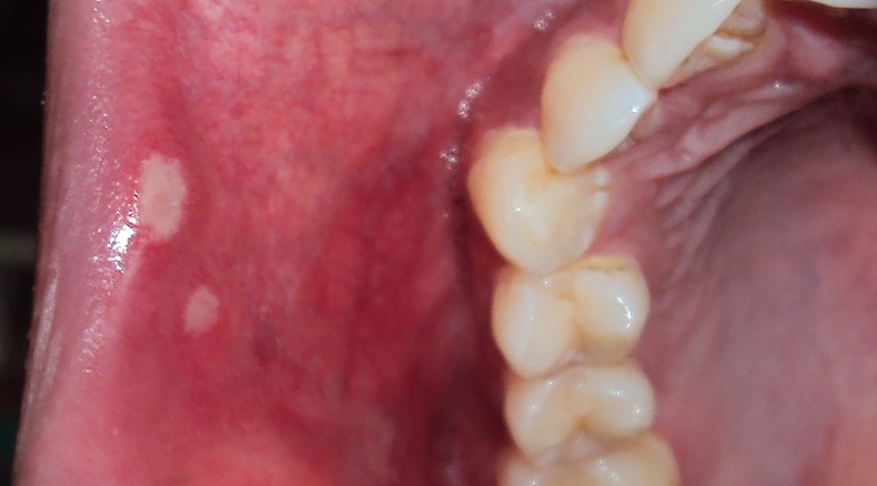
Apthous ulcer on upper labial mucosa.
Investigations
Haemoglobin percentage, total lymphocyte count, differential lymphocyte count (neutrophil, lymphocytes, eosinophils, monocytes, basophills, erythrocyte sedimentation rate, bleeding time, clotting time, plasma glucose random and ELISA tests were done. The haemogram results were within normal limits.
Treatment
A diode laser was used (power output of 0.10 W, 3 J/cm2), in pulsed, non-contact mode, for 1 min per point in single sitting for each of the two ulcers (figure 2). The patient had no discomfort during the laser session. No local or general anaesthesia was given to the patient (figure 3).
Figure 2.

Diode laser 940 nm.
Figure 3.
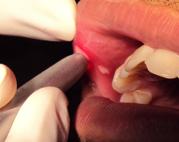
Intraoperative.
Outcome and follow-up
Immediately after therapy, the patient reported no further burning sensation. The healing process was checked 1, 4 and 7 days after therapy and the final evaluation was done 2 months after therapy. The ulcers healed 4 days after therapy (figure 4). No recurrence was present in a follow-up period of 2 months.
Figure 4.

Follow-up (4 days after therapy).
Discussion
The patient with RAS reported severe pain and burning sensation. The conventional strategy is to reduce the pain; however, none of the treatments used to date were effective.4
The mechanism by which diode laser therapy can reduce pain is not fully understood. Some of the explanations for the analgesic effect of laser therapy are as follows: blockage of action potential generation and conduction of nociceptive signals in primary afferent neuron, increase in amount of natural analgesics such as opioid peptides, decrease in the release of chemical substances such as histamine and blockage of acetyl choline, reduction in synthesis of bradykinin and prostaglandin E2, improvement in local microcirculation and supply of oxygen to hypoxic cells and mediation of synaptic gate transfer substance.5
Learning points.
940 nm diode laser therapy relieves pain and burning sensation immediately.
Diode laser therapy is easy and safe, without any potential side effects.
Eliminates the potential adverse effects caused by the drugs.
Footnotes
Competing interests: None.
Patient consent: Obtained.
Provenance and peer review: Not commissioned; externally peer reviewed.
References
- 1.Colvard M, Kuo P. Managing apthous ulcers: laser treatment applied. J Am Dent Assoc 1991;2013:51–3 [DOI] [PubMed] [Google Scholar]
- 2.De Souza TOF, et al. Clinical evaluation of low-level laser treatment for recurring apthous stomatitis. Photomed Laser Surg 2010;2013(s2):1–8 [DOI] [PubMed] [Google Scholar]
- 3.Tezel A, Kara C, Balkaya V, et al. An evaluation of different treatments for recurrent apthous stomatitis and patient perceptions: Nd:YAG laser versus medication. Photomed Laser Surg 2009;2013:101–6 [DOI] [PubMed] [Google Scholar]
- 4.Sharon-Buller A, Sela M. CO2 -laser treatment of ulcerative lesions. Oral Surg Oral Med Oral Pathol Oral Radiol Endod 2004;2013:332–4 [DOI] [PubMed] [Google Scholar]
- 5.Zand N, et al. Relieving pain in minor apthous stomatitis by a single session of non-thermal carbon dioxide laser irradiation. Lasers Med Sci 2009;2013:515–20 [DOI] [PubMed] [Google Scholar]