



Abstract
Toxoplasma is a leading cause of posterior uveitis in immunocompetent patients manifesting as a focal posterior retinochoroiditis. The clinical diagnosis of ocular toxoplasmosis is usually straightforward. There is typically a fluffy white retinal lesion which may lie adjacent to a pigmented chorioretinal scar and a prominent vitreous, or additionally, anterior chamber cellular reaction. Several unusual presentations in ocular toxoplasmosis have been reported, including: papillitis, neuroretinitis, retrobulbar neuritis, retinal detachment and macular oedema. This is a case of presumed primary toxoplasma papillitis in a 14-year-old child with complete absence of vitritis at presentation that made the diagnosis challenging. This evolved into neuroretinitis that resolved upon introducing antitoxoplasma antibiotics.
Background
Toxoplasma papillitis and neuroretinitis are rare and separate entities of ocular toxoplasmosis. Diagnosis of toxoplasma papillitis can be challenging to make as some of these patients lack the typical appearance of toxoplasma chorioretinitis and have foci of inflammation at the immediate edge or within the superficial nerve head. However, the presence of variable vitreous inflammation may be a marker for ocular toxoplasmosis and any degree of vitreous inflammation should prompt a suspicion of toxoplasmosis.
This case demonstrates that the absence of vitreous inflammation does not exclude a diagnosis of ocular toxoplasmosis.
Case presentation
A healthy 14-year-old boy presented with a 1-week history of progressive loss of vision of the right eye. There was no ophthalmic or medical history. He lives with his mother and has a pet cat. On examination he was afebrile and neurological examination was normal. There was no lymphadenopathy. On ocular examination, visual acuity in the right eye was counting fingers (CF) and in the left eye 6/6. A right relative afferent pupillary defect was present. Both eyes were white. Slit lamp examination of the right eye showed a few keratic precipitates on the cornea. There were 2+ cells in the anterior chamber. Posterior segment examination of the right eye revealed a binocular indirect ophthalmoscopy (BIO) score of 0 with no vitreous haze. The optic disc was grossly swollen with a focus of a fluffy white lesion on the inferotemporal margin, sheathing of adjacent arcade vessels and a gross elevation of the peripapillary and posterior pole (figure 1). The peripheral retina was normal. Ocular examination of the left eye was entirely normal. The following laboratory studies were normal: full blood count, erythrocyte sedimentation rate, urea and electrolytes and urine analysis. MRI brain scan and orbit were normal. Patient was admitted with a presumed diagnosis of central retinal vasculitis involving the optic disc and started on intravenous methylprednisolone 1 g/day for 3 days. Three days later, the diagnosis was revisited and toxoplasma screen and cat scratch panel were requested.
Figure 1.
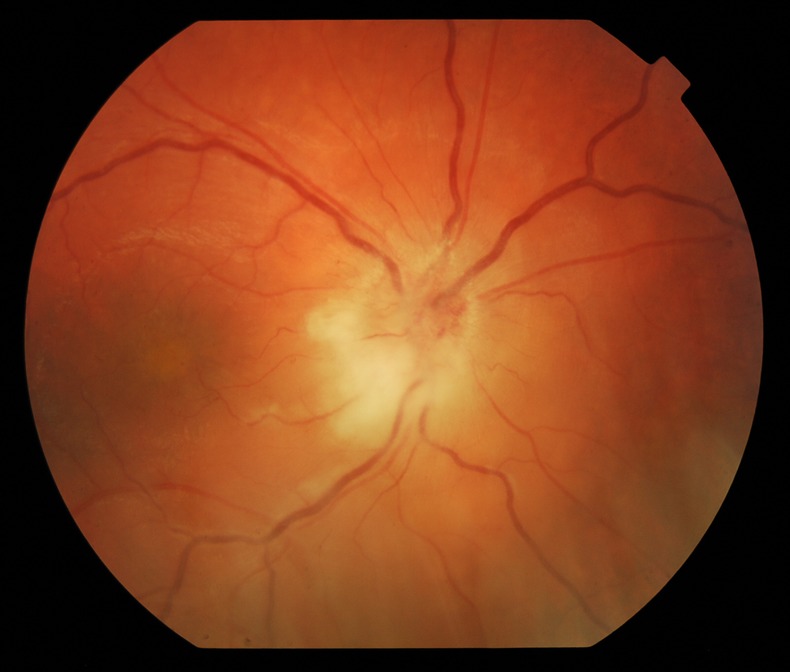
Grossly swollen optic nerve head with a focus of a fluffy white lesion on the inferotemporal margin.
A week later the visual acuity was still CF. The anterior chamber cells had settled, but a new vitritis presented with cells and a BIO score of 2. Optic disc appearance had improved with resolution of the neurosensory detachment but still an inferotemporal area of active retinitis adjacent to the disc and the new appearance of incomplete macular star was noted (figure 2).
Figure 2.
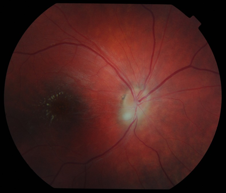
New appearance of incomplete macular star suggesting neuroretinitis.
Toxoplasma dye test showed IgG positivity at 30 IU/ml but negative IgM. Cat scratch serology was negative.
Treatment
Patient was started on the following oral medications: azithromycin 250 mg daily, pyrimethamine 100 mg loading followed by 50 mg daily and Prednisolone 40 mg daily for 1 week, reduced by 10 mg weekly.
Outcome and follow-up
After 10 days of therapy, visual acuity improved slightly to 6/60; with complete recovery of anterior chamber and vitreous inflammation. Pyrimethamine and azithromycin were continued for 6 weeks and steroid treatment was tapered to stop over 8 weeks.
Six months later, the right eye has improved to 6/9. Fundus examination showed complete resolution of the retinitis and a scar with hyperpigmented perimeter. The incomplete macular star had resolved with residual retinal pigment hyper/hypopigmentary changes (figure 3).
Figure 3.
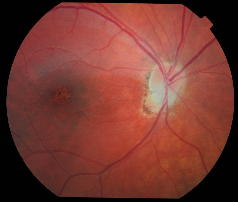
Complete resolution of the retinitis with a pigmented scar. The macular star resolved with retinal pigment epithelial changes.
Discussion
Clinical findings are usually the basis by which the diagnosis of ocular toxoplasmosis is made. The ocular lesion usually presents as a focal necrotising chorioretinitis. Recurrences are common as satellite lesions adjacent to an old atrophic scar but this could be absent in cases with primary acquired disease.
Folk and Lobes1 reported six patients with severe papillitis secondary to toxoplasmosis; nearly all had incorrect initial diagnoses.
The most indicative finding of active ocular toxoplasmosis is vitritis of any intensity. We reviewed the literature1 2 for similar cases of presumed toxoplasma papillitis and found 11 case reports of patients all of which had vitreous inflammation at presentation. Unusually, our patient did not have any signs of vitritis at presentation, which made the diagnosis challenging. One could argue that vitreous inflammation might have been mild and disguised in the presence of anterior chamber cells; however, two senior consultant ophthalmologists have examined the patient and could confirm the complete absence of vitritis.
Although toxoplasmosis is the most common cause of retinitis in children, the differential diagnosis of toxoplasmosis in this age group includes a number of diseases that may lead to posterior uveitis, such as viral infections with herpes simplex and zoster causing acute retinal necrosis (ARN). Bacterial infections such as syphilis and cat scratch disease as well as non-infectious causes as Behçet's disease and lymphoma should also be considered. Retinal lesions in ARN are usually found in the periphery with vitritis. However, primary acquired ocular toxoplasmosis may mimic ARN in immunocompromised patients and also in old age. Steroid treatment without appropriate antimicrobial cover would have been problematic with a diagnosis of ARN as it would have irreversibly caused full thickness retinal necrosis. Syphilis was not considered due to the patient age and history at time of diagnosis and cat scratch serology was negative.
A positive serology for IgG in our case only supports the clinical diagnosis.3 However, laboratory confirmation of a clinical diagnosis of toxoplasmosis is not achieved in up to 40% of cases.4 Nevertheless, the fact that the patient responded well to antitoxoplasma treatment made ocular toxoplasmosis the most likely diagnosis. If our patient had not responded to the treatment, a vitreous tap would have been considered for combined PCR to detect Toxoplasma gondii nucleic acids and Goldmann-Witmer coefficient analysis for intraocular antibody production.5
In clinical practice, corticosteroids are usually given in combination with antibiotics to reduce the inflammatory reaction and to minimise tissue damage, especially with optic disc or macular involvement. However, the use of corticosteroids can lead to the development of fulminant ocular toxoplasmosis if given without antibiotic cover. While some authors6 concluded that corticosteroids should be given only with a shield of antiparasitic drugs, others7 are not particularly concerned about giving corticosteroids to patients with Toxoplasma retinochoroiditis without antimicrobial cover at the initial phase because T gondii are slowly dividing organisms and the laboratory results of these tests usually return in few days.
In summary, we report a case of presumed primary acquired ocular toxoplasmosis presenting with papillitis and no vitritis at presentation. Findings suggestive for ocular toxoplasmosis in this case include uveitis, fluffy white inflammation in the optic nerve head and peripapillary area, and gradual resolution of retinitis over 6 weeks with antitoxoplasma treatment and positive toxoplasmosis serology in a child.
Learning points.
Ocular toxoplasmosis is one of the most common causes of posterior uveitis.
Toxoplasmosis should always be sought in cases of papillitis.
Vitreous inflammation is the most suggestive finding of ocular toxoplasmosis. However, a diagnosis of ocular toxoplasmosis should still be considered even in the complete absence of vitritis.
Visual outcome is strongly influenced by optic disc or macular involvement.
Visual prognosis is generally good and a visual field defect is a common sequelae.
Footnotes
Contributors: This case report was written and researched by MAM, with guidance of MV. The case was managed by MV who had final approval of the version to be published.
Competing interests: None.
Patient consent: Obtained.
Provenance and peer review: Not commissioned; externally peer reviewed.
References
- 1.Folk JC, Lobes LA. Presumed toxoplasmic papillitis. Ophthalmology 1984;2013:64–7 [DOI] [PubMed] [Google Scholar]
- 2.Fish RH, Hoskins JC, Kline LB. Toxoplasma neuroretinitis. Ophthalmology 1993;2013:1177–82 [DOI] [PubMed] [Google Scholar]
- 3.Rahi A, Tabarra K. Laboratory investigations in posterior uveitis. Int Ophthalmol Clin 1995;2013:65–6 [DOI] [PubMed] [Google Scholar]
- 4.Garweg JG, de Groot-Mijnes JD, Montoya JG. Diagnostic approach to ocular toxoplasmosis. Ocul Immunol Inflamm 2011;2013:456. [DOI] [PMC free article] [PubMed] [Google Scholar]
- 5.De Groot-Mijnes JD, Rothova A, Van Loon AM, et al. Polymerase chain reaction and Goldmann-Witmer coefficient analysis are complimentary for the diagnosis of infectious uveitis. Am J Ophthalmol 2006;2013:313–18 [DOI] [PubMed] [Google Scholar]
- 6.Bosch-Driessen EH, Rothova A. Sense and nonsense of corticosteroid administration in the treatment of ocular toxoplasmosis. Commentary. Br J Ophthalmol 1998;2013:858–60 [DOI] [PMC free article] [PubMed] [Google Scholar]
- 7.McDonald HR. Diagnostic and therapeutic challenges. Retina 2007;2013:385–90 [DOI] [PubMed] [Google Scholar]