Abstract
Immune suppressed organ transplant recipients suffer increased morbidity and mortality from primary cutaneous SCC. We studied tumor microenvironment in transplant-associated SCC (TSCC), immune-competent SCC and normal skin by IHC, IF and RT-PCR on surgical discard. We determined T cell polarization in TSCC and SCC by intracellular cytokine staining of T cell crawl outs from human skin explants. We studied the effects of IL-22, an inducer of keratinocyte proliferation, on SCC proliferation in vitro. SCC and TSCC are both associated with significantly higher numbers of CD3+ and CD8+ T cells compared to normal skin. TSCC showed a higher proportion of Foxp3+ T regs to CD8+ T cells compared to SCC and a lower percentage of IFN-γ producing CD4+ T cells. TSCC, however, had a higher percentage of IL-22 producing CD8+ T cells compared to SCC. TSCC showed more diffuse Ki67 and IL-22 receptor (IL-22R) expression by IHC. IL-22 induced SCC proliferation in vitro despite serum starvation. Diminished cytotoxic T cell function in TSCC due to decreased CD8/T-reg ratio may permit tumor progression. Increased IL-22 and IL-22R expression could accelerate tumor growth in transplant patients. IL-22 may be an attractive candidate for targeted therapy of SCC without endangering allograft survival.
Introduction
Cutaneous squamous cell carcinoma (SCC) is the second most common human cancer; in the great majority of cases, excision with clear margins provides cure. In immune suppressed solid organ transplant recipients (OTRs) however, the incidence of SCC is more than 100 times greater than the general population [1]. Furthermore, transplant associated SCCs (TSCCs) are particularly aggressive and OTRs are more susceptible to recurrence and metastasis [2]. Some transplant recipients can develop hundreds of rapidly growing SCCs, resulting in massive local tissue damage. Extensive body surface area involvement also renders surgery, the primary treatment modality, difficult and disfiguring. In the absence of surgery, there are no medical treatments available for SCCs in OTRs, resulting in significant morbidity and mortality shortly after transplantation [2], [3]. Thus, there is a critical need for targeted medical treatments for these aggressive cancers in this patient population.
The immune microenvironment associated with SCC is dynamic, comprised of opposing forces driving tumor promotion and tumor suppression [4], [5], [6], [7], [8]. Regulatory T cells (T regs) and macrophage-derived angiogenic factors may directly support proliferation and invasion by SCC [9], while CD8+ cytotoxic cells and other factors in the adaptive and innate arms of the immune system can protect the host.
We are particularly interested in IL-22 producing T cells in the SCC microenvironment. IL-22 is traditionally thought to be produced by CD4+ helper T lymphocytes (Th) including Th1, Th17, and Th22, however a subset of CD8+ cytotoxic T cells (Tc22) have also been shown to produce this cytokine [10], [11], [12], [13]. IL-22 is involved in inflammatory and wound healing processes and mediates its effects via a heterodimeric receptor that is highly expressed within various tissues [14]. Epithelial cells of the skin and other organs such as the respiratory and digestive tracts are its primary targets. Binding of IL-22 to its receptor results in activation of signaling pathways that lead to induction of genes involved in cell cycle progression and prevention of apoptosis [15]. In psoriasis, a benign inflammatory skin disease characterized by hyperproliferative keratinocytes, IL-22 induces inflammation, mediates keratinocyte proliferation, and inhibits keratinocyte terminal differentiation [16], [17], [18].
In contrast, the role of IL-22 in proliferation and progression of human skin cancers like SCC remains undefined. In the present study, we aimed to establish the role of IL-22 in SCCs in both immune competent and transplant recipients and to evaluate the immune microenvironment for the numbers and polarization states of tumor-associated T cells. We directed our attention to differences between SCC and TSCC in order to gain insight into the mechanisms that drive their vastly disparate clinical behaviors.
Our results show TSCCs, are more proliferative, exhibit a distinct T cell mediated response favoring tumor growth and T cell polarization that favors production of IL-22, and show more diffuse expression of IL-22R. Such findings suggest a model that may account for their clinical presentation; therapeutic intervention directed towards IL-22 could provide a new treatment modality for these highly aggressive and sometimes fatal forms of SCCs.
Results
Transplant Associated SCC (TSCC) is More Proliferative than SCC from Immune Competent Patients
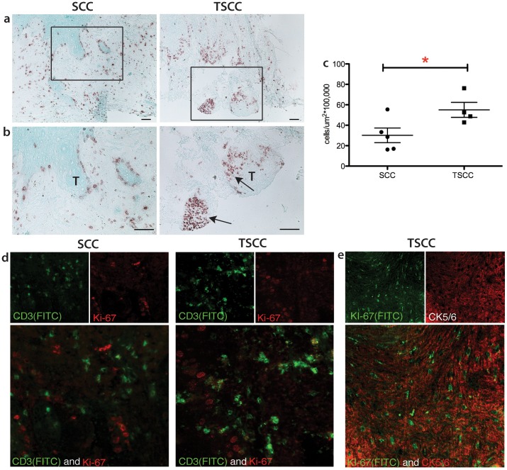
Solid organ transplant recipients are at increased risk for developing cutaneous SCC. SCCs in this group of patients tend to be more numerous, more aggressive and also has increased propensity to grow more rapidly. [19]. Transplant patients included in the study presented met criteria for catastrophic carcinomatosis defined by Berg and Otley in 2002 [3] to include (1) severe field disease; or (2) >10 SCCs excised in a one year time period, or history of in transit, regional or distant metastatic disease. Thus, these patients represented transplant recipients with the most severe skin cancer burden. All tumors specimens evaluate were obtained from AJCC Stage 1 primary cutaneous SCC. [20] We performed immunohistochemical staining for Ki-67, a marker for proliferation, to assess the proliferative rate of SCC versus TSCC. Both SCC and TSCC showed significant proliferative activity (Figure 1a and b). We found that Ki-67 expression is increased approximately 2-fold in TSCC as compared to SCC (55.08±7.3 cells/µm2×105 versus 30.12±7.1 cells/µm2×105 mean ± SEM p<0.05; Figure 1c). Notably, the pattern of Ki-67 expression was also different between these two groups. In SCC, Ki-67+ cells were present along the periphery of tumor nests, i.e. the leading edge of the carcinoma. In contrast, transplant associated SCCs not only showed positivity at the invasive edge; they also demonstrated Ki-67 positivity in aggregates within the tumor (arrows). Double label immunofluorescence showed Ki-67 was positive for the nucleus of cytokeratin 5/6 (CK5/6) positive cancer cells but not CD3 positive cells (Figure 1e and 1d); suggesting proliferating cells are keratinocytes rather than immune cells.
Figure 1. Transplant associated SCC (TSCC) shows diffuse Ki-67 staining and increased numbers of Ki-67+ cells.
Representative immunohistochemistry at (a) 10X and (b) 20X, with (c) mean cell count values of Ki-67+ cells in SCC (n = 5) and TSCC (n = 5). T indicates tumor. Only cells along or within tumors were counted. Asterisks (*) indicate statistical significance, where *P<0.05. Bar = 100 µm. (d) Ki-67 (red) did not colocalize with CD3 (green) in SCC and TSCC. (e) Intracytoplasmic staining of CK5/6 (red) colocalizes with intranuclear staining of KI-67(green).
SCC and TSCC Microenvironments are Characterized by Significantly Higher Numbers of CD3+ and CD8+ T Cells Compared to Normal Skin
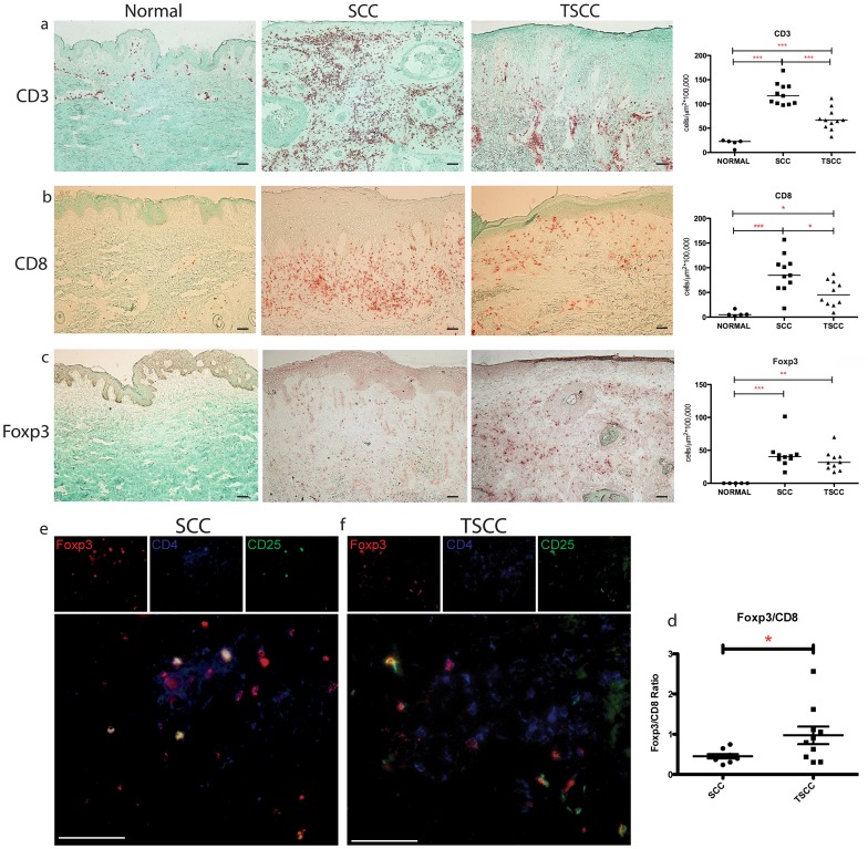
As host immunity can regulate tumor behavior, we set out to characterize the number, type, and distribution of tumor-associated T cells in the SCC and TSCC microenvironment (Figure 2). We found significantly greater numbers of CD3+ T cells associated with SCC and TSCC compared to normal skin (SCC 122.44±7.30 cells/µm2×105 and TSCC 68.73±7.38 cells/µm2×105 vs. Normal 22.56±0.73 cells/µm2×105, mean ± SEM, p<0.01; Figure 2a). There were also significantly greater numbers of CD8+ T cells associated with SCC and TSCC compared to normal skin (SCC 95.70±9.92 cells/µm2×105 and TSCC 48.22±8.38 cells/µm2×105 vs. Normal 6.88±2.56 cells/µm2×105, mean ± SEM, p<0.05; Figure 2b). Both CD3+ and CD8+ T cells were more abundant in SCC compared to TSCC. We also observed that CD3+ and CD8+ T cells predominantly aggregated in the peritumoral regions while relatively few T cells were located within tumor nests.
Figure 2. TSCC shows fewer CD8+ T cells and increased Foxp3/CD8 ratio.
Representative immunohistochemistry and summary data with median cell count values of (a) CD3+ T cells (b) CD8+ T cells (c) Foxp3+ T cells in normal skin (n = 5), SCC (n = 10) and TSCC (n = 10). Each dot represents one patient. Asterisks (*) indicate significance, where *P<0.05, **P<0.01 and ***P<0.005. Bar = 100 µm. Triple label immunofluorescence confirms the presence of CD4+CD25+Foxp3+ T regulatory cells (see arrows) in SCC (e) and TSCC (f). Images are presented in pseudo color: Foxp3 (red), CD4 (blue) and CD25 (green), located above merged image. Red and green overlapping cells appear yellow in color; red and blue appear purple; and green and blue appear aqua; cells labeled with all three stains appear white.
The FoxP3+:CD8+ T Cell Ratio is Higher in TSCC
We wanted to assess whether T-regs are present in TSCC and SCC. We performed IHC for Forkhead box P3 (Foxp3), a known marker for T regs, on human SCC and TSCC. Representative images are shown (Figure 2c). Cell counts for Foxp3 showed significantly increased number of FoxP3+ cells in both SCC and TSCC compared with normal skin (TSCC 34.32±4.89 cells/µm2×105 and SCC 43.72±6.96 cells/µm2×105 vs. none in normal skin, p<0.01). Triple label immunofluorescence confirmed the presence of CD4+CD25+Foxp3+ cells, in both SCC and TSCC tissue (Figure 2e and f). While the absolute numbers of FoxP3+ cells associated with SCC were similar to TSCC, the proportion of FoxP3+cells r to cytotoxic (CD8+) T cells was significantly increased (∼2 fold) in TSCC (TSCC 0.97±0.22 vs. SCC 0.45±0.05, p<0.05; Figure 2d), indicating a tumor permissive environment in TSCC.
Tc22 Cells are Increased and Th1 Cells are Decreased in TSCC
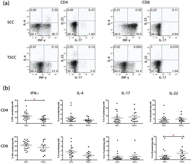
We were interested in T cell polarization in TSCC vs. SCC. Th1 cells, of which IFN-γ+ secreting CD4+ cells are a prototypic example, are known to play a role in anti-tumor immunity [21]. In contrast, IL-22 producing T cells (Th22 or Tc22) can support keratinocyte proliferation and potentially accelerate tumor growth. TSCC has been associated with a Th2 phenotype while keratinocyte proliferation in psoriasis has been linked to Th17 and Th22 phenotype. Thus, Th1, Th2, Th17, Th22 and CD8+ IFN-γ and IL-22 producing T cells might all be important in the TSCC environment. Thus, we evaluated the percentages of Th1 (IFN-γ+/IL-17−), Th2 (IL-4+), Th17 (IL-17+) and Th22 (IL-22+/IL-17−) CD4+ and CD8+ cells from 20 SCC and 12 TSCC explants We collected T cell “crawl-outs” from SCC explants. While, the percentages of Th2 and Th17 cells were comparable in SCC and TSCC, we found that TSCC was associated with an increased percentage of IL-22 producing CD8+ T cells (TSCC 4.2% ±1.2% vs. SCC 2.2% ±0.75%, p<0.05) and decreased numbers of CD4+ Th1 T cells (TSCC 15.1% ±2.3% vs. SCC 25.1% ±3.2%, p<0.05; Figure 3). The decreased percentage of Th1 cells may skew the balance of pro- and anti-tumor forces toward a “tumor permissive” environment. Increased IL-22 producing cells might contribute to enhanced proliferation of TSCC.
Figure 3. TSCC shows increased Tc22 and decreased Th1 polarization.
T cell “crawl outs” were activated and intracellular cytokines stained. Live CD3+CD4+ and CD3+CD8+ cells were gated, and then frequencies of the cells producing indicated cytokines were analyzed. (a) Representative dot plot analysis of IFN-γ, IL-4, IL-17, and IL-22 expression in CD4+ and CD8+ T cells from SCC specimens. Numbers indicate percent gated cells. (b) Summary results from 20 SCC and 12 TSCC patients. The Mann-Whitney U-test was used for the statistical comparison between two groups. Asterisks (*) indicate statistical significance (p<0.05).
The Expression of IL-22 and Related Cytokines is Increased in SCCs, TSCCs and their Adjacent Peritumoral Skin
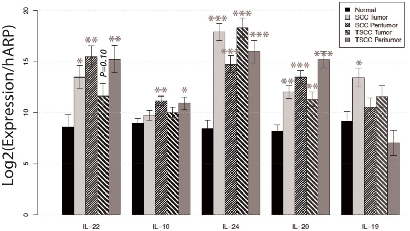
To further assess the expression of IL-22 within the tissue, we performed reverse transcriptase-PCR (RT-PCR) on mRNA extracted from SCC, TSCC, their respective adjacent non-tumor bearing skin and normal skin from healthy volunteers. RT-PCR analysis showed that mean IL-22 mRNA expression was increased approximately 30-fold in SCC and 8-fold in TSCC compared to normal skin (p<0.05; Figure 4). Even more striking, there was significantly increased expression of IL-22 in the peritumoral regions of SCC and TSCC compared to normal skin. IL-22 mRNA was increased 111-fold in SCC and 97-fold in TSCC peritumoral skin. In general, we found that the entire family of IL-10 cytokines including IL-10, IL-19, IL-20, IL-22 and IL-24 was increased in tumor compared to normal skin.
Figure 4. IL-22 expression is increased in SCC, TSCC, and juxtatumoral skin.
The relative mRNA expression of IL-22 relative to Human Acidic Ribosomal Protein (HARP) in normal (n = 9), SCC (n = 9), SCC peritumoral (n = 9), TSCC (n = 7), and TSCC peritumoral tissue (n = 7). Data expressed as mean relative mRNA expression ± standard error. Asterisks (*) indicate statistical significance, where *P<0.05.
IL-22 Receptor and pSTAT-3 are Upregulated in TSCC and SCC
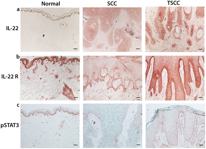
Binding of IL-22 to its receptor results in tyrosine phosphorylation of JAK and subsequent activation of downstream signal-transducer-and-activator (STAT) molecules, of which STAT-3 is the principal mediator [22], [23], [24]. Particularly, STAT-3 has been implicated in both the initiation and promotion stages of cutaneous carcinogenesis, regulating keratinocyte survival and proliferation following ultraviolet irradiation [25], [26], [27], [28].To investigate if this pathway is upregulated in SCCs, we stained SCC (n = 5), TSCC (n = 5), and normal skin (n = 5) for IL-22, IL-22 receptor and phosphorylated STAT-3 (pSTAT-3). Representative images are shown (Figure 5). We found diffuse expression of IL-22 in transplant SCC and SCC from immune competent patients. We also found increased expression of IL-22 receptor at the leading edge of the invasive front in SCC (Figure 5a); this was in contrast to TSCC, which showed diffuse IL-22 receptor expression (Figure 5b). Diffuse IL-22 receptor expression mirrored diffuse expression of Ki67 expression in TSCC (Figure 1). pSTAT-3 is increased in all tumor samples compared to normal skin (Figure 5c).
Figure 5. IL-22, IL-22R and downstream regulator pSTAT3 are upregulated in SCC and TSCC.
Expression of (a) IL-22, (b) IL-22 Receptor (IL22R) and (c) downstream molecule Phosphorylated Signal-transducer-and-activator of transcriptase 3 (pSTAT3) were all increased in tumor tissue compared to normal skin by immunohistochemistry. Bar = 100 µm.
IL-22 Enhances the Proliferation of Human Cutaneous SCC
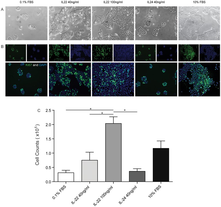
Since IL-22 drives proliferation in normal keratinocytes in culture, we wanted to investigate whether IL-22 can induce proliferation of SCCs. A431 SCC cells were cultured for 48 hours with complete medium (CM) or under serum starvation conditions with or without the indicated cytokines. Culture with CM without IL-22 or IL-24 resulted in robust proliferation of A431 cells. When A431 were serum starved (0.1%FBS) they did not proliferate. Addition of IL-22 (40 or 100 ng/ml) rescue of serum starved A431 cells. A431 cells formed larger colonies when treated with IL-22 compared to the cells in 0.1% FBS or IL-24 as visualized with light microscopy (Figure 6A). Ki67 staining confirmed the hyperproliferative state of A431 cells cultured with IL-22 (Figure 6B). Ki67+ cells were predominantly localized at the periphery of the colony in slow growth conditions, whereas those cells were also found within and throughout the colony in rapid proliferation conditions. These observations correspond to our findings with TSCC (Figure 1) where we found Ki67+ cells within tumor nests. Finally, addition of IL-22 at 100 ng/ml resulted in significant increase of proliferation of A431 cells by approximately 8-fold compared to growth of serum starved cells (p<0.05). These results suggest that IL-22 might play a role in driving SCC proliferation in transplant patients.
Figure 6. IL 22 increases the proliferation of human cutaneous SCC in vitro.
A431 cells were cultured in full media (10% FBS) or in serum starvation media (0.1% FBS) with or without the addition of the indicated cytokines for 72 hours. (a) Cells cultured in full media, and in starvation media supplemented with IL-22 (40 ng/ml and 100 ng/ml) show considerably greater proliferative behavior with increased colony formation when compared to those grown in starvation media alone or supplemented with IL-24 (40 ng/ml). (b) Representative images of IF staining using the proliferation marker Ki-67 (green) and the nuclear stain DAPI (blue). Cells grown in full media, as well as those treated with IL-22 (40 ng/ml and 100 ng/ml) show an increased number of proliferating nuclei when compared to those grown in starvation media alone or supplemented with IL-24 (40 ng/ml). Additionally, they demonstrate a more disorganized pattern of proliferation, with KI67+ cells no longer limited to the periphery, but rather seen throughout the tumor colonies. (c) Cell counts were performed after 72 hours of cultivation in the indicated conditions. The addition of 100 ng/ml IL-22 to the starvation media (0.1% FBS) resulted in a hyperproliferation of tumor cells, yielding significantly greater cell numbers when compared to those grown in serum starvation alone, or serum starvation supplemented with IL-22 (40 ng/ml) or IL-24 (40 ng/ml) (one-way ANOVA, p<0.001).
Discussion
Over 250,000 organ transplant recipients (OTRs) currently live in the United States and approximately 30,000 transplant surgeries will be performed this year [29]. As these patients live longer, morbidity may result not from the transplant itself, but from other disease processes. The increased incidence of skin cancer, especially squamous cell carcinoma (SCC), is well-known [1], [19], [30], [31], [32], [33]. However, few studies to date have attempted to explain the more aggressive course of SCCs in the transplant population in specific terms. In this report, we endeavored to do that by examining the differences between SCCs in immune competent patients and in transplant recipients at the cellular level. Our results demonstrated evidence of increased proliferative activity in TSCC compared to SCC and an altered immune microenvironment. These findings enable us to propose a model that may help explain the rapidly growing and aggressive nature of TSCC.
A general assessment of the tumors’ relative proliferative activity was first performed using Ki-67. This marker has previously been shown to correlate with tumor grade, and can help identify aggressive carcinomas [34], and is a strong prognostic indicator for melanoma [35]. We discovered a two-fold increase in Ki-67 positivity in TSCC compared to SCC, which corresponds to high risk clinical behavior. Unexpectedly, we also found a distinct pattern of Ki-67 expression in TSCCs. Previous reports described Ki-67positivity along the periphery of SCC tumor nests [36]. However, our IHC staining of TSCC showed Ki-67 positivity not only at the invasive edge but also within the tumor nodule. Double label immunofluorescence for keratinocyte markers CK5/6 and Ki-67 confirmed that the proliferating cells were indeed SCC cells (Figure 1e). These findings suggest that more proliferation occurs in tumors from transplant patients than in immune-competent patients. Furthermore, a lack of co-localization of Ki-67 with pan-T cell marker CD3 may indicate that T cells are not proliferating locally.
A significant finding in our current study is that TSCCs have significantly increased IL-22 producing CD8+ cytotoxic T cells (Tc) compared to SCCs in the immune competent group. While IL-22 has been implicated in psoriasis [10], [37], [38], [39], a benign hyperproliferative keratinocyte disease that shares gene expression patterns with SCC [40], mounting evidence suggests that IL-22 may also be implicated in malignant processes. IL-22 has been shown to support the growth of mantle cell lymphoma [41], anaplastic large cell lymphoma [42], hepatocellular carcinoma [43] and colon carcinoma through STAT3 activation [44], [45]. Most recently, IL-22 demonstrated pro-tumoral activity in human pancreatic cancer cell line via inducing the expression of vascular endothelial growth factor (VEGF) and anti-apoptotic factor Bcl-XL [46]. The function of IL-22 in cutaneous SCC, however, has not been identified yet. Our findings of increased gene expression of IL-22 and its related family of cytokines in tumor tissue, as well as increased protein expression of IL-22, IL-22R, and the downstream modulator pSTAT-3, all reinforce the importance of the IL-22 pathway in TSCCs. Differences between the SCC and TSCC microenvironments may translate exponentially when one considers the difference in disease burden between the two groups. An immune competent patient ordinarily presents with a single SCC whereas transplant patients with catastrophic SCC present with tens to hundreds of lesions comprising upward of 50% body surface area. In this clinical presentation, increased IL-22 production by Tc22 cells and increased IL-22 receptor density set the stage for sustained accelerated carcinomatosis in transplant patients with catastrophic disease.
Our functional studies provide evidence that IL-22 augments the proliferation of a human cutaneous SCC cell line (A431) in culture in a dose-dependent manner. In fact, we observed IL-22-driven proliferation of SCC cells was most pronounced under starvation conditions (Figure 6). This supports the hypothesis that IL-22 in the tumor microenvironment might drive SCC proliferation under high metabolic demand associated with tumor growth and diminished enrichment of the environment secondary to tissue necrosis.
IL-22 may also have other effects in addition to its role in proliferation. One study revealed IL-22 can augment production of immunosuppressive cytokines, and diminish T cell production of interferon (IFN)-γ, the prototypical Th1 cytokine [46]. Th1 cells support Tc and NK cell anti-tumor responses, and in some cases drive anti-tumor immunity in the absence of Tc cells. Thus, Th1 and Tc responses are key mediators of anti-tumor immunity and are likely important in generating responses against SCC. Although we have not directly tested IL-22’s effect on IFN-γ production in SCC, our results also reveal a decrease in percentage of IFN-γ producing T cells in transplant recipients.
Within the tumor milieu, other factors may additionally contribute to the aggressive nature of TSCC. Typically, anti-tumor response involves processing tumor-associated antigens and the subsequent generation of Th and cytotoxic Tc cells [6], [8]. CD4+ T cells prime tumor-specific Tc cells so that these CD8+ T cells can directly lyse tumor cells [47]. Since T cell mediated immunity is central to controlling tumor growth, we characterized and compared tumor-associated T cells in SCC, TSCC and normal skin. Our results show fewer T cells, particularly CD8+ T cells, in our TSCC samples compared to SCC samples, supporting the recent report that decreased numbers of tumor-infiltrating CD8+ T cells is associated with aggressive tumor phenotypes of lymph node metastasis [48].
We also found more Foxp3+ cells in tumor tissue compared to normal skin. Foxp3+ include T reg cells which are known to promote immune tolerance [49]. While crucial for preventing autoimmunity [50], T regs may also suppress beneficial anti-tumor immunity [51], [52] and aid in evasion of immune surveillance. Specifically, they can regulate the immune response by suppressing the proliferation and cytokine production of effector T cells [53], [54]. Studies have also shown that an increased presence of T regs is correlated with poorer prognosis and decreased survival rates in gastric [51], breast [55] and ovarian carcinomas [56], [57]. Notably, we showed that TSCCs contain a higher ratio of T regs to Tc cells compared to SCC. This disruption of the T reg to Tc cell balance may result in a compromised ability to mount an anti-tumor response, contributing to the aggressive nature of TSCC.
A question that remains to be addressed is what causes the disparate T cell and cytokine expression profiles between SCC and TSCC. An obvious difference between these patients is iatrogenic immunosuppression. Although the nature of this study does not allow correlation of results with particular drugs and dosages, in general, OTRs are placed on high-dose immunosuppressive drugs, such as cyclosporine A and azathioprine. A number of studies have suggested immunosuppressive medications contribute to an increased risk for skin cancer through both direct carcinogenic effects as well as decreased immunosurveillance [58], [59]. However, the effects and degree of immune cell alterations by these drugs and their mechanisms of action remain an active area of research. In general, the incidence of skin cancer is proportional to the level of immune suppression, as CD4 counts are significantly lower in OTR with SCC versus those patients without such malignancy [59].
An important implication of our study is in the treatment of aggressive SCCs, especially in the transplant population. Currently, many transplant recipients exhibit catastrophic carcinomatosis that is beyond conventional surgery. Since there are also no effective medical treatments for these patients, one option is to remove the patient from immune suppression. Although possible in renal transplant patients, this also necessitates a return to dialysis as the graft often fails and this drastically diminishes their life expectancy over time [2], [3]. In addition, this strategy cannot be used with heart, heart-lung, or liver transplant recipients [3]. Prior efforts at immune based treatments have also been largely unsuccessful because such strategies are centered on using myeloid dendritic cells (mDC) to induce Th1 type responses. In transplant patients in particular, Th1 induction is associated with graft rejection, rendering this approach unsuitable for SCCs in OTRs. Our previous studies showed that mDCs from SCC do not stimulate T cell proliferation [60], thus making the success of this approach less attractive. Therefore, we believe targeting the IL-22 pathway may prove to be a useful therapy for TSCCs, while sparing the transplanted organ by continuing suppression of the autoimmune response.
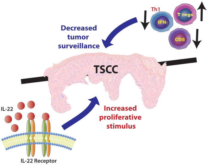
In summary, our findings provide strong evidence that TSCC has a unique immune microenvironment that is conducive to tumor proliferation as shown schematically in Figure 7. The aggressive nature of TSCC may result from at least two opposing forces: a) increased T regs and decreased CD8+ T cells, leading to decreased immune surveillance, and b) increased exposure to IL-22, which may provide a proliferative stimulus and accelerate tumor growth. Although further work elucidating the role of IL-22 in SCC proliferation and invasion is warranted, our study sheds light upon the possible role of IL-22 in tumorigenesis in vivo and provides important information about tumor immunity in transplant recipients. Furthermore, this study suggests that targeting the IL-22 pathway may be an important, life-saving therapeutic approach for aggressive SCCs in the transplant population.
Figure 7. The proposed model of accelerated development of TSCC.
An increased proportion of T regs, combined with decreased numbers of CD8+ and IFN-γ producing T cells, leads to decreased tumor surveillance. Greater percentage of IL-22 producing T cells suggests an increased proliferation stimulus. The overall imbalance could explain the rapidly proliferative nature of TSCC. IL-22 blockade may be an attractive candidate for targeted SCC therapy, especially in the transplant population.
Materials and Methods
Institutional Review Board approval was obtained before enrolling patients to participate in this study. Institutional Review Board approval at NYU Langone Rockefeller and Weill- Cornell was obtained before enrolling patients to participate in this study. Written informed consent was obtained before their participation, and the study was performed with strict adherence to the Declaration of Helsinki Principles.
Skin Samples used in the Study
For immunohistochemistry and immunofluorescence, cutaneous stage 1 SCC and transplant-associated SCC samples were obtained during Mohs micrographic surgery. Tumors were obtained from sun-exposed regions of the face, head and neck. Normal specimens were obtained by 3-mm punch biopsies from non-sun exposed areas of donors without skin cancer. For flow cytometry, tumor samples were obtained from debulking prior to Mohs micrographic surgery (MMS).
Immunohistochemistry
Standard procedures were used for immunohistochemistry as previously described [61]. Briefly, frozen tissue sections of normal skin (n = 5) and SCCs (n = 5–10) and TSCC (n = 10) were stained with the following antibodies: CD3, CD8 (BD Pharmingen, San Diego, CA diluted at 1∶100), Foxp3 (Abcam, Cambridge, MA diluted 1∶40), IL-22 (R&D systems; 1∶25), IL-22 Receptor (Prosci; 1∶250), KI-67 (Santa Cruz Biotechnology; 1∶50), pSTAT3 (Santa Cruz Biotechnology; 1∶50). Biotin-labeled horse anti-mouse antibody (Vector Laboratories, Burlingame, CA) was amplified with avidin-biotin complex (Vector Laboratories) and developed with chromogen 3-amino-9-ethylcarbazole (Sigma Aldrich, St. Louis, MO). Counterstaining was carried out with light green (Sigma-Aldrich). Appropriate isotype controls were performed with each immunohistochemistry experiment. Positive cells were counted in the dermis around SCC tumor nests using NIH IMAGE J software (Bethesda, MD), and cell counts per unit area (µm2×100,000) were determined [62].
Immunofluorescence
Standard procedures were used for immunofluorescence as previously described (Fuentes-Duculan et al, 2010). Frozen skin sections from SCC and TSCC lesions (n = 3–5) and normal skin (n = 3) were fixed with acetone and blocked with 10% normal goat serum (Vector laboratories) for 30 minutes. Primary antibodies Foxp3, Ki-67, Cytokeratin 5/6 (Table S1) were incubated overnight at 4°C and amplified with the appropriate secondary antibody (goat anti-mouse IgG1 conjugated to Alexa Fluor 488 or 568) for 30 minutes at room temperature. Sections were then blocked with 10% normal mouse serum for 30 minutes and the second primary antibodies (CD4(647), CD25(FITC), CD3(FITC), Ki-67(FITC)) were incubated at 4°C overnight and amplified with goat anti-FITC conjugated to Alexa Fluor 488 the next day. Images were acquired using appropriate filters of a Zeiss Axioplan 2 widefield fluorescence microscope with a Plan Neofluar 20×0.7 numerical aperture lens (Carl Zeiss Microimaging Inc., Thornwood, NY) and a Hamamatsu Orca ER CCD camera (Hamamatsu, Bridgewater, NJ) controlled by MetaVue software (MDS Analytical Technologies, Downington, PA). Images in each figure are presented both as single color stains (green and red or green, red and blue) located above the merged image, so that localization of two markers on similar or different cells can be appreciated. Cells that co-express the two markers (green and red) are yellow in color, while cells that co-express the three markers (green, red and blue) appear as white. A white line denotes the dermoepidermal junction. Dermal collagen fibers gave green autofluorescence, and antibodies conjugated with a fluorochrome often gave background epidermal fluorescence. Size bar = 100 µm.
Skin Preparation and Flow Cytometry
SCC (n = 20) and TSCC (n = 12) specimens were obtained by Mohs micrographic surgery. Surgical discard for ex-vivo T cell phenotype analysis was obtained from 12 tumors from 6 transplant recipients. Each of these was a renal transplant recipient. Four of the 6 were on a regimen that included cyclosporine. Five were on more than one drug. The mean age at transplant was 36. Mean time on immune suppression at time of surgery was 21.6 years. Data are summarized in Table S1. Samples were cultured overnight in 2.4 U/ml Dispase II (Roche Diagnostics, Mannheim, Germany) at 4°C to separate the epidermis and dermis. T cells were obtained by culturing the dermis in RPMI 1640 (Invitrogen) supplemented with 5% pooled human serum (Mediatech Inc., Manassas, VA), 0.1% gentamicin (Invitrogen), and 1% 1 M HEPES buffer (Sigma Aldrich, St Louis, MO) for 48 hours at 37°C and allowing them to spontaneously “crawl-out” into culture. Thereafter, to obtain cells emigrated from the dermis, the supernatants were collected and filtered with 40 µm pore nylon cell strainers. Emigrated cells were activated for 4 hours using 25 ng/ml phorbol myristate acetate and 2 µg/ml ionomycin, in the presence of 10 µg/ml brefeldin A (all from Sigma Aldrich, St Louis, MO). EDTA (2 mM; Fisher Scientific, Pittsburg, PA) was added for 10 minutes on ice to stop activation. Cells were then incubated in aqua marina live/dead dye (Invitrogen) on ice for 30 minutes for dead cell discrimination and subsequently fixed with 4% paraformaldehyde (BD Biosciences) on ice for 20 minutes. The cells were permeabilized in FACSPerm (BD Biosciences), blocked in 1∶50 mouse serum (BD Biosciences), and incubated for 30 minutes on ice with the following anti-human, mouse monoclonal antibodies: CD3-Pacific Blue (eBioscience), CD4-Phycoerythrin-Cy7, CD8-PerCp-Cy5.5, IFN-γ-Alexa Fluor 700 (BD Pharmingen), IL-4-Phycoerythrin (BD Pharmingen), IL-17-Alexa Fluor 488 (eBioscience), and IL-22-Allophycocyanin (R&D Systems). Detailed information of antibodies used is described in Table S1. After incubation, cells were washed twice and collected. Samples were acquired using an LSR-II flow cytometer (BD Biosciences) and analyzed with FlowJo software (TreeStar Inc., Ashland, OR). Live CD3+CD4+ and CD3+CD8+ cells were first gated and then the frequencies of the cells producing indicated cytokines were analyzed.
RNA Isolation
Total RNA isolation was carried out as described earlier [60], [61] Briefly, SCC (n = 9) and TSCC (n = 7) tumor samples were removed at Mohs micrographic surgery, and patient-matched, site-matched peritumoral skin were obtained at the time of repair after clear margins were achieved. Normal skin was obtained from normal volunteers (n = 9). All samples were snap-frozen and stored in liquid nitrogen. Individual frozen samples were placed in 1 ml of room temperature RLT Lysis buffer with 1% β-mercaptoethanol (Qiagen, Valencia, CA) and immediately homogenized at full power for 30 seconds using a PowerGen 1000 homogenizer (Fisher Scientific, Pittsburgh, PA). Homogenates were sonicated on ice for 20 seconds at full power. DNA was removed with on-column DNase digestion using an RNase-free DNase Set (Qiagen). RNA was isolated using the RNeasy Mini Kit (Qiagen) according to manufacturer’s recommendations. Total RNA concentration and purity was evaluated using an Ultraspec 2100 prospectrophotometer (Amersham Biosciences).
RT-PCR
The primer and probe used for IL-10 (Hs00961622_m1), IL-19(Hs00604657_m1), IL-20 (Hs00218888_m1), IL-22 (Hs01574154_m1) and IL-24 (Hs01114274_m1) were from Applied Biosystems (Foster City, CA). The sequences of the primers and probe for human acidic ribosomal protein (HARP) are: HARP-forward, CGCTGCTGAACATGCTCAA, HARP-ß reverse, TGTCGAACACCTGCTGGATG; HARP-probe, 6-FAM TCCCCCTTCTCCTTTGGGCTGG-TAMRA (GenBank accession no. NM-001002). The RT-PCR reaction was carried out using 10 ng total RNA and EZ PCR Core Reagents (Applied Biosystems) according to the manufacturer’s directions. The samples were amplified and quantified on an Applied Biosystems PRISM 7900 HT using the following thermal cycler conditions: 2 minutes at 50°C, 30 minutes at 60°C, 5 minutes 95°C; and 40 cycles of 15 seconds at 95°C followed by 60 seconds at 60°C. Each sample and gene was normalized to the hARP gene, a housekeeping gene.
Cell Culture and Cell Staining
A Cutaneous SCC cell line A-431 (ATCC) was cultured in DMEM supplemented with 0.1% FBS (starvation medium) at 37°C for 48 hours to synchronize cellular proliferation at G0 phase. Subsequently, cells were grown in one of the following five conditions: 1) 0.1% FBS 2) 0.1% FBS +40 ng/mL IL-22 3) 0.1% FBS +100 ng/mL IL-22 4) 0.1% FBS +40 ng/mL IL-24 and 5) 10% FBS (Full growth Media). Both IL-22 and IL-24 were reconstituted in PBS prior to administration according to the manufacturer’s protocol. After 72 hours, the cells were collected for either immunofluorescence staining or cell counting. Immunofluorescence procedure was similar to that outlined in the paragraph above, with the use of proliferation marker Ki-67 as the primary antibody conjugated to Alexa Fluor 488. DAPI was used to counterstain cell nuclei. Images were presented as both single color stains (green or blue) as well as a merged image in order to highlight the degree and distribution of proliferating cells. For cell counting, cells were harvested by trypsinization and counted using an automated cell counter (Countess Invitrogen Life Technologies).
Statistical Analysis
Unless otherwise specified, statistical analyses were performed using GraphPad Prism software. Comparisons of cell counts were performed using a two-tailed, Student’s t-test, with p<0.05 considered significant. The Mann-Whitney U-test was used for the statistical comparison of flow cytometry data. Averaged results of multiple experiments are presented as the arithmetic mean ± SEM. RT-PCR values were normalized to hARP and the log2-transformed. Here we have repeated measures experiments with repeated measures (Tissue, LS and NL) within two groups of patients defined by Immune Status (Immune Compromised or Not). A mixed effect model was used with fixed effect Tissue+ImmStatus:Tissue and random intercept for patients within Immune Status (1|ImmStatus/Patient).
Supporting Information
Antibodies used for flow cytometry.
(PDF)
Transplant patient characteristics.
(PDF)
Funding Statement
This work was supported in part by the Dana Foundation Human Immunology Consortium Grant (JAC, JSP). HM, MSF, CQFW, and JGK were supported by the Milstein Medical Program and in part by the National Institutes of Health (NIH) grant 8 KL2 TR000151. JFD is supported in part by grant # 8 UL1 TR000043 from the National Center for Research Resources, NIH. JG is supported by the NIH grant 8 UL1b TR000043 from National Center for Advancing Translational Sciences. The funders had no role in study design, data collection and analysis, decision to publish, or preparation of the manuscript.
References
- 1. Lindelof B, Sigurgeirsson B, Gabel H, Stern RS (2000) Incidence of skin cancer in 5356 patients following organ transplantation. Br J Dermatol 143: 513–519. [PubMed] [Google Scholar]
- 2. Carucci JA (2004) Cutaneous oncology in organ transplant recipients: meeting the challenge of squamous cell carcinoma. J Invest Dermatol 123: 809–816. [DOI] [PubMed] [Google Scholar]
- 3.Berg D, Otley CC (2002) Skin cancer in organ transplant recipients: Epidemiology, pathogenesis, and management. J Am Acad Dermatol 47: 1–17; quiz 18–20. [DOI] [PubMed]
- 4. Kretz-Rommel A, Qin F, Dakappagari N, Ravey EP, McWhirter J, et al. (2007) CD200 expression on tumor cells suppresses antitumor immunity: new approaches to cancer immunotherapy. J Immunol 178: 5595–5605. [DOI] [PubMed] [Google Scholar]
- 5. Mellman I, Coukos G, Dranoff G (2011) Cancer immunotherapy comes of age. Nature 480: 480–489. [DOI] [PMC free article] [PubMed] [Google Scholar]
- 6. Rosenberg SA (2005) Cancer immunotherapy comes of age. Nature clinical practice Oncology 2: 115. [DOI] [PMC free article] [PubMed] [Google Scholar]
- 7. Tamai H, Watanabe S, Zheng R, Deguchi K, Cohen PA, et al. (2008) Effective treatment of spontaneous metastases derived from a poorly immunogenic murine mammary carcinoma by combined dendritic-tumor hybrid vaccination and adoptive transfer of sensitized T cells. Clin Immunol 127: 66–77. [DOI] [PubMed] [Google Scholar]
- 8. Topalian SL, Weiner GJ, Pardoll DM (2011) Cancer immunotherapy comes of age. Journal of clinical oncology : official journal of the American Society of Clinical Oncology 29: 4828–4836. [DOI] [PMC free article] [PubMed] [Google Scholar]
- 9. Pettersen JS, Fuentes-Duculan J, Suarez-Farinas M, Pierson KC, Pitts-Kiefer A, et al. (2011) Tumor-associated macrophages in the cutaneous SCC microenvironment are heterogeneously activated. J Invest Dermatol 131: 1322–1330. [DOI] [PMC free article] [PubMed] [Google Scholar]
- 10. Wolk K, Witte E, Witte K, Warszawska K, Sabat R (2010) Biology of interleukin-22. Seminars in immunopathology 32: 17–31. [DOI] [PubMed] [Google Scholar]
- 11. Res PC, Piskin G, de Boer OJ, van der Loos CM, Teeling P, et al. (2010) Overrepresentation of IL-17A and IL-22 producing CD8 T cells in lesional skin suggests their involvement in the pathogenesis of psoriasis. PLoS One 5: e14108. [DOI] [PMC free article] [PubMed] [Google Scholar]
- 12. Chung Y, Yang X, Chang SH, Ma L, Tian Q, et al. (2006) Expression and regulation of IL-22 in the IL-17-producing CD4+ T lymphocytes. Cell Res 16: 902–907. [DOI] [PubMed] [Google Scholar]
- 13.Nograles KE, Zaba LC, Shemer A, Fuentes-Duculan J, Cardinale I, et al.. (2009) IL-22-producing “T22” T cells account for upregulated IL-22 in atopic dermatitis despite reduced IL-17-producing TH17 T cells. J Allergy Clin Immunol 123: 1244–1252 e1242. [DOI] [PMC free article] [PubMed]
- 14. Xie MH, Aggarwal S, Ho WH, Foster J, Zhang Z, et al. (2000) Interleukin (IL)-22, a novel human cytokine that signals through the interferon receptor-related proteins CRF2–4 and IL-22R. J Biol Chem 275: 31335–31339. [DOI] [PubMed] [Google Scholar]
- 15. Pan H, Hong F, Radaeva S, Gao B (2004) Hydrodynamic gene delivery of interleukin-22 protects the mouse liver from concanavalin A-, carbon tetrachloride-, and Fas ligand-induced injury via activation of STAT3. Cell Mol Immunol 1: 43–49. [PubMed] [Google Scholar]
- 16. Wolk K, Witte E, Wallace E, Docke WD, Kunz S, et al. (2006) IL-22 regulates the expression of genes responsible for antimicrobial defense, cellular differentiation, and mobility in keratinocytes: a potential role in psoriasis. Eur J Immunol 36: 1309–1323. [DOI] [PubMed] [Google Scholar]
- 17. Boniface K, Guignouard E, Pedretti N, Garcia M, Delwail A, et al. (2007) A role for T cell-derived interleukin 22 in psoriatic skin inflammation. Clin Exp Immunol 150: 407–415. [DOI] [PMC free article] [PubMed] [Google Scholar]
- 18. Zheng Y, Danilenko DM, Valdez P, Kasman I, Eastham-Anderson J, et al. (2007) Interleukin-22, a T(H)17 cytokine, mediates IL-23-induced dermal inflammation and acanthosis. Nature 445: 648–651. [DOI] [PubMed] [Google Scholar]
- 19.Krynitz B, Edgren G, Lindelof B, Baecklund E, Brattstrom C, et al.. (2012) Risk of skin cancer and other malignancies in kidney, liver, heart and lung transplant recipients 1970 to 2008-A Swedish population-based study. Int J Cancer. [DOI] [PubMed]
- 20. Warner CL, Cockerell CJ (2011) The new seventh edition American Joint Committee on Cancer staging of cutaneous non-melanoma skin cancer: a critical review. American journal of clinical dermatology 12: 147–154. [DOI] [PubMed] [Google Scholar]
- 21. Ikeda H, Old LJ, Schreiber RD (2002) The roles of IFN gamma in protection against tumor development and cancer immunoediting. Cytokine Growth Factor Rev 13: 95–109. [DOI] [PubMed] [Google Scholar]
- 22. Dumoutier L, Van Roost E, Ameye G, Michaux L, Renauld JC (2000) IL-TIF/IL-22: genomic organization and mapping of the human and mouse genes. Genes Immun 1: 488–494. [DOI] [PubMed] [Google Scholar]
- 23. Dumoutier L, Louahed J, Renauld JC (2000) Cloning and characterization of IL-10-related T cell-derived inducible factor (IL-TIF), a novel cytokine structurally related to IL-10 and inducible by IL-9. J Immunol 164: 1814–1819. [DOI] [PubMed] [Google Scholar]
- 24. Levy DE, Darnell JE Jr (2002) Stats: transcriptional control and biological impact. Nat Rev Mol Cell Biol 3: 651–662. [DOI] [PubMed] [Google Scholar]
- 25. Chan KS, Sano S, Kiguchi K, Anders J, Komazawa N, et al. (2004) Disruption of Stat3 reveals a critical role in both the initiation and the promotion stages of epithelial carcinogenesis. J Clin Invest 114: 720–728. [DOI] [PMC free article] [PubMed] [Google Scholar]
- 26. Sano S, Chan KS, Kira M, Kataoka K, Takagi S, et al. (2005) Signal transducer and activator of transcription 3 is a key regulator of keratinocyte survival and proliferation following UV irradiation. Cancer Res 65: 5720–5729. [DOI] [PubMed] [Google Scholar]
- 27. Chan KS, Sano S, Kataoka K, Abel E, Carbajal S, et al. (2008) Forced expression of a constitutively active form of Stat3 in mouse epidermis enhances malignant progression of skin tumors induced by two-stage carcinogenesis. Oncogene 27: 1087–1094. [DOI] [PubMed] [Google Scholar]
- 28. Kim DJ, Angel JM, Sano S, DiGiovanni J (2009) Constitutive activation and targeted disruption of signal transducer and activator of transcription 3 (Stat3) in mouse epidermis reveal its critical role in UVB-induced skin carcinogenesis. Oncogene 28: 950–960. [DOI] [PMC free article] [PubMed] [Google Scholar]
- 29.UNOS (2012) Database. wwwUNOSORG.
- 30. Euvrard S, Kanitakis J, Claudy A (2003) Skin cancers after organ transplantation. N Engl J Med 348: 1681–1691. [DOI] [PubMed] [Google Scholar]
- 31. Jensen P, Hansen S, Moller B, Leivestad T, Pfeffer P, et al. (1999) Skin cancer in kidney and heart transplant recipients and different long-term immunosuppressive therapy regimens. J Am Acad Dermatol 40: 177–186. [DOI] [PubMed] [Google Scholar]
- 32.Zwald FO, Brown M (2011) Skin cancer in solid organ transplant recipients: advances in therapy and management: part II. Management of skin cancer in solid organ transplant recipients. J Am Acad Dermatol 65: 263–279; quiz 280. [DOI] [PubMed]
- 33.Zwald FO, Brown M (2011) Skin cancer in solid organ transplant recipients: advances in therapy and management: part I. Epidemiology of skin cancer in solid organ transplant recipients. J Am Acad Dermatol 65: 253–261; quiz 262. [DOI] [PubMed]
- 34. Aune G, Stunes AK, Tingulstad S, Salvesen O, Syversen U, et al. (2011) The proliferation markers Ki-67/MIB-1, phosphohistone H3, and survivin may contribute in the identification of aggressive ovarian carcinomas. Int J Clin Exp Pathol 4: 444–453. [PMC free article] [PubMed] [Google Scholar]
- 35. Ladstein RG, Bachmann IM, Straume O, Akslen LA (2010) Ki-67 expression is superior to mitotic count and novel proliferation markers PHH3, MCM4 and mitosin as a prognostic factor in thick cutaneous melanoma. BMC Cancer 10: 140. [DOI] [PMC free article] [PubMed] [Google Scholar]
- 36. Patel GK, Yee CL, Terunuma A, Telford WG, Voong N, et al. (2012) Identification and characterization of tumor-initiating cells in human primary cutaneous squamous cell carcinoma. J Invest Dermatol 132: 401–409. [DOI] [PMC free article] [PubMed] [Google Scholar]
- 37. Zhang N, Pan HF, Ye DQ (2011) Th22 in inflammatory and autoimmune disease: prospects for therapeutic intervention. Molecular and cellular biochemistry 353: 41–46. [DOI] [PubMed] [Google Scholar]
- 38. Witte E, Witte K, Warszawska K, Sabat R, Wolk K (2010) Interleukin-22: a cytokine produced by T, NK and NKT cell subsets, with importance in the innate immune defense and tissue protection. Cytokine & growth factor reviews 21: 365–379. [DOI] [PubMed] [Google Scholar]
- 39. Jabbari A, Johnson-Huang LM, Krueger JG (2011) Role of the immune system and immunological circuits in psoriasis. Giornale italiano di dermatologia e venereologia : organo ufficiale, Societa italiana di dermatologia e sifilografia 146: 17–30. [PubMed] [Google Scholar]
- 40. Haider AS, Peters SB, Kaporis H, Cardinale I, Fei J, et al. (2006) Genomic analysis defines a cancer-specific gene expression signature for human squamous cell carcinoma and distinguishes malignant hyperproliferation from benign hyperplasia. J Invest Dermatol 126: 869–881. [DOI] [PubMed] [Google Scholar]
- 41. Gelebart P, Zak Z, Dien-Bard J, Anand M, Lai R (2011) Interleukin 22 signaling promotes cell growth in mantle cell lymphoma. Transl Oncol 4: 9–19. [DOI] [PMC free article] [PubMed] [Google Scholar]
- 42. Bard JD, Gelebart P, Anand M, Amin HM, Lai R (2008) Aberrant expression of IL-22 receptor 1 and autocrine IL-22 stimulation contribute to tumorigenicity in ALK+ anaplastic large cell lymphoma. Leukemia 22: 1595–1603. [DOI] [PMC free article] [PubMed] [Google Scholar]
- 43. Jiang R, Tan Z, Deng L, Chen Y, Xia Y, et al. (2011) Interleukin-22 promotes human hepatocellular carcinoma by activation of STAT3. Hepatology 54: 900–909. [DOI] [PubMed] [Google Scholar]
- 44. Nagalakshmi ML, Rascle A, Zurawski S, Menon S, de Waal Malefyt R (2004) Interleukin-22 activates STAT3 and induces IL-10 by colon epithelial cells. Int Immunopharmacol 4: 679–691. [DOI] [PubMed] [Google Scholar]
- 45. Ziesche E, Bachmann M, Kleinert H, Pfeilschifter J, Muhl H (2007) The interleukin-22/STAT3 pathway potentiates expression of inducible nitric-oxide synthase in human colon carcinoma cells. J Biol Chem 282: 16006–16015. [DOI] [PubMed] [Google Scholar]
- 46. Curd LM, Favors SE, Gregg RK (2012) Pro-tumour activity of interleukin-22 in HPAFII human pancreatic cancer cells. Clin Exp Immunol 168: 192–199. [DOI] [PMC free article] [PubMed] [Google Scholar]
- 47. Toes RE, Ossendorp F, Offringa R, Melief CJ (1999) CD4 T cells and their role in antitumor immune responses. J Exp Med 189: 753–756. [DOI] [PMC free article] [PubMed] [Google Scholar]
- 48.Kim ST, Jeong H, Woo OH, Seo JH, Kim A, et al.. (2012) Tumor-infiltrating Lymphocytes, Tumor Characteristics, and Recurrence in Patients With Early Breast Cancer. Am J Clin Oncol. [DOI] [PubMed]
- 49. Yu P, Fu YX (2006) Tumor-infiltrating T lymphocytes: friends or foes? Lab Invest 86: 231–245. [DOI] [PubMed] [Google Scholar]
- 50. Rutella S, Lemoli RM (2004) Regulatory T cells and tolerogenic dendritic cells: from basic biology to clinical applications. Immunol Lett 94: 11–26. [DOI] [PubMed] [Google Scholar]
- 51. Beyer M, Schultze JL (2006) Regulatory T cells in cancer. Blood 108: 804–811. [DOI] [PubMed] [Google Scholar]
- 52. Beyer M, Kochanek M, Giese T, Endl E, Weihrauch MR, et al. (2006) In vivo peripheral expansion of naive CD4+CD25high FoxP3+ regulatory T cells in patients with multiple myeloma. Blood 107: 3940–3949. [DOI] [PubMed] [Google Scholar]
- 53. Thornton AM, Shevach EM (1998) CD4+CD25+ immunoregulatory T cells suppress polyclonal T cell activation in vitro by inhibiting interleukin 2 production. J Exp Med 188: 287–296. [DOI] [PMC free article] [PubMed] [Google Scholar]
- 54. Ng WF, Duggan PJ, Ponchel F, Matarese G, Lombardi G, et al. (2001) Human CD4(+)CD25(+) cells: a naturally occurring population of regulatory T cells. Blood 98: 2736–2744. [DOI] [PubMed] [Google Scholar]
- 55. Bates GJ, Fox SB, Han C, Leek RD, Garcia JF, et al. (2006) Quantification of regulatory T cells enables the identification of high-risk breast cancer patients and those at risk of late relapse. J Clin Oncol 24: 5373–5380. [DOI] [PubMed] [Google Scholar]
- 56. Wolf D, Wolf AM, Rumpold H, Fiegl H, Zeimet AG, et al. (2005) The expression of the regulatory T cell-specific forkhead box transcription factor FoxP3 is associated with poor prognosis in ovarian cancer. Clin Cancer Res 11: 8326–8331. [DOI] [PubMed] [Google Scholar]
- 57. Curiel TJ, Coukos G, Zou L, Alvarez X, Cheng P, et al. (2004) Specific recruitment of regulatory T cells in ovarian carcinoma fosters immune privilege and predicts reduced survival. Nat Med 10: 942–949. [DOI] [PubMed] [Google Scholar]
- 58. Guba M, Graeb C, Jauch KW, Geissler EK (2004) Pro- and anti-cancer effects of immunosuppressive agents used in organ transplantation. Transplantation 77: 1777–1782. [DOI] [PubMed] [Google Scholar]
- 59. Ulrich C, Kanitakis J, Stockfleth E, Euvrard S (2008) Skin cancer in organ transplant recipients–where do we stand today? Am J Transplant 8: 2192–2198. [DOI] [PubMed] [Google Scholar]
- 60. Bluth MJ, Zaba LC, Moussai D, Suarez-Farinas M, Kaporis H, et al. (2009) Myeloid dendritic cells from human cutaneous squamous cell carcinoma are poor stimulators of T-cell proliferation. J Invest Dermatol 129: 2451–2462. [DOI] [PMC free article] [PubMed] [Google Scholar]
- 61. Kaporis HG, Guttman-Yassky E, Lowes MA, Haider AS, Fuentes-Duculan J, et al. (2007) Human basal cell carcinoma is associated with Foxp3+ T cells in a Th2 dominant microenvironment. J Invest Dermatol 127: 2391–2398. [DOI] [PubMed] [Google Scholar]
- 62. Fuentes-Duculan J, Suarez-Farinas M, Zaba LC, Nograles KE, Pierson KC, et al. (2010) A subpopulation of CD163-positive macrophages is classically activated in psoriasis. J Invest Dermatol 130: 2412–2422. [DOI] [PMC free article] [PubMed] [Google Scholar]
Associated Data
This section collects any data citations, data availability statements, or supplementary materials included in this article.
Supplementary Materials
Antibodies used for flow cytometry.
(PDF)
Transplant patient characteristics.
(PDF)


