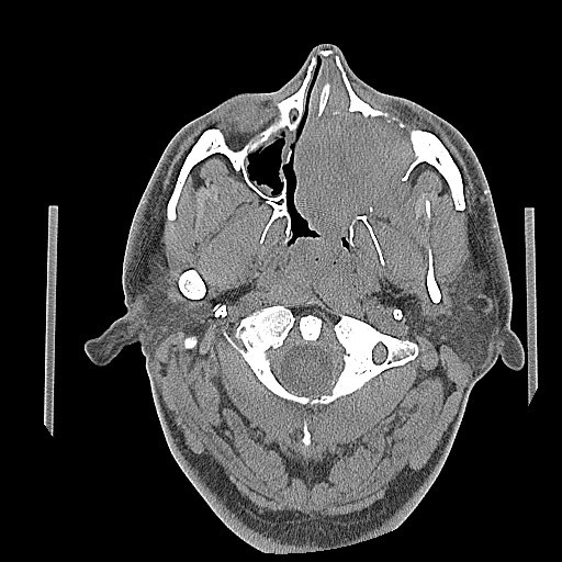
Figure 1.

CT image of the brain, axial view – There is a large expansile soft tissue mass centered within the left maxillary sinus with extensive osseous dehiscence. Tumor extends into the left orbital floor, nasal cavity, left nasopharynx, left pterygopalatine fossa, left premaxillary space, and left infratemporal fossa.
