Abstract
To establish a safer and more effective vaccine against pneumococcal respiratory infections, current knowledge regarding the antigens common among pneumococcal strains and improvements to the system for delivering these antigens across the mucosal barrier must be integrated. We developed a pneumococcal vaccine that combines the advantages of pneumococcal surface protein A (PspA) with a nontoxic intranasal vaccine delivery system based on a nanometer-sized hydrogel (nanogel) consisting of a cationic cholesteryl group-bearing pullulan (cCHP). The efficacy of the nanogel-based PspA nasal vaccine (cCHP-PspA) was tested in murine pneumococcal airway infection models. Intranasal vaccination with cCHP-PspA provided protective immunity against lethal challenge with Streptococcus pneumoniae Xen10, reduced colonization and invasion by bacteria in the upper and lower respiratory tracts, and induced systemic and nasal mucosal Th17 responses, high levels of PspA-specific serum immunoglobulin G (IgG), and nasal and bronchial IgA antibody responses. Moreover, there was no sign of PspA delivery by nanogel to either the olfactory bulbs or the central nervous system after intranasal administration. These results demonstrate the effectiveness and safety of the nanogel-based PspA nasal vaccine system as a universal mucosal vaccine against pneumococcal respiratory infection.
INTRODUCTION
The use of polysaccharide-based injectable multivalent pneumococcal conjugate vaccines (PCV7, -10, and -13) has diminished the number of fatal infections due to pneumococci expressing the particular polysaccharides present in the vaccine (1–3). However, Streptococcus pneumoniae remains a problematic pathogen (4, 5) because of the large number of different capsular polysaccharides associated with virulent disease in humans. In particular, nonvaccine strains are emerging pathogens that result in morbidity and mortality due to pneumococcal diseases, including pneumonia and meningitis (6–8).
Clinical demand to overcome these problems has prompted the preclinical development of universal serotype-independent pneumococcal vaccines that are based on a surface protein common to all strains. Pneumococcal surface protein A (PspA), a pneumococcal virulence factor (9–13), is genetically variable (14) but highly cross-reactive (9, 10). PspA is commonly expressed by all capsular serotypes of S. pneumoniae (15) and is classified into 3 families (family 1, clades 1 and 2; family 2, clades 3 through 5; and family 3, clade 6) according to sequence similarities (14). Given that parenteral immunization with PspA induces cross-reactive neutralizing immune responses in mice (16–18) and humans (19), using PspA as a serotype-independent common antigen for the development of pneumococcal vaccines seems to be an ideal strategy.
Pneumococcal infection is generally preceded by colonization of the upper airway (20, 21). Nasal carriage of pneumococci is the primary source for spread of the infection among humans (22, 23). Therefore, an optimal vaccine strategy to prevent and control the spread of pneumococcal disease would induce protective immunity against both colonization and invasive disease. Several studies have confirmed the efficacy of PspA as a nasal vaccine antigen by coadministering PspA with a mucosal adjuvant such as cholera toxin (CT) or cholera toxin subunit B (CTB) to mice (24–26). The mice subsequently mount antigen-specific immune responses in not only the systemic compartment but also the respiratory mucosal compartment (24, 25, 27), where bacterial colonization occurs (20). PspA-specific secretory immunoglobulin A (sIgA) antibodies induced by intranasal immunization with PspA and an adjuvant (i.e., a plasmid expressing Flt3 ligand cDNA) provide protection against pneumococcal colonization (28). In addition, studies in mice have revealed that this protection is mediated by antigen-specific interleukin 17A (IL-17A)-secreting CD4+ T cells induced by intranasal immunization with pneumococcal whole-cell antigen (29, 30).
Therefore, the intranasal vaccination route is an improved route for preventing colonization of the nasal cavity by pneumococci. A leading obstacle to the practical use of nasal vaccine with a protein-based pneumococcal antigen is the need to coadminister a toxin-based mucosal adjuvant (e.g., CT) for effective induction of antigen-specific immune responses (31, 32). However, the use of such toxin-based adjuvants is undesirable in humans, as it carries the concern that the toxin may reach the central nervous system (CNS) or redirect the vaccine antigen into the CNS through the olfactory nerve in the nasal cavity (33, 34). To bypass these concerns, we recently developed a nasal vaccine delivery system based on a non-toxin-based mucosal antigen carrier, a cationic cholesteryl pullulan (cCHP) nanogel (35).
Here we show the efficacy of a nanogel-based nasal pneumococcal vaccine in which PspA is incorporated into a cCHP nanogel (cCHP-PspA). We also characterized the cCHP-PspA-induced PspA-specific Th17 and antibody responses against S. pneumoniae. Mice immunized with nasal cCHP-PspA were protected from lethal challenge with S. pneumoniae and had fewer pneumococci on their respiratory mucosae. These results suggest that a nontoxic nasal vaccine comprising nanogel-based PspA offers a practical and effective strategy against pneumococcal infection by preventing both nasal colonization and invasive diseases.
MATERIALS AND METHODS
Mice.
Female BALB/c mice (aged 6 to 7 weeks) were purchased from SLC (Shizuoka, Japan). All of the mice were housed with ad libitum food and water on a standard 12-h–12-h light-dark cycle. All experiments were performed in accordance with the guidelines provided by the Animal Care and Use committees of the University of Tokyo and were approved by the Animal Committee of the Institute of Medical Science of the University of Tokyo.
Recombinant PspA.
Recombinant PspA of S. pneumoniae Rx1, which belongs to PspA family 1, clade 2 (14), was prepared as described previously, with slight modifications (26). Briefly, a plasmid encoding PspA/Rx1 (pUAB055; amino acids 1 through 302) (GenBank accession no. M74122) was used to transform Escherichia coli BL21(DE3) cells. This construct contains amino acids 1 through 302 of the PspA protein from strain Rx1 plus a 6×His tag at the C terminus (26). The sonicated cell supernatant was loaded onto a DEAE-Sepharose column (BD Healthcare, Piscataway, NJ) and a nickel affinity column (Qiagen, Valencia, CA). This was followed by gel filtration on a Sephadex G-100 column (BD Healthcare).
Preparation of cCHP-recombinant PspA complex for intranasal vaccination.
A cCHP nanogel (size, ∼40 nm) generated from a cationic cholesteryl group-bearing pullulan was used for all experiments. The cCHP-PspA complex for each immunization was prepared by mixing 7.5 μg PspA with cCHP at a 3:1 molecular ratio (volume, 18 μl per mouse) and incubating the mixture for 1 h at 45°C. Before the complex was used in in vivo studies, the fluorescence resonance energy transfer (FRET) of fluorescein isothiocyanate (FITC)-PspA and a tetramethyl rhodamine isothiocyanate (TRITC)-cCHP nanogel was measured with a fluorescence spectrometer (model FP-6500; Jasco, Easton, MD) as described previously (37). FRET analyses confirmed that the cCHP nanogel appropriately formed nanoparticles after the incorporation of PspA (see Fig. S1 in the supplemental material). Dynamic light scattering analysis showed that the cCHP nanogel maintained the same nanoscale size (32.8 ± 0.2 nm) even after the incorporation of PspA. Lipopolysaccharide (LPS) contamination of purified PspA and cCHP (<10 endotoxin units/mg protein) was measured with a Limulus test (Wako, Osaka, Japan).
Immunization.
Once weekly for 3 consecutive weeks, female BALB/c mice were immunized intranasally with cCHP-PspA, PspA plus CT (1 μg; List Biological Laboratory, Campbell, CA), PspA alone, or phosphate-buffered saline (PBS) only. Some experiments included an irrelevant antigen as a control; in these studies, mice were immunized intranasally with a complex of cCHP nanogel and a recombinant nontoxic receptor-binding fragment of Clostridium botulinum type A neurotoxin subunit antigen Hc (cCHP-BoHc/A) (35). Serum, nasal wash fluid (NW), and bronchoalveolar lavage fluid (BALF) samples were harvested 1 week after the last immunization. For NWs, 200 μl sterile PBS was flushed through the posterior choanae (38). BALF was harvested by instilling 1 ml of sterile PBS through a blunt needle placed in the trachea (38).
Bacterial strain.
We used the kanamycin-resistant pneumococcal strain S. pneumoniae Xen10 (Caliper Life Sciences, MA), derived from the wild-type strain A66.1, which expresses PspA of family 1, clades 1 and 2 (39). S. pneumoniae Xen10 carries a stable copy of the modified Photorhabdus luminescens lux operon at a single integration site on the bacterial chromosome (40). The virulence of S. pneumoniae Xen10 is comparable to that of the parent strain (40, 41). For challenge studies, S. pneumoniae 3JYP3670, which expresses PspA of family 2, clade 4, was used (10). All of the S. pneumoniae strains were grown in brain heart infusion (BHI) broth at 37°C in 5% CO2.
Pneumococcal infection model.
To evaluate the efficacy of intranasal vaccination with cCHP-PspA, mice were challenged 1 week after the last immunization. The cell densities of exponentially growing S. pneumoniae Xen10 cultured at 37°C in BHI broth were estimated from the optical density at 600 nm (OD600); cells were pelleted and then diluted with PBS. Lethal (2 × 10⁵ CFU) and sublethal (2 × 10⁴ CFU) challenge doses diluted in 50 μl sterile PBS were administered intranasally to isoflurane-anesthetized mice. Mice were restrained vertically for 5 min to ensure inhalation of the organisms into the trachea. In addition, mice were inoculated intranasally with a lethal challenge dose (5 × 10⁴ CFU) of strain 3JYP3670 in the same way as that for strain Xen10. Nasal passages and lung tissues were homogenized in 500 μl sterile PBS for 1 min, and the numbers of bacterial colonies were determined by plating samples on LB agar plates containing kanamycin (200 μl/ml).
In vivo imaging of immunized and challenged mice.
Bioluminescence of bacteria was monitored for 1 min 24, 48, and 72 h after lethal challenge by using an Ivis charge-coupled device (CCD) camera (Xenogen, Alameda, CA). Total photon emission from the entire thorax of each mouse was quantified by using the LivingImage software package (Xenogen). The results are provided as numbers of photons/s/cm2/sr.
Antibody titer and subclass analysis.
Antibody titers were determined by using enzyme-linked immunosorbent assay (ELISA) as described previously, with slight modifications (25). In brief, samples (2-fold serial dilutions) were loaded into individual wells, and the plate was coated with 1 μg/ml recombinant PspA and incubated. Goat anti-mouse IgA, IgG, IgG1, IgG2a, IgG2b, IgG3, and IgM (dilution factor, 1:4,000) conjugated with horseradish peroxidase were used as secondary antibodies. Reactions were visualized by using the TMB microwell peroxidase substrate system (XPL, Gaithersburg, MD). The endpoint titer is expressed as the reciprocal log2 of the last dilution that gave an OD450 that was 0.1 unit greater than that of the negative control.
PspA-specific CD4+ T cell responses.
By using anti-CD4 microbeads (Miltenyi Biotec, Sunnyvale, CA) according to the manufacturer's instructions, CD4+ T cells were isolated from the spleens and cervical lymph nodes (CLNs) of mice intranasally immunized with cCHP-PspA, PspA alone, or PBS only. The purified CD4+ T cells were resuspended at 1 × 10⁶ cells/ml in RPMI 1640 (Cellgro, Mediatech, Washington, DC) supplemented with 10 mM HEPES, 50 μM 2-mercaptoethanol, 100 U/ml penicillin, 100 μl/ml streptomycin, and 10% fetal calf serum and then cocultured with irradiated (2,000 rad) splenic antigen-presenting cells (2 × 10⁶ cells/ml) from naïve BALB/c mice for 5 days at 37°C in 5% CO2 in the presence of 1 μg/ml PspA. Cytokine levels in CD4+ T cell culture supernatants were determined by using cytokine-specific DuoSet ELISA kits (R&D Systems, Minneapolis, MN) according to the manufacturer's instructions.
Radioisotope counting assay.
To trace the distribution of PspA after intranasal immunization, PspA was labeled with indium chloride (Nihon Medi-Physics, Tokyo, Japan) anhydride (Dojindo, Kumamoto, Japan) via N-terminal and ε-Lys amino groups, using diethylenetriaminepentaacetic acid as described previously (42). 111In-labeled PspA was administered alone or as a complex with cCHP nanogel. The radioisotope counts in the nasal passage, olfactory bulbs, and brain 10 min and 1, 6, 12, 24, and 48 h after instillation were estimated with a γ-counter (Wizard model 1480; PerkinElmer, Waltham, MA). The results are provided as standardized uptake values (SUVs), calculated as radioisotope counts (cpm) per gram of tissue divided by the ratio of the injected dose (1 × 10⁶ cpm) to body weight (in grams).
Flow cytometric analysis.
Mice were immunized intranasally with FITC-PspA in cCHP nanogel, FITC-PspA alone, or PBS only; 6 h later, mononuclear cells were prepared from the nasal passages of each group by mechanical dissociation through 70-μm nylon mesh, as described previously (38, 43). Isolated cells were stained with phycoerythrin (PE)-Cy7-conjugated anti-CD11c (BD Bioscience) and analyzed by flow cytometry. The percentage of PspA+ cells in the CD11c+ fractions was calculated for each experimental group.
Data analysis.
Data are expressed as means ± standard deviations (SD). Statistical analysis for most comparisons among groups was performed with Tukey's t test; differences were considered statistically significant when the P value was <0.05. For survival data, the Fisher exact test was used to compare the numbers of alive versus dead mice in the cCHP-PspA, PspA-CT, and PBS-only groups with those in the PspA-only group.
RESULTS
Intranasal vaccination with cCHP-PspA induces protective immunity against lethal challenge with S. pneumoniae.
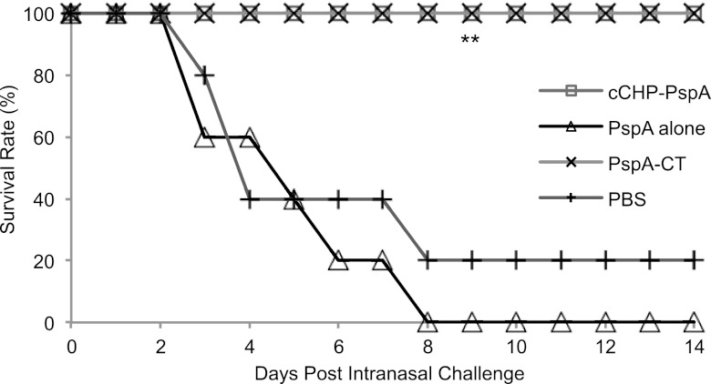
To evaluate whether intranasal cCHP-PspA vaccination induces protective immunity against pneumococcal challenge, we vaccinated mice with cCHP-PspA, PspA-CT, PspA alone, or PBS only. One week after the last immunization, we lethally challenged vaccinated mice with the virulent strain S. pneumoniae Xen10 (2 × 10⁵ CFU), which is S. pneumoniae A66.1 rendered bioluminescent by the integration of a modified lux operon into its chromosome (40). The PspA expression level of strain Xen10 was confirmed to be comparable to that of the parent strain (see Fig. S2 in the supplemental material). We then evaluated survival rates after lethal challenge over a 2-week period. The survival rate of the cCHP-PspA-vaccinated group was 100%, as was that for PspA-CT-vaccinated mice (Fig. 1). In contrast, most of the mice intranasally immunized with PspA alone (survival rate, 0%) or with PBS (20% survival) died within 8 days of challenge with S. pneumoniae Xen10 (Fig. 1). The survival rates of the groups immunized with cCHP-PspA or PspA-CT were higher and were statistically significant compared to that of the group immunized with PspA alone (P < 0.01). The results from the PspA-only and PBS-only groups did not differ (P > 0.05). In addition, immunization with the irrelevant antigen BoHc/A incorporated into cCHP (cCHP-BoHc/A) (35) did not protect mice from challenge with S. pneumoniae Xen10 (see Fig. S3). Because PspA family 2 (clades 3 through 5) and family 1 (clades 1 and 2) constitute 94 to 99% of clinical isolates of pneumococci (14, 44–49), we also challenged mice with the strain 3JYP3670, which expresses PspA belonging to clade 4 of family 2 (10). Unlike mice inoculated with cCHP-BoHc/A, PspA alone, or PBS only, mice nasally immunized with cCHP-PspA were protected from lethal challenge with 3JYP3670 (PspA of clade 4) (10), as was the case with Xen10 expressing PspA of clades 1 and 2 (see Fig. S4).
Fig 1.
Intranasal vaccination with cCHP-PspA induced protective immunity against pneumococci. One week after the final immunization, mice were challenged with S. pneumoniae Xen10 (2 × 10⁵ CFU/mouse), and survival was monitored. Data are representative of three independent experiments, and each group consisted of 5 mice. P values were calculated by using the Fisher exact test to compare the numbers of alive versus dead mice in each group with the result obtained for the PspA-only group. **, P < 0.01 compared with the group immunized with PspA alone. Abbreviations: cCHP, cationic cholesteryl group-bearing pullulan; CT, cholera toxin; PspA, pneumococcal surface protein A.
Intranasal vaccination with cCHP-PspA enhances bacterial clearance from BALF and the lung.
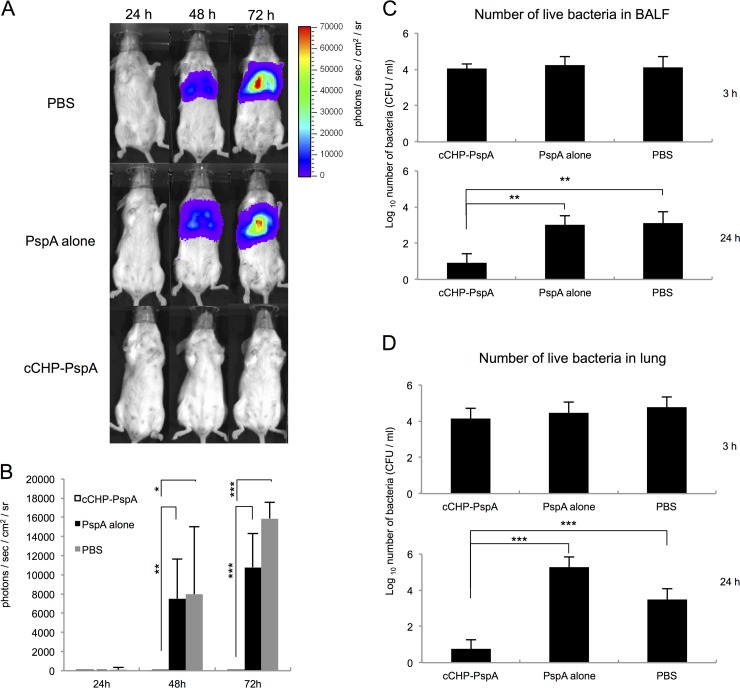
To assess whether intranasal immunization with cCHP-PspA prevented pulmonary infection with pneumococci, we performed in vivo bioluminescence imaging of S. pneumoniae Xen10 after lethal challenge (2 × 10⁵ CFU) of mice intranasally vaccinated with cCHP-PspA, PspA alone, or PBS. The lungs of mice immunized with PspA alone or with PBS only (control group) showed high-intensity photon signals in a pattern consistent with that of full-blown lung infection (Fig. 2A). In contrast, the lungs of mice immunized with cCHP-PspA lacked bioluminescence, indicating the absence of pulmonary infection. Forty-eight and 72 h after infection, photon counts of the cCHP-PspA-vaccinated group were significantly lower than those of the other two groups (Fig. 2B).
Fig 2.
In vivo imaging revealed no sign of pneumococcal infection in the lungs of mice immunized intranasally with cCHP-PspA; these mice also showed enhanced bacterial clearance from the BALF and lung. Images (A) and average photon counts (B) show bioluminescence due to S. pneumoniae Xen10 in each group of mice infected intranasally with S. pneumoniae Xen10 (2 × 10⁵ CFU/mouse) and imaged 24, 48, and 72 h after infection. (C and D) One week after the final immunization, mice were challenged with a sublethal dose (2 × 10⁴ CFU/mouse) of S. pneumoniae Xen10. BALF and lung tissues were collected, and the numbers of S. pneumoniae Xen10 organisms 3 and 24 h after challenge were determined. Data are representative of three independent experiments, and each group consisted of 5 mice. *, P < 0.05; **, P < 0.01; ***, P < 0.001. Abbreviations: BALF, bronchoalveolar lavage fluid; cCHP, cationic cholesteryl-group-bearing pullulan; PspA, pneumococcal surface protein A.
To investigate whether intranasal immunization with cCHP-PspA hastened bacterial clearance from the lung, we counted the bacteria in the BALF and lung tissues of mice intranasally vaccinated with cCHP-PspA, PspA alone, or PBS and sublethally challenged with S. pneumoniae Xen10 (2 × 10⁴ CFU). Three hours after challenge, bacterial numbers in BALF (Fig. 2C) and lung tissue (Fig. 2D) did not differ among the three vaccination groups. However, 24 h after challenge, the bacterial counts in the BALF and lung homogenates from the cCHP-PspA-vaccinated groups were significantly lower (about 100-fold) than those for the mice immunized with PspA alone or PBS only (Fig. 2C and D).
Intranasal vaccination with cCHP-PspA reduces bacterial colonization in the nasal cavity.
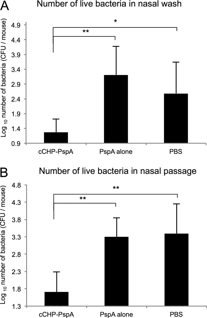
We next examined whether intranasal cCHP-PspA immunization affected nasal carriage of pneumococci in mice challenged with S. pneumoniae Xen10. Three days after challenge, bacterial numbers in NWs (Fig. 3A) and nasal passages (Fig. 3B) of mice immunized with the cCHP-PspA nasal vaccine were decreased significantly (approximately 100-fold) compared to those for the two control groups.
Fig 3.
Intranasal vaccination with cCHP-PspA reduced bacterial colonization of the nasal cavity. One week after the final immunization, mice were challenged with a sublethal dose (2 × 10⁴ CFU/mouse) of Streptococcus pneumoniae Xen10. Nasal washes and tissues were collected, and the numbers of S. pneumoniae Xen10 3 days after infection were determined. Data are representative of three independent experiments, and each group consisted of 5 mice. *, P < 0.05; **, P < 0.01. Abbreviations: cCHP, cationic cholesteryl group-bearing pullulan; PspA, pneumococcal surface protein A.
Intranasal vaccination with cCHP-PspA induces strong Th17 and Th2 responses.
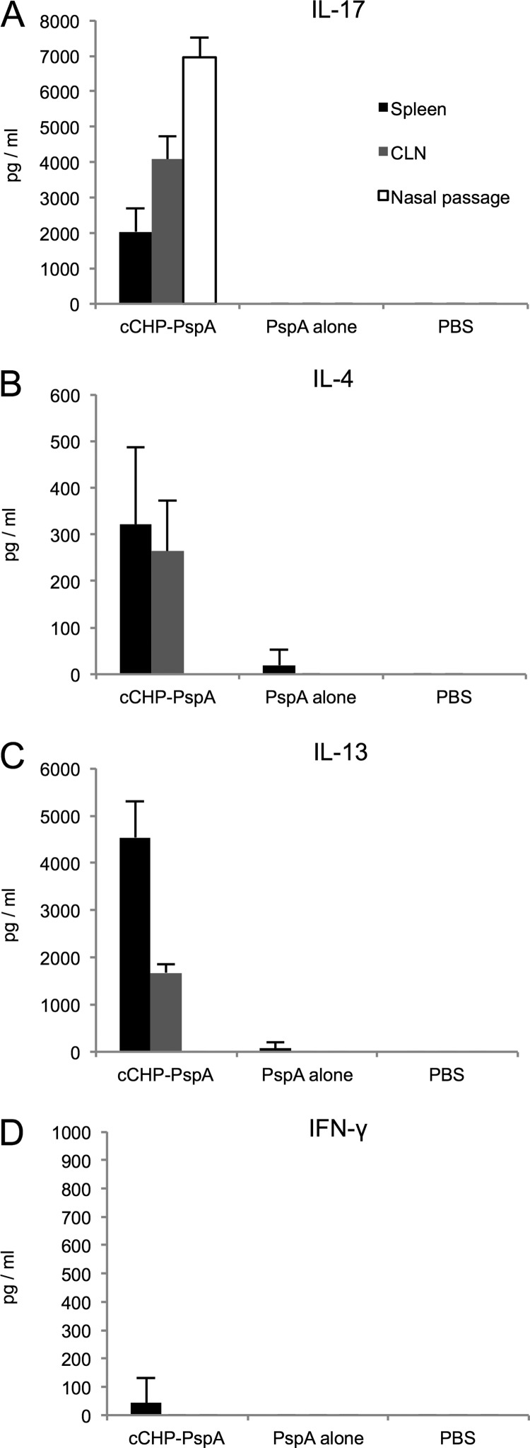
We then examined the type of immune responses elicited by intranasal cCHP-PspA vaccination. Compared with PspA alone or PBS, cCHP-PspA induced higher levels of IL-17 in CD4+ T cells from the spleen, CLNs, and nasal passages (Fig. 4A). The cCHP-PspA-vaccinated group produced high levels of IL-4 and IL-13, the hallmark cytokines of a Th2-type immune response, but only scant amounts of gamma interferon (Fig. 4B to D). These results show the potential of a cCHP-PspA nasal vaccine as an advanced pneumococcal vaccine that can induce a Th17 response together with a Th2-type immune response.
Fig 4.
CD4+ T cells from cCHP-PspA-immunized mice produce Th17- and Th2-type immune responses. Cytokines produced by CD4+ T cells isolated from the spleens, cervical lymph nodes, and nasal passages of mice immunized with cCHP-PspA, PspA alone, or PBS only were analyzed. Data are representative of five independent experiments, and each group consisted of 5 mice. Abbreviations: cCHP, cationic cholesteryl-group-bearing pullulan; CLN, cervical lymph node; IFN-γ, gamma interferon; IL, interleukin; PspA, pneumococcal surface protein A.
Intranasal vaccination with cCHP-PspA induces high levels of systemic antibodies.
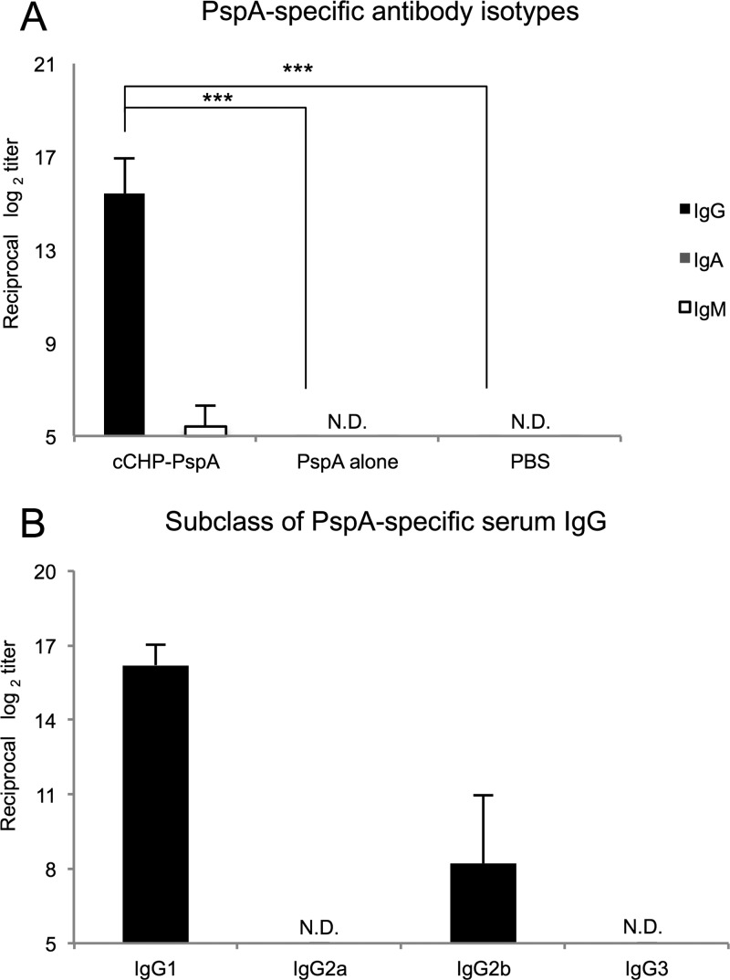
To address whether intranasal administration of cCHP-PspA induced PspA-specific antibody responses, we examined the serum titers of PspA-specific antibodies. PspA-specific IgG responses in the systemic compartment were significantly higher in mice immunized with intranasal cCHP-PspA than in those given PspA only (Fig. 5A). Unlike the predominant IgG response, IgM and IgA titers in the serum samples were very low (Fig. 5A).
Fig 5.
Intranasal vaccination with cCHP-PspA induced high levels of systemic antibodies. The data show the PspA-specific serum IgG level (A) and subclass analysis for IgG1, IgG2a, IgG2b, and IgG3 (B) for each immunized group (cCHP-PspA, PspA alone, or PBS only). Titers of PspA-specific IgG in sera were measured on day 7 after final immunization. Data are representative of three independent experiments, and each group consisted of 5 mice. N.D., not detected by ELISA with samples diluted 1:32. ***, P < 0.001. Abbreviations: cCHP, cationic cholesteryl group-bearing pullulan; Ig, immunoglobulin; PspA, pneumococcal surface protein A.
Intranasal immunization with cCHP-PspA induced primarily IgG1 antibodies, followed by IgG2b antibodies (Fig. 5B). This pattern indicated skewing toward a Th2-type response and was consistent with the cytokine profiles of the culture supernatants from antigen-stimulated CD4+ T cells prepared from the same mice (Fig. 4B and C).
Intranasal vaccination with cCHP-PspA induces high levels of mucosal antigen-specific sIgA antibodies.
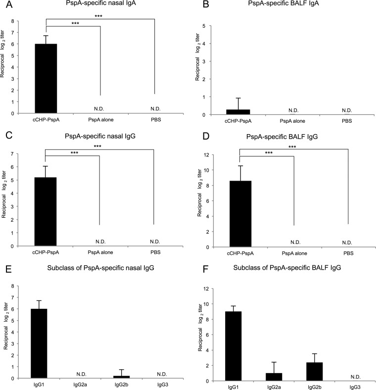
We next examined whether vaccinated mice also produced mucosal antigen-specific Ig responses. Intranasal vaccination with cCHP-PspA induced PspA-specific mucosal IgA antibodies in the nasal secretions (Fig. 6A). In addition, BALF samples from mice intranasally vaccinated with cCHP-PspA contained PspA-specific IgA antibodies (Fig. 6B), and PspA-specific IgG antibodies were detected at high titers in both the NWs and BALF of mice intranasally immunized with cCHP-PspA (Fig. 6C and D). The nasal and BALF antigen-specific IgGs induced by intranasal immunization with cCHP-PspA were primarily of the IgG1 and IgG2b subclasses (Fig. 6E and F), similar to the Ig responses in the systemic compartment (Fig. 5B). Taken together, these results further support the benefit of cCHP-based nanogel as an effective nasal vaccine delivery vehicle for the induction of PspA-specific systemic and mucosal antibody responses against S. pneumoniae.
Fig 6.
Intranasal vaccination with cCHP-PspA induced strong PspA-specific secretory IgA and IgG responses. Titers of nasal (A and C) and bronchial (B and D) IgA and IgG induced by intranasal immunization with PspA alone or PspA mixed with cCHP are shown. Titers of PspA-specific IgA and IgG in nasal washes and BALFs were measured on day 7 after final immunization. Intranasal cCHP-PspA vaccination induced high levels of IgG1 and IgG2b in mucosal secretions of the upper (E) and lower (F) airways. Data are representative of five independent experiments, and each group consisted of 5 mice. N.D., not detected in undiluted samples. ***, P < 0.001. Abbreviations: BALF, bronchoalveolar lavage fluid; cCHP, cationic cholesteryl-group-bearing pullulan; Ig, immunoglobulin; PspA, pneumococcal surface protein A.
cCHP delivers PspA to dendritic cells (DCs) without CNS accumulation of PspA.
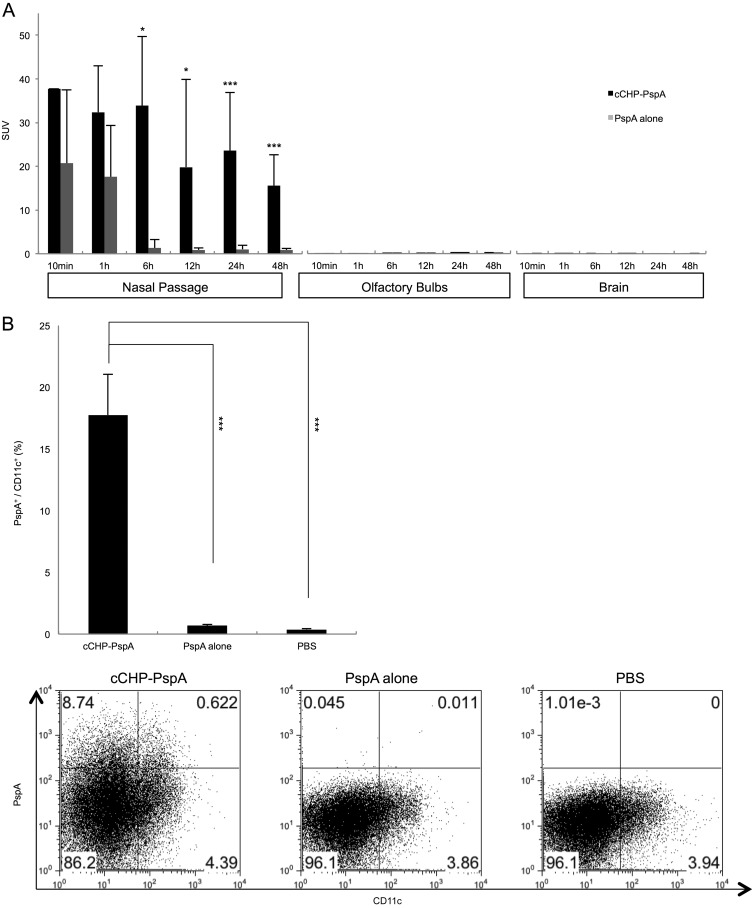
The potential for antigen deposition and accumulation in the CNS through the olfactory fossa is one of the great concerns surrounding the use of nasal vaccines (33, 34, 50). To address this important concern, we instilled 111In-labeled PspA alone or in complex with cCHP into the nasal cavities of mice. Beginning 6 h after administration, the nasal passages of mice treated with 111In-labeled cCHP-PspA had higher SUVs than did those of mice treated with 111In-labeled PspA alone, but there was no accumulation of 111In-labeled PspA in the olfactory bulbs or brain throughout the 48-h observation period (Fig. 7A).
Fig 7.
Intranasal vaccination with cCHP-PspA induced no accumulation of PspA in the central nervous system (A) but enhanced the efficiency of uptake of PspA by dendritic cells in the nasal passages (B). (A) 111In-labeled PspA was administered intranasally with or without cCHP nanogel, and the radioisotope counts (SUVs) in the nasal passages, olfactory bulbs, and brain were estimated 10 min and 1, 6, 12, 24, and 48 h after instillation. (B) Dendritic cells in the nasal passages of mice immunized intranasally with cCHP-PspA, PspA alone, or PBS were analyzed by flow cytometry 6 h after immunization. Data are representative of three independent experiments, and each group consisted of 5 mice. *, P < 0.05; ***, P < 0.001. Abbreviations: cCHP, cationic cholesteryl group-bearing pullulan; PspA, pneumococcal surface protein A.
The cCHP vaccine delivery system enabled prolonged antigen exposure at the nasal epithelium, allowing continuous antigen uptake by nasal DCs located in the epithelial layer and lamina propria of the nasal passages for the initiation of antigen-specific immune responses. Whereas 17.8% of the DCs located in the nasal passages had taken up PspA in the mice intranasally immunized with cCHP-PspA, only 0.7% of nasal DCs contained PspA antigen in mice that had been immunized intranasally with PspA alone (Fig. 7B). These results further support the concept that the cCHP-PspA vaccine formulation is an attractive inhalant delivery vehicle that effectively delivers and sustains antigen at the nasal epithelium for continuous antigen uptake by DCs without antigen deposition in the CNS.
DISCUSSION
We showed that cCHP-PspA-vaccinated mice survived a lethal challenge with S. pneumoniae (Fig. 1; see Fig. S4 in the supplemental material), whereas mice vaccinated with cCHP complexed with an irrelevant antigen (BoHc/A) did not (see Fig. S3 and S4). Importantly, compared with those of mice inoculated with control constructs, the respiratory tracts of mice immunized with intranasal cCHP-PspA had less colonization and invasion by pneumococcal organisms (Fig. 2 and 3). Intranasal administration of cCHP-PspA resulted in enhanced PspA-specific Th17 responses (Fig. 4A) and mucosal IgA and systemic IgG antibody responses (Fig. 5 and 6), all of which are involved in establishing protective immunity against pneumococci (10, 28–30). To our knowledge, the current study is the first to show the efficacy of a nasal vaccine not only for inducing protective immune responses but also for preventing nasal colonization by use of a single protein antigen (PspA) without adding any biologically active adjuvant.
The precise mechanisms underlying the efficacy of cCHP-PspA as a nasal vaccine against S. pneumoniae lung infection remain to be elucidated. However, we speculate that serum and BALF IgGs, the main isotype of antibody induced by the cCHP-PspA nasal vaccine in the lower respiratory compartment (Fig. 5A and 6D), play key roles in survival against lethal challenge with S. pneumoniae, given that antibody titers of PspA-specific IgA in the BALF were low (Fig. 6B) and therefore might contribute only minimally to protection against invasive diseases. This hypothesis is supported by the results of a previous study (28) in which IgA−/− mice immunized with intranasal PspA-adjuvant (i.e., a plasmid expressing Flt3 ligand cDNA) mounted a protective immune response against lethal challenge with S. pneumoniae. Our current study shows that the cCHP-PspA nasal vaccine effectively induced antigen-specific sIgA antibodies in the upper airways (Fig. 6A). Immunization of IgA−/− mice with intranasal PspA-adjuvant did not prevent pneumococcal colonization of the nasal cavity (28). In light of the findings of the previous study (28) and our current one, serum antigen-specific IgG antibodies are crucial to preventing invasive disease associated with clinical signs, whereas antigen-specific sIgA antibodies are essential for preventing colonization of the upper respiratory tract by S. pneumoniae.
In addition to the essential role of sIgA in protection from nasopharyngeal colonization by pneumococci, IL-17A-producing CD4+ T cells play an important role in preventing pneumococcal nasal colonization in mice immunized with intranasal pneumococcal whole-cell antigen (29, 30). Recent studies have found that IL-17 promotes multiple aspects of humoral immunity by enhancing B cell proliferation and isotype switching (51), B cell recruitment to the respiratory mucosa, and expression of the polymeric immunoglobulin receptor on the airway epithelium (52). In the current study, we found that intranasal immunization with cCHP-PspA generated Th17 cells in the nasal passages, draining lymph nodes, and systemic compartment (Fig. 4A). Therefore, our findings suggest that intranasal immunization with cCHP-PspA induces both humoral and cellular immune responses, which are required for protective immunity against pneumococcal colonization and invasive disease. In addition to their essential role in antipneumococcal immunity (29, 30), Th17 responses are a hallmark of autoimmunity (53). Therefore, future studies should carefully examine whether the Th17 responses induced by intranasal immunization with cCHP-PspA are associated with any adverse effects.
As one might expect, the protective immunity induced by nasal cCHP-PspA was not observed when an irrelevant antigen, BoHc/A, was incorporated into cCHP (cCHP-BoHc/A) (35) and used as a nasal vaccine (see Fig. S3 and S4 in the supplemental material). Moreover, mice immunized intranasally with cCHP-PspA (PspA of clades 1 and 2) were protected against challenge with pneumococcal strain 3JYP3670, which expresses PspA of clade 4 (10), whereas mice immunized with cCHP-BoHc/A, PspA alone, or PBS were not (see Fig. S4). These findings highlight the potential advantage of nasal vaccination of cCHP-PspA in inducing antigen-specific protective immunity with subtype cross-reactivity.
Note that cCHP lacks any biologically active adjuvant effect because it cannot activate immune cells by itself (35). The nanogel formulation had no effect on the expression of costimulatory molecules on nasal DCs (see Fig. S5 in the supplemental material), which are supposed to already express high steady-state levels of costimulatory molecules in the mucosal environment in response to numerous inhaled antigens. Our current and previous studies have shown that antigens are released from the nanogel and are taken up efficiently by DCs in the nasal mucosa (Fig. 7B) (35). These studies suggest that cCHP nanogel is an effective carrier that has strong chaperone-like activity, enabling the delivery of PspA across the nasal mucosal epithelial cell layer for subsequent uptake by DCs and initiation of antigen-specific immune responses.
In summary, this study introduced a promising nanometer-sized carrier-based pneumococcal nasal vaccine that incorporates cCHP nanogel and the pneumococcal serotype-independent protein antigen PspA. The antigen-specific immune responses induced by this vaccine effectively protected mice against the respiratory pathogen S. pneumoniae. Our results confirmed that cCHP nanogel is a promising candidate carrier of a protein antigen for a mucosal vaccine that induces humoral and cellular immune responses against PspA to combat colonization and invasion of the airways by respiratory pathogens.
Supplementary Material
ACKNOWLEDGMENTS
This study was supported by a grant-in-aid from the Research Fellowship of the Japan Society for the Promotion of Science (JSPS) (I.G.K., A.S., and T.N.); by programs of special coordination funds for promoting science and technology, a grant-in-aid for scientific research on priority areas, and a grant-in-aid for scientific research from the Ministry of Education, Culture, Sports, Science, and Technology of Japan (J.K. and H.K.); by the Ministry of Health, Labor, and Welfare of Japan (J.K., Y.Y., and H.K.); by the New Energy and Industrial Technology Development Organization (NEDO) (Y.Y. and H.K.); by the Young Researcher Overseas Visits Program for Vitalizing Brain Circulation of the Japan Society for the Promotion of Science (J.K., H.K., and Y.Y.); by the Program for Promotion of Basic and Applied Researches for Innovations in Bio-Oriented Industry (BRAIN) (T.N., S. Sato, and J.K.); by the Yakult Bio-Science Foundation (J.K.); and by the Global Center of Excellence Program “Center of Education and Research for Advanced Genome-Based Medicine—For Personalized Medicine and the Control of Worldwide Infectious Diseases” (H.K.).
We declare that we have no conflicts of interest.
Footnotes
Published ahead of print 4 March 2013
Supplemental material for this article may be found at http://dx.doi.org/10.1128/IAI.00240-13.
REFERENCES
- 1. Dinleyici EC, Yargic ZA. 2008. Pneumococcal conjugated vaccines: impact of PCV-7 and new achievements in the postvaccine era. Expert Rev. Vaccines 7:1367–1394 [DOI] [PubMed] [Google Scholar]
- 2. Rose M, Zielen S. 2009. Impact of infant immunization programs with pneumococcal conjugate vaccine in Europe. Expert Rev. Vaccines 8:1351–1364 [DOI] [PubMed] [Google Scholar]
- 3. Principi N, Esposito S. 2012. Use of the 13-valent pneumococcal conjugate vaccine in infants and young children. Expert Opin. Biol. Ther. 12:641–648 [DOI] [PubMed] [Google Scholar]
- 4. Huang SS, Johnson KM, Ray GT, Wroe P, Lieu TA, Moore MR, Zell ER, Linder JA, Grijalva CG, Metlay JP, Finkelstein JA. 2011. Healthcare utilization and cost of pneumococcal disease in the United States. Vaccine 29:3398–3412 [DOI] [PubMed] [Google Scholar]
- 5. Thigpen MC, Whitney CG, Messonnier NE, Zell ER, Lynfield R, Hadler JL, Harrison LH, Farley MM, Reingold A, Bennett NM, Craig AS, Schaffner W, Thomas A, Lewis MM, Scallan E, Schuchat A. 2011. Bacterial meningitis in the United States, 1998–2007. N. Engl. J. Med. 364:2016–2025 [DOI] [PubMed] [Google Scholar]
- 6. Weinberger DM, Malley R, Lipsitch M. 2011. Serotype replacement in disease after pneumococcal vaccination. Lancet 378:1962–1973 [DOI] [PMC free article] [PubMed] [Google Scholar]
- 7. Hsu HE, Shutt KA, Moore MR, Beall BW, Bennett NM, Craig AS, Farley MM, Jorgensen JH, Lexau CA, Petit S, Reingold A, Schaffner W, Thomas A, Whitney CG, Harrison LH. 2009. Effect of pneumococcal conjugate vaccine on pneumococcal meningitis. N. Engl. J. Med. 360:244–256 [DOI] [PMC free article] [PubMed] [Google Scholar]
- 8. Singleton RJ, Hennessy TW, Bulkow LR, Hammitt LL, Zulz T, Hurlburt DA, Butler JC, Rudolph K, Parkinson A. 2007. Invasive pneumococcal disease caused by nonvaccine serotypes among Alaska native children with high levels of 7-valent pneumococcal conjugate vaccine coverage. JAMA 297:1784–1792 [DOI] [PubMed] [Google Scholar]
- 9. Briles DE, Hollingshead SK, Nabors GS, Paton JC, Brooks-Walter A. 2000. The potential for using protein vaccines to protect against otitis media caused by Streptococcus pneumoniae. Vaccine 19(Suppl 1):S87–S95 [DOI] [PubMed] [Google Scholar]
- 10. Briles DE, Hollingshead SK, King J, Swift A, Braun PA, Park MK, Ferguson LM, Nahm MH, Nabors GS. 2000. Immunization of humans with recombinant pneumococcal surface protein A (rPspA) elicits antibodies that passively protect mice from fatal infection with Streptococcus pneumoniae bearing heterologous PspA. J. Infect. Dis. 182:1694–1701 [DOI] [PubMed] [Google Scholar]
- 11. Briles DE, Hollingshead SK, Paton JC, Ades EW, Novak L, van Ginkel FW, Benjamin, WH 2003. Immunizations with pneumococcal surface protein A and pneumolysin are protective against pneumonia in a murine model of pulmonary infection with Streptococcus pneumoniae. J. Infect. Dis. 188:339–348 [DOI] [PubMed] [Google Scholar]
- 12. Briles DE, Tart RC, Swiatlo E, Dillard JP, Smith P, Benton KA, Ralph BA, Brooks-Walter A, Crain MJ, Hollingshead SK, McDaniel LS. 1998. Pneumococcal diversity: considerations for new vaccine strategies with emphasis on pneumococcal surface protein A (PspA). Clin. Microbiol. Rev. 11:645–657 [DOI] [PMC free article] [PubMed] [Google Scholar]
- 13. Olafsdottir TA, Lingnau K, Nagy E, Jonsdottir I. 2012. Novel protein-based pneumococcal vaccines administered with the Th1-promoting adjuvant IC31 induce protective immunity against pneumococcal disease in neonatal mice. Infect. Immun. 80:461–468 [DOI] [PMC free article] [PubMed] [Google Scholar]
- 14. Hollingshead SK, Becker R, Briles DE. 2000. Diversity of PspA: mosaic genes and evidence for past recombination in Streptococcus pneumoniae. Infect. Immun. 68:5889–5900 [DOI] [PMC free article] [PubMed] [Google Scholar]
- 15. Crain MJ, Waltman WD, 2nd, Turner JS, Yother J, Talkington DF, McDaniel LS, Gray BM, Briles DE. 1990. Pneumococcal surface protein A (PspA) is serologically highly variable and is expressed by all clinically important capsular serotypes of Streptococcus pneumoniae. Infect. Immun. 58:3293–3299 [DOI] [PMC free article] [PubMed] [Google Scholar]
- 16. McDaniel LS, Sheffield JS, Delucchi P, Briles DE. 1991. PspA, a surface protein of Streptococcus pneumoniae, is capable of eliciting protection against pneumococci of more than one capsular type. Infect. Immun. 59:222–228 [DOI] [PMC free article] [PubMed] [Google Scholar]
- 17. Tart RC, McDaniel LS, Ralph BA, Briles DE. 1996. Truncated Streptococcus pneumoniae PspA molecules elicit cross-protective immunity against pneumococcal challenge in mice. J. Infect. Dis. 173:380–386 [DOI] [PubMed] [Google Scholar]
- 18. Xin W, Li Y, Mo H, Roland KL, Curtiss R. 2009. PspA family fusion proteins delivered by attenuated Salmonella enterica serovar Typhimurium extend and enhance protection against Streptococcus pneumoniae. Infect. Immun. 77:4518–4528 [DOI] [PMC free article] [PubMed] [Google Scholar]
- 19. Nabors GS, Braun PA, Herrmann DJ, Heise ML, Pyle DJ, Gravenstein S, Schilling M, Ferguson LM, Hollingshead SK, Briles DE, Becker RS. 2000. Immunization of healthy adults with a single recombinant pneumococcal surface protein A (PspA) variant stimulates broadly cross-reactive antibodies to heterologous PspA molecules. Vaccine 18:1743–1754 [DOI] [PubMed] [Google Scholar]
- 20. Gray BM, Converse GM, 3rd, Dillon HC., Jr 1980. Epidemiologic studies of Streptococcus pneumoniae in infants: acquisition, carriage, and infection during the first 24 months of life. J. Infect. Dis. 142:923–933 [DOI] [PubMed] [Google Scholar]
- 21. Faden H, Duffy L, Wasielewski R, Wolf J, Krystofik D, Tung Y. 1997. Relationship between nasopharyngeal colonization and the development of otitis media in children. J. Infect. Dis. 175:1440–1445 [DOI] [PubMed] [Google Scholar]
- 22. Leiberman A, Dagan R, Leibovitz E, Yagupsky P, Fliss DM. 1999. The bacteriology of the nasopharynx in childhood. Int. J. Pediatr. Otorhinolaryngol. 49(Suppl 1):S151–S153 [DOI] [PubMed] [Google Scholar]
- 23. Hoge CW, Reichler MR, Dominguez EA, Bremer JC, Mastro TD, Hendricks KA, Musher DM, Elliott JA, Facklam RR, Breiman RF. 1994. An epidemic of pneumococcal disease in an overcrowded, inadequately ventilated jail. N. Engl. J. Med. 331:643–648 [DOI] [PubMed] [Google Scholar]
- 24. Wu HY, Nahm MH, Guo Y, Russell MW, Briles DE. 1997. Intranasal immunization of mice with PspA (pneumococcal surface protein A) can prevent intranasal carriage, pulmonary infection, and sepsis with Streptococcus pneumoniae. J. Infect. Dis. 175:839–846 [DOI] [PubMed] [Google Scholar]
- 25. Yamamoto M, Briles DE, Yamamoto S, Ohmura M, Kiyono H, McGhee JR. 1998. A nontoxic adjuvant for mucosal immunity to pneumococcal surface protein A. J. Immunol. 161:4115–4121 [PubMed] [Google Scholar]
- 26. Briles DE, Ades E, Paton JC, Sampson JS, Carlone GM, Huebner RC, Virolainen A, Swiatlo E, Hollingshead SK. 2000. Intranasal immunization of mice with a mixture of the pneumococcal proteins PsaA and PspA is highly protective against nasopharyngeal carriage of Streptococcus pneumoniae. Infect. Immun. 68:796–800 [DOI] [PMC free article] [PubMed] [Google Scholar]
- 27. Oma K, Zhao J, Ezoe H, Akeda Y, Koyama S, Ishii KJ, Kataoka K, Oishi K. 2009. Intranasal immunization with a mixture of PspA and a Toll-like receptor agonist induces specific antibodies and enhances bacterial clearance in the airways of mice. Vaccine 27:3181–3188 [DOI] [PubMed] [Google Scholar]
- 28. Fukuyama Y, King JD, Kataoka K, Kobayashi R, Gilbert RS, Oishi K, Hollingshead SK, Briles DE, Fujihashi K. 2010. Secretory-IgA antibodies play an important role in the immunity to Streptococcus pneumoniae. J. Immunol. 185:1755–1762 [DOI] [PubMed] [Google Scholar]
- 29. Lu YJ, Gross J, Bogaert D, Finn A, Bagrade L, Zhang Q, Kolls JK, Srivastava A, Lundgren A, Forte S, Thompson CM, Harney KF, Anderson PW, Lipsitch M, Malley R. 2008. Interleukin-17A mediates acquired immunity to pneumococcal colonization. PLoS Pathog. 4:e1000159 doi:10.1371/journal.ppat.1000159 [DOI] [PMC free article] [PubMed] [Google Scholar]
- 30. Malley R. 2005. CD4+ T cells mediate antibody-independent acquired immunity to pneumococcal colonization. Proc. Natl. Acad. Sci. U. S. A. 102:4848–4853 [DOI] [PMC free article] [PubMed] [Google Scholar]
- 31. Xu-Amano J, Kiyono H, Jackson RJ, Staats HF, Fujihashi K, Burrows PD, Elson CO, Pillai S, McGhee JR. 1993. Helper T cell subsets for immunoglobulin A responses: oral immunization with tetanus toxoid and cholera toxin as adjuvant selectively induces Th2 cells in mucosa associated tissues. J. Exp. Med. 178:1309–1320 [DOI] [PMC free article] [PubMed] [Google Scholar]
- 32. Freytag LC, Clements JD. 2005. Mucosal adjuvants. Vaccine 23:1804–1813 [DOI] [PubMed] [Google Scholar]
- 33. Mutsch M, Zhou W, Rhodes P, Bopp M, Chen RT, Linder T, Spyr C, Steffen R. 2004. Use of the inactivated intranasal influenza vaccine and the risk of Bell's palsy in Switzerland. N. Engl. J. Med. 350:896–903 [DOI] [PubMed] [Google Scholar]
- 34. van Ginkel FW, Jackson RJ, Yuki Y, McGhee JR. 2000. Cutting edge: the mucosal adjuvant cholera toxin redirects vaccine proteins into olfactory tissues. J. Immunol. 165:4778–4782 [DOI] [PubMed] [Google Scholar]
- 35. Nochi T, Yuki Y, Takahashi H, Sawada S, Mejima M, Kohda T, Harada N, Kong IG, Sato A, Kataoka N, Tokuhara D, Kurokawa S, Takahashi Y, Tsukada H, Kozaki S, Akiyoshi K, Kiyono H. 2010. Nanogel antigenic protein-delivery system for adjuvant-free intranasal vaccines. Nat. Mater. 9:572–578 [DOI] [PubMed] [Google Scholar]
- 36. Reference deleted.
- 37. Ayame H, Morimoto N, Akiyoshi K. 2008. Self-assembled cationic nanogels for intracellular protein delivery. Bioconjug. Chem. 19:882–890 [DOI] [PubMed] [Google Scholar]
- 38. Kurono Y, Yamamoto M, Fujihashi K, Kodama S, Suzuki M, Mogi G, McGhee JR, Kiyono H. 1999. Nasal immunization induces Haemophilus influenzae-specific Th1 and Th2 responses with mucosal IgA and systemic IgG antibodies for protective immunity. J. Infect. Dis. 180:122–132 [DOI] [PubMed] [Google Scholar]
- 39. Darrieux M, Miyaji EN, Ferreira DM, Lopes LM, Lopes AP, Ren B, Briles DE, Hollingshead SK, Leite LC. 2007. Fusion proteins containing family 1 and family 2 PspA fragments elicit protection against Streptococcus pneumoniae that correlates with antibody-mediated enhancement of complement deposition. Infect. Immun. 75:5930–5938 [DOI] [PMC free article] [PubMed] [Google Scholar]
- 40. Francis KP, Yu J, Bellinger-Kawahara C, Joh D, Hawkinson MJ, Xiao G, Purchio TF, M. Caparon G, Lipsitch M, Contag PR. 2001. Visualizing pneumococcal infections in the lungs of live mice using bioluminescent Streptococcus pneumoniae transformed with a novel gram-positive lux transposon. Infect. Immun. 69:3350–3358 [DOI] [PMC free article] [PubMed] [Google Scholar]
- 41. Kadurugamuwa JL, Modi K, Coquoz O, Rice B, Smith S, Contag PR, Purchio T. 2005. Reduction of astrogliosis by early treatment of pneumococcal meningitis measured by simultaneous imaging, in vivo, of the pathogen and host response. Infect. Immun. 73:7836–7843 [DOI] [PMC free article] [PubMed] [Google Scholar]
- 42. Michel RB, Andrews PM, Castillo ME, Mattes MJ. 2005. In vitro cytotoxicity of carcinoma cells with 111In-labeled antibodies to HER-2. Mol. Cancer Ther. 4:927–937 [DOI] [PubMed] [Google Scholar]
- 43. Fukuyama S, Hiroi T, Yokota Y, Rennert PD, Yanagita M, Kinoshita N, Terawaki S, Shikina T, Yamamoto M, Kurono Y, Kiyono H. 2002. Initiation of NALT organogenesis is independent of the IL-7R, LTβR, and NIK signaling pathways but requires the Id2 gene and CD3−CD4+CD45+ cells. Immunity 17:31–40 [DOI] [PubMed] [Google Scholar]
- 44. Hollingshead SK. 2006. Pneumococcal surface protein A (PspA) family distribution among clinical isolates from adults over 50 years of age collected in seven countries. J. Med. Microbiol. 55:215–221 [DOI] [PubMed] [Google Scholar]
- 45. Ren B, Szalai AJ, Hollingshead SK, Briles DE. 2004. Effects of PspA and antibodies to PspA on activation and deposition of complement on the pneumococcal surface. Infect. Immun. 72:114–122 [DOI] [PMC free article] [PubMed] [Google Scholar]
- 46. Beall B, Gherardi G, Facklam RR, Hollingshead SK. 2000. Pneumococcal PspA sequence types of prevalent multiresistant pneumococcal strains in the United States and of internationally disseminated clones. J. Clin. Microbiol. 38:3663–3669 [DOI] [PMC free article] [PubMed] [Google Scholar]
- 47. Brandileone M. 2004. Typing of pneumococcal surface protein A (PspA) in Streptococcus pneumoniae isolated during epidemiological surveillance in Brazil: towards novel pneumococcal protein vaccines. Vaccine 22:3890–3896 [DOI] [PubMed] [Google Scholar]
- 48. Mollerach M, Regueira M, Bonofiglio L, Callejo R, Pace J, Di Fabio JL, Hollingshead SK, Briles DE. 2004. Invasive Streptococcus pneumoniae isolates from Argentinian children: serotypes, families of pneumococcal surface protein A (PspA) and genetic diversity. Epidemiol. Infect. 132:177–184 [DOI] [PMC free article] [PubMed] [Google Scholar]
- 49. Vela Coral MC, Fonseca N, Castaneda E, Di Fabio JL, Hollingshead SK, Briles DE. 2001. Pneumococcal surface protein A of invasive Streptococcus pneumoniae isolates from Colombian children. Emerg. Infect. Dis. 7:832–836 [DOI] [PMC free article] [PubMed] [Google Scholar]
- 50. Dubin PJ, Kolls JK. 2009. Interleukin-17A and interleukin-17F: a tale of two cytokines. Immunity 30:9–11 [DOI] [PubMed] [Google Scholar]
- 51. Doreau A, Belot A, Bastid J, Riche B, Trescol-Biemont MC, Ranchin B, Fabien N, Cochat P, Pouteil-Noble C, Trolliet P, Durieu I, Tebib J, Kassai B, Ansieau S, Puisieux A, Eliaou JF, Bonnefoy-Berard N. 2009. Interleukin 17 acts in synergy with B cell-activating factor to influence B cell biology and the pathophysiology of systemic lupus erythematosus. Nat. Immunol. 10:778–785 [DOI] [PubMed] [Google Scholar]
- 52. Jaffar Z, Ferrini ME, Herritt LA, Roberts K. 2009. Cutting edge: lung mucosal Th17-mediated responses induce polymeric Ig receptor expression by the airway epithelium and elevate secretory IgA levels. J. Immunol. 182:4507–4511 [DOI] [PMC free article] [PubMed] [Google Scholar]
- 53. Bettelli E, Oukka M, Kuchroo VK. 2007. Th17 cells in the circle of immunity and autoimmunity. Nat. Immunol. 8:345–350 [DOI] [PubMed] [Google Scholar]
Associated Data
This section collects any data citations, data availability statements, or supplementary materials included in this article.


