

Abstract
In the title compound, C13H13NO3, the isoxazole ring is approximately coplanar with the phenyl ring, the dihedral angle between their planes being 7.37 (19)°. In the crystal, centrosymmetrically related molecules are linked into dimers by pairs of C—H⋯O hydrogen bonds, generating a ring of graph-set motif R 2 2(10).
Related literature
For the biological activity of isoxazole derivatives, see: Angibaud et al. (2003 ▶). For the structure of a related compound, see: Yao & Deng (2008 ▶). For the synthesis of 3-phenylisoxazole-5-carboxylic acid, see: Liu et al. (2006 ▶).
Experimental
Crystal data
C13H13NO3
M r = 231.24
Monoclinic,

a = 4.6311 (10) Å
b = 16.596 (4) Å
c = 15.897 (3) Å
β = 98.321 (4)°
V = 1208.9 (5) Å3
Z = 4
Mo Kα radiation
μ = 0.09 mm−1
T = 296 K
0.36 × 0.28 × 0.17 mm
Data collection
Bruker APEXII CCD diffractometer
Absorption correction: multi-scan (SADABS; Bruker, 2008 ▶) T min = 0.968, T max = 0.984
6039 measured reflections
2169 independent reflections
1511 reflections with I > 2σ(I)
R int = 0.037
Refinement
R[F 2 > 2σ(F 2)] = 0.046
wR(F 2) = 0.126
S = 1.03
2169 reflections
156 parameters
H-atom parameters constrained
Δρmax = 0.11 e Å−3
Δρmin = −0.20 e Å−3
Data collection: APEX2 (Bruker, 2008 ▶); cell refinement: SAINT (Bruker, 2008 ▶); data reduction: SAINT; program(s) used to solve structure: SHELXS97 (Sheldrick, 2008 ▶); program(s) used to refine structure: SHELXL97 (Sheldrick, 2008 ▶); molecular graphics: SHELXTL (Sheldrick, 2008 ▶); software used to prepare material for publication: publCIF (Westrip, 2010 ▶).
Supplementary Material
Crystal structure: contains datablock(s) I, global. DOI: 10.1107/S1600536813009392/rz5055sup1.cif
Structure factors: contains datablock(s) I. DOI: 10.1107/S1600536813009392/rz5055Isup2.hkl
Supplementary material file. DOI: 10.1107/S1600536813009392/rz5055Isup3.cml
Additional supplementary materials: crystallographic information; 3D view; checkCIF report
Table 1. Hydrogen-bond geometry (Å, °).
| D—H⋯A | D—H | H⋯A | D⋯A | D—H⋯A |
|---|---|---|---|---|
| C8—H8⋯O2i | 0.93 | 2.37 | 3.277 (2) | 166 |
Symmetry code: (i)  .
.
Acknowledgments
This work was supported financially by the National Natural Science Foundation of China (21172262).
supplementary crystallographic information
Comment
Isoxazole derivatives, as useful intermediates in organic synthesis, show widespread biological activities, and are employed as antiviral drugs, antibacteria reagents, fungicide, anti-inflammatory agents, analgesics, antidepressants, anticonvulsants and pesticides (Angibaud et al., 2003). In the molecule of the title compound (Fig. 1), the dihedral angle between the phenyl and the isoxazole rings is 7.37 (19)°. The bond lengths within the isoxazole ring [C7—N1 = 1.306 (2) Å, N1—O1 = 1.402 (18) Å, O1—C9 = 1.345 (2) Å, C9—C8 = 1.327 (2) Å and C8—C7 = 1.409 (2) Å] are in agreement with those reported by Yao & Deng (2008) for 5-amino-3-(4-pyridyl)isoxazole [C7—N1 = 1.316 (18) Å, N1—O1 = 1.429 (14) Å, O1—C9 = 1.353 (17) Å, C9—C8 = 1.368 (19) Å and C8—C7 = 1.400 (19) Å]. In the crystal, centrosymmetrically related molecules are linked into dimers by C—H···O hydrogen bonds (Table 1), generating a ring of graph-set motif < i>R22(10).
Experimental
3-Phenylisoxazole-5-carboxylic acid (10 mmol, 1.95 g; Liu et al., 2006) was dissolved in 100 ml dichloromethane, then thionyl chloride(12 mmol, 1.43 g)was dropped into the solution and stirred for 20 minutes in ice bath. The solvent was removed under reduced pressure and the mixture was used for the next step without further purification. 2-Propanol (20 mmol, 1.5 ml) was added subsequently and the mixture stirred for 6 h at room temperature. The resulting residue was purified as a white solid (1.96 g, 85% yield). Recrystallization in ethyl acetate gave fine colourless crystals suitable for X-ray study. All chemicals were purchased by Sigma Aldrich Germany.
Refinement
All H atoms were placed in idealized positions and allowed to ride on the respective parent atom with C—H = 0.93–0.98 Å and with Uiso(H) = of 1.2Ueq(C) or 1.5Ueq(C) for methyl H atoms.
Figures
Fig. 1.

The molecular structure of the title compound, with displacement ellipsoids drawn at the 30% probability level.
Fig. 2.
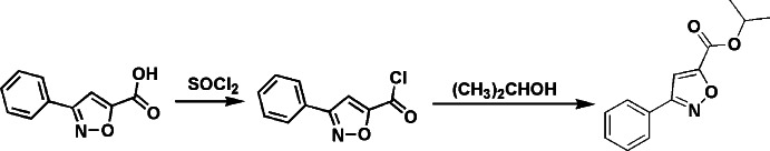
The formation of the title compound.
Crystal data
| C13H13NO3 | F(000) = 488 |
| Mr = 231.24 | Dx = 1.271 Mg m−3Dm = 1.270 Mg m−3Dm measured by not measured |
| Monoclinic, P21/n | Mo Kα radiation, λ = 0.71073 Å |
| Hall symbol: -P 2yn | Cell parameters from 1178 reflections |
| a = 4.6311 (10) Å | θ = 2.5–21.2° |
| b = 16.596 (4) Å | µ = 0.09 mm−1 |
| c = 15.897 (3) Å | T = 296 K |
| β = 98.321 (4)° | Block, colourless |
| V = 1208.9 (5) Å3 | 0.36 × 0.28 × 0.17 mm |
| Z = 4 |
Data collection
| Bruker APEXII CCD diffractometer | 2169 independent reflections |
| Radiation source: fine-focus sealed tube | 1511 reflections with I > 2σ(I) |
| Graphite monochromator | Rint = 0.037 |
| φ and ω scans | θmax = 25.1°, θmin = 1.8° |
| Absorption correction: multi-scan (SADABS; Bruker, 2008) | h = −4→5 |
| Tmin = 0.968, Tmax = 0.984 | k = −19→19 |
| 6039 measured reflections | l = −18→17 |
Refinement
| Refinement on F2 | Primary atom site location: structure-invariant direct methods |
| Least-squares matrix: full | Secondary atom site location: difference Fourier map |
| R[F2 > 2σ(F2)] = 0.046 | Hydrogen site location: inferred from neighbouring sites |
| wR(F2) = 0.126 | H-atom parameters constrained |
| S = 1.03 | w = 1/[σ2(Fo2) + (0.0611P)2 + 0.0883P] where P = (Fo2 + 2Fc2)/3 |
| 2169 reflections | (Δ/σ)max < 0.001 |
| 156 parameters | Δρmax = 0.11 e Å−3 |
| 0 restraints | Δρmin = −0.20 e Å−3 |
Special details
| Geometry. All e.s.d.'s (except the e.s.d. in the dihedral angle between two l.s. planes) are estimated using the full covariance matrix. The cell e.s.d.'s are taken into account individually in the estimation of e.s.d.'s in distances, angles and torsion angles; correlations between e.s.d.'s in cell parameters are only used when they are defined by crystal symmetry. An approximate (isotropic) treatment of cell e.s.d.'s is used for estimating e.s.d.'s involving l.s. planes. |
| Refinement. Refinement of F2 against ALL reflections. The weighted R-factor wR and goodness of fit S are based on F2, conventional R-factors R are based on F, with F set to zero for negative F2. The threshold expression of F2 > σ(F2) is used only for calculating R-factors(gt) etc. and is not relevant to the choice of reflections for refinement. R-factors based on F2 are statistically about twice as large as those based on F, and R- factors based on ALL data will be even larger. |
Fractional atomic coordinates and isotropic or equivalent isotropic displacement parameters (Å2)
| x | y | z | Uiso*/Ueq | ||
| N1 | 1.1576 (4) | 0.66232 (10) | 0.15016 (9) | 0.0630 (5) | |
| O1 | 1.0131 (3) | 0.60739 (8) | 0.19660 (7) | 0.0607 (4) | |
| O2 | 0.4550 (3) | 0.46825 (8) | 0.12822 (8) | 0.0746 (5) | |
| O3 | 0.6627 (3) | 0.50549 (7) | 0.25802 (7) | 0.0576 (4) | |
| C1 | 0.9997 (5) | 0.69977 (13) | −0.07802 (12) | 0.0712 (6) | |
| H1 | 0.8549 | 0.6615 | −0.0930 | 0.085* | |
| C2 | 1.0757 (6) | 0.75230 (15) | −0.13884 (14) | 0.0828 (7) | |
| H2 | 0.9802 | 0.7496 | −0.1944 | 0.099* | |
| C3 | 1.2896 (6) | 0.80792 (14) | −0.11749 (17) | 0.0844 (7) | |
| H3 | 1.3391 | 0.8433 | −0.1584 | 0.101* | |
| C4 | 1.4315 (5) | 0.81193 (13) | −0.03635 (16) | 0.0845 (7) | |
| H4 | 1.5793 | 0.8496 | −0.0222 | 0.101* | |
| C5 | 1.3566 (5) | 0.76046 (12) | 0.02457 (14) | 0.0681 (6) | |
| H5 | 1.4541 | 0.7636 | 0.0799 | 0.082* | |
| C6 | 1.1380 (4) | 0.70408 (10) | 0.00457 (11) | 0.0521 (5) | |
| C7 | 1.0454 (4) | 0.65230 (10) | 0.07063 (10) | 0.0491 (4) | |
| C8 | 0.8306 (4) | 0.59153 (11) | 0.06221 (10) | 0.0530 (5) | |
| H8 | 0.7213 | 0.5728 | 0.0124 | 0.064* | |
| C9 | 0.8187 (4) | 0.56698 (10) | 0.14107 (10) | 0.0492 (5) | |
| C10 | 0.6272 (4) | 0.50799 (11) | 0.17418 (11) | 0.0525 (5) | |
| C11 | 0.4715 (5) | 0.45151 (12) | 0.29747 (12) | 0.0629 (5) | |
| H11 | 0.2788 | 0.4501 | 0.2627 | 0.075* | |
| C12 | 0.4454 (7) | 0.48786 (17) | 0.38172 (15) | 0.1041 (10) | |
| H12A | 0.6342 | 0.4896 | 0.4158 | 0.156* | |
| H12B | 0.3159 | 0.4558 | 0.4099 | 0.156* | |
| H12C | 0.3692 | 0.5416 | 0.3738 | 0.156* | |
| C13 | 0.6001 (6) | 0.36909 (14) | 0.30211 (16) | 0.0981 (8) | |
| H13A | 0.6165 | 0.3505 | 0.2458 | 0.147* | |
| H13B | 0.4765 | 0.3331 | 0.3280 | 0.147* | |
| H13C | 0.7901 | 0.3705 | 0.3355 | 0.147* |
Atomic displacement parameters (Å2)
| U11 | U22 | U33 | U12 | U13 | U23 | |
| N1 | 0.0788 (12) | 0.0650 (10) | 0.0460 (9) | −0.0149 (9) | 0.0112 (8) | 0.0018 (7) |
| O1 | 0.0757 (9) | 0.0651 (8) | 0.0408 (7) | −0.0141 (7) | 0.0070 (6) | 0.0003 (6) |
| O2 | 0.0940 (11) | 0.0756 (9) | 0.0512 (8) | −0.0266 (8) | 0.0007 (8) | −0.0020 (7) |
| O3 | 0.0695 (9) | 0.0626 (8) | 0.0410 (7) | −0.0099 (7) | 0.0085 (6) | 0.0042 (6) |
| C1 | 0.0870 (16) | 0.0752 (14) | 0.0524 (12) | −0.0040 (12) | 0.0136 (11) | 0.0074 (11) |
| C2 | 0.1036 (19) | 0.0902 (17) | 0.0575 (13) | 0.0099 (15) | 0.0219 (13) | 0.0172 (12) |
| C3 | 0.111 (2) | 0.0674 (15) | 0.0847 (18) | 0.0077 (14) | 0.0463 (16) | 0.0227 (13) |
| C4 | 0.1026 (19) | 0.0696 (15) | 0.0871 (17) | −0.0156 (13) | 0.0332 (15) | 0.0076 (13) |
| C5 | 0.0797 (15) | 0.0618 (12) | 0.0650 (13) | −0.0042 (11) | 0.0182 (11) | 0.0010 (10) |
| C6 | 0.0606 (12) | 0.0479 (10) | 0.0505 (11) | 0.0045 (9) | 0.0171 (9) | 0.0010 (8) |
| C7 | 0.0570 (11) | 0.0483 (10) | 0.0427 (10) | 0.0027 (9) | 0.0095 (8) | −0.0019 (8) |
| C8 | 0.0630 (12) | 0.0541 (11) | 0.0409 (10) | −0.0005 (9) | 0.0043 (9) | −0.0014 (8) |
| C9 | 0.0592 (11) | 0.0474 (10) | 0.0402 (10) | 0.0009 (9) | 0.0047 (8) | −0.0036 (8) |
| C10 | 0.0662 (12) | 0.0493 (10) | 0.0417 (10) | 0.0033 (9) | 0.0068 (9) | 0.0004 (8) |
| C11 | 0.0712 (13) | 0.0637 (13) | 0.0550 (11) | −0.0105 (10) | 0.0132 (10) | 0.0104 (9) |
| C12 | 0.147 (3) | 0.107 (2) | 0.0693 (16) | −0.0330 (18) | 0.0530 (17) | −0.0100 (14) |
| C13 | 0.122 (2) | 0.0687 (15) | 0.107 (2) | 0.0054 (15) | 0.0270 (17) | 0.0262 (14) |
Geometric parameters (Å, º)
| N1—C7 | 1.306 (2) | C5—H5 | 0.9300 |
| N1—O1 | 1.4022 (18) | C6—C7 | 1.468 (2) |
| O1—C9 | 1.345 (2) | C7—C8 | 1.409 (2) |
| O2—C10 | 1.198 (2) | C8—C9 | 1.327 (2) |
| O3—C10 | 1.3197 (19) | C8—H8 | 0.9300 |
| O3—C11 | 1.463 (2) | C9—C10 | 1.469 (3) |
| C1—C6 | 1.377 (3) | C11—C13 | 1.489 (3) |
| C1—C2 | 1.384 (3) | C11—C12 | 1.490 (3) |
| C1—H1 | 0.9300 | C11—H11 | 0.9800 |
| C2—C3 | 1.361 (3) | C12—H12A | 0.9600 |
| C2—H2 | 0.9300 | C12—H12B | 0.9600 |
| C3—C4 | 1.362 (3) | C12—H12C | 0.9600 |
| C3—H3 | 0.9300 | C13—H13A | 0.9600 |
| C4—C5 | 1.373 (3) | C13—H13B | 0.9600 |
| C4—H4 | 0.9300 | C13—H13C | 0.9600 |
| C5—C6 | 1.381 (3) | ||
| C7—N1—O1 | 105.89 (14) | C7—C8—H8 | 127.6 |
| C9—O1—N1 | 107.68 (12) | C8—C9—O1 | 110.58 (16) |
| C10—O3—C11 | 117.22 (15) | C8—C9—C10 | 130.77 (17) |
| C6—C1—C2 | 120.2 (2) | O1—C9—C10 | 118.60 (15) |
| C6—C1—H1 | 119.9 | O2—C10—O3 | 124.97 (18) |
| C2—C1—H1 | 119.9 | O2—C10—C9 | 122.10 (16) |
| C3—C2—C1 | 120.1 (2) | O3—C10—C9 | 112.92 (16) |
| C3—C2—H2 | 119.9 | O3—C11—C13 | 108.73 (18) |
| C1—C2—H2 | 119.9 | O3—C11—C12 | 105.69 (16) |
| C2—C3—C4 | 120.2 (2) | C13—C11—C12 | 114.28 (19) |
| C2—C3—H3 | 119.9 | O3—C11—H11 | 109.3 |
| C4—C3—H3 | 119.9 | C13—C11—H11 | 109.3 |
| C3—C4—C5 | 120.1 (2) | C12—C11—H11 | 109.3 |
| C3—C4—H4 | 120.0 | C11—C12—H12A | 109.5 |
| C5—C4—H4 | 120.0 | C11—C12—H12B | 109.5 |
| C4—C5—C6 | 120.7 (2) | H12A—C12—H12B | 109.5 |
| C4—C5—H5 | 119.6 | C11—C12—H12C | 109.5 |
| C6—C5—H5 | 119.6 | H12A—C12—H12C | 109.5 |
| C1—C6—C5 | 118.61 (18) | H12B—C12—H12C | 109.5 |
| C1—C6—C7 | 120.53 (18) | C11—C13—H13A | 109.5 |
| C5—C6—C7 | 120.80 (17) | C11—C13—H13B | 109.5 |
| N1—C7—C8 | 111.06 (15) | H13A—C13—H13B | 109.5 |
| N1—C7—C6 | 120.10 (16) | C11—C13—H13C | 109.5 |
| C8—C7—C6 | 128.77 (16) | H13A—C13—H13C | 109.5 |
| C9—C8—C7 | 104.79 (16) | H13B—C13—H13C | 109.5 |
| C9—C8—H8 | 127.6 | ||
| C7—N1—O1—C9 | −0.11 (18) | N1—C7—C8—C9 | 0.9 (2) |
| C6—C1—C2—C3 | 0.7 (3) | C6—C7—C8—C9 | −176.01 (17) |
| C1—C2—C3—C4 | 0.4 (4) | C7—C8—C9—O1 | −0.9 (2) |
| C2—C3—C4—C5 | −0.8 (4) | C7—C8—C9—C10 | 176.46 (18) |
| C3—C4—C5—C6 | 0.1 (3) | N1—O1—C9—C8 | 0.68 (19) |
| C2—C1—C6—C5 | −1.4 (3) | N1—O1—C9—C10 | −177.06 (15) |
| C2—C1—C6—C7 | 175.88 (18) | C11—O3—C10—O2 | −1.7 (3) |
| C4—C5—C6—C1 | 0.9 (3) | C11—O3—C10—C9 | 176.98 (15) |
| C4—C5—C6—C7 | −176.28 (18) | C8—C9—C10—O2 | 5.0 (3) |
| O1—N1—C7—C8 | −0.5 (2) | O1—C9—C10—O2 | −177.82 (17) |
| O1—N1—C7—C6 | 176.73 (14) | C8—C9—C10—O3 | −173.73 (18) |
| C1—C6—C7—N1 | −173.13 (18) | O1—C9—C10—O3 | 3.5 (2) |
| C5—C6—C7—N1 | 4.0 (3) | C10—O3—C11—C13 | 84.7 (2) |
| C1—C6—C7—C8 | 3.5 (3) | C10—O3—C11—C12 | −152.16 (19) |
| C5—C6—C7—C8 | −179.33 (18) |
Hydrogen-bond geometry (Å, º)
| D—H···A | D—H | H···A | D···A | D—H···A |
| C8—H8···O2i | 0.93 | 2.37 | 3.277 (2) | 166 |
Symmetry code: (i) −x+1, −y+1, −z.
Footnotes
Supplementary data and figures for this paper are available from the IUCr electronic archives (Reference: RZ5055).
References
- Angibaud, P., Bourdrez, X., Devine, A., End, D. W., Freyne, E., Ligny, Y., Muller, P., Mannens, G., Pilatte, I., Poncelet, V., Skrzat, S., Smets, G., Van Dun, J., Van Remoortere, P., Venet, M. & Wouters, W. (2003). Bioorg. Med. Chem. Lett. 13, 1543–1548. [DOI] [PubMed]
- Bruker (2008). APEX2, SAINT and SADABS Bruker AXS Inc., Madison, Wisconsin, USA.
- Liu, L.-J., Yong, J.-P., Dai, X.-J., Jia, J., Wang, X.-Z. & Wang, J.-W. (2006). Chem. J. Chin. Univ. 27, 1669–1672.
- Sheldrick, G. M. (2008). Acta Cryst. A64, 112–122. [DOI] [PubMed]
- Westrip, S. P. (2010). J. Appl. Cryst. 43, 920–925.
- Yao, Z. & Deng, J.-C. (2008). Acta Cryst. E64, o131. [DOI] [PMC free article] [PubMed]
Associated Data
This section collects any data citations, data availability statements, or supplementary materials included in this article.
Supplementary Materials
Crystal structure: contains datablock(s) I, global. DOI: 10.1107/S1600536813009392/rz5055sup1.cif
Structure factors: contains datablock(s) I. DOI: 10.1107/S1600536813009392/rz5055Isup2.hkl
Supplementary material file. DOI: 10.1107/S1600536813009392/rz5055Isup3.cml
Additional supplementary materials: crystallographic information; 3D view; checkCIF report