Abstract
Introduction
Entry of blood circulating agents into the brain is highly selectively con-trolled by specific transport machineries at the blood brain barrier (BBB), whose excellent barrier restrictiveness make brain drug delivery and targeting very challenging.
Methods
Essential information on BBB cellular microenvironment were reviewed and discussed towards impacts of BBB on brain drug delivery and targeting.
Results
Brain capillary endothelial cells (BCECs) form unique biological structure and architecture in association with astrocytes and pericytes, in which microenvironment the BCECs express restrictive tight junctional complexes that block the paracellular inward/outward traverse of biomolecules/compounds. These cells selectively/specifically control the transportation process through carrier and/or receptor mediated transport machineries that can also be exploited for the delivery of pharmaceuticals into the brain. Intelligent molecular therapies should be designed using such transport machineries for the efficient delivery of designated drugs into the brain. For better clinical outcomes, these smart pharmaceuticals should be engineered as seamless nanosystems to provide simultaneous imaging and therapy (multimodal theranostics).
Conclusion
The exceptional functional presence of BBB selectively controls inward and outward transportation mechanisms, thus advanced smart multifunctional nanomedicines are needed for the effective brain drug delivery and targeting. Fully understanding the biofunctions of BBB appears to be a central step for engineering of intelligent seamless therapeutics consisting of homing device for targeting, imaging moiety for detecting, and stimuli responsive device for on-demand liberation of therapeutic agent.
Keywords: Blood-Brain Barrier; Brain Capillary Endothelial Cells; Brain Tumor; Drug Delivery; Drug Targeting; Nanomedicines,Theranostics
Introduction
Since Broman suggested the cerebral capillary endothelial cells' contribution in the physical barrier function of the blood-brain barrier (BBB), it is now clear that BBB protects brain “the holly central dogma” from undesired blood circulating agents. Such protection is mainly based upon the cellular architecture of brain capillary endothelial cells (BCECs), but not the astrocytic end feet or pericytes, even though these cells are in close collaborations with BCECs. Such concept has already been raised from a study supported by electron microscopic cytochemical using 40 kDa horseradish peroxidase (HRP) to visualize the BBB after systemic injections (Reese and Karnovsky 1967).
The permeability of BBB appears to be regulated by a range of intricate transport machineries located at the membrane of the BCECs which are deemed to be responsive to autocrine and paracrine stimulations to present their selective barrier functionalities (Rubin and Staddon 1999).
Although the pericyte and astrocyte cells support the BCECs physically and by sharing the capillary basement membrane with the endothelium (Armulik et al 2010, Correale and Villa 2009, Abbott et al 2006), it is the tight junctional characteristics that make BBB impermeable to many compounds (Krizbai and Deli 2003). While the pericyte and astrocyte cells appear to be involved in the modulation/regulation of permeability restrictiveness of BBB, the BCECs show different features in comparison with peripheral endothelial cells in part due to intercommunication with astrocytes and pericytes. Thus, structurally and functionally, one can envisage BBB as brain capillary endothelial cells (BCECs) with the physical and paracrine interactions between the ECs, the pericytes, and the astrocytes (Liebner et al 2011, Correale and Villa 2009, Abbott et al 2006).
Induction of BBB may be termed as “directive” and “im-permissive” events; nevertheless the ability of BCECs to form a restrictive barrier between blood and brain is not completely inherent solely to the BCECs and rather it seems to be induced harmonically by the surrounding microenvironment including associated cells (astrocytes and pericytes) (Liebner et al 2011, Correale and Villa 2009, Abbott et al 2006), extracellular matrix (Robert and Robert 1998) and hormonal biomolecules (Banks 2010). It appears that the BCECs possess an intrinsic potential to form barrier, however some exogenous stimulus should be in place for the formation of its full phenotype and achievement of its ultimate fate. Fig. 1 represents the schematic illustration of BCECs.
Fig. 1 .
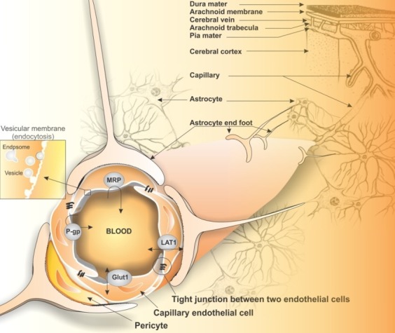
Schematic representation of microenvironment of brain capillary endothelial cells in association with astrocytes and pericytes. Star shape astrocytes in communication with both brain capillaries and neurons via end-foot. The ultra-structural characteristics such as tight junction of the brain capillary endothelial cells (BCECs) differentiate them from other peripheral capillaries. Specialized transport machineries are also involved at the luminal section of BCECs showing vesicular trafficking path used for traverse necessary macromolecules from blood to brain and vice versa.
As illustrated in Fig. 1, such intricate characteristics of BBB make brain drug delivery and targeting such a challenging field.
Our prime aim in this review is to provide basic overviews on the structure and functions of BCECs in relation to drug delivery and targeting hurdles to brain tumors. We will discuss the more recent advancement regarding multimodal and multifunctional nanomedicines and theranostics, which can circumvent BCECs restrictive barrier functions or even exploit the cellular molecular capacity of BBB (e.g., transport machineries) to get into brain central dogma for imaging and therapy of brain tumors such as glioblastomamultiforme (GBM).
BBB junctional complexes and cell-to-cell interactions
Structurally, the BBB is composed of four main cellular elements, including: 1) endothelial cells (ECs), 2) astrocyte end-feet, 3) microglial cells, and 4) pericytes. These cells are in close direct/indirect communications that make transport across the BBB as a selectively controlled process which is physically controlled via tight junction and physiologically through cell surface transport systems and enzymes (Correale and Villa 2009). Stable cell-to-cell interactions are required to keep the structural integrity of tissues. Dynamic changes in cell-to-cell adhesion will participate in the morphogenesis of developing tissues. In this regard, adhesion mechanisms are highly regulated during tissue morphogenesis and related to the processes of cell motility and cell migration. Regarding junctional bio-structures, the cell junctions at BBB site, can be classified into three functional groups, including: 1) tight junctions (TJs), 2) anchoring (adherent) junctions (AJs), and 3) gap (communication) junctions (GJs). Of these junctions, the TJs seal cells together in cell sheet, the AJs attach cells to their neighbors or to the extra-cellular matrix mechanically, and the GJs mediate the passage of chemicals or electrical signals from one interacting cell to its partner (Omidi and Gumbleton 2005).
Because of crucial role of TJs in restrictive function of BBB, these of note structures are briefly discussed. Fig. 2 represents the TJs and its complexity with other proteins at the BBB site. These TJs generate a rate-limiting restrictive barrier to paracellular diffusion of solutes between the brain microvasculature endothelial cells, in which they are the most apical elements of the junctional complexes. Morphologically, TJs form a continuous intricate network of interconnected proteins that are together arranged as a series of multiple barriers. It should be noticed that disruption of the BBB is a consistent phenomenon that may take place in the development of several CNS diseases, including brain tumors. In most cases, such pathological conditions are associated with an increase in the microvascular permeability, vasogenic edema, swollen astrocyte end feet, and BBB disruption (Nico and Ribatti 2012).
Fig. 2 .
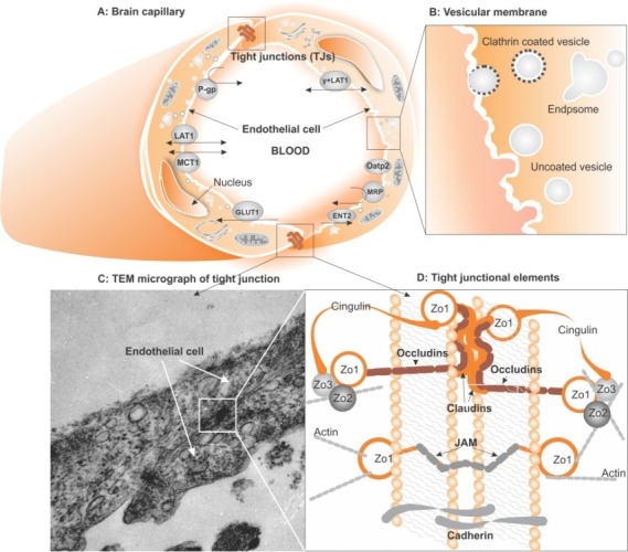
Schematic illustration of brain capillary endothelial cells (BCECs) and transmission electron microscopy (TEM) micrograph of tight junctions (TJ) and associated components. A) Schematic representation of two elongated brain capillary endothelial cells expressing apical transporters. B) Vesicular membrane of BCECs showing coated and uncoated vesicles. C) TEM micrograph of BCECs showing TJs as dense proteinaceous domains. D) Tight junctional interactions of two endothelial cells, showing embedded proteins within a cholesterol-enriched region of the plasma membrane. Claudins are multigene family that form the backbone of TJ strands by making dimers and binding homotypically to claudins on adjacent cells to produce the primary seal of the TJ. Claudin 1, 3 and 5 present at the BBB. Occludin functions as a dynamic regulatory protein causing increased electrical resistance across the membrane and decreased paracellular permeability.
Tight junctional complexes of BBB
The tight junctions between the brain microvasculature endothelial cells are very similar to the tight junction complexes of the epithelia cells than to the tight junctions of the endothelial in peripheral blood vessels. While TJs at the BBB site share many characteristics with epithelial tight junctions, there also display pivotal differences. The TJs at the BBB site, however, are highly sensitive to ambient factors resulting in disruption of the TJs at the BBB site. For example, the disruption of TJs at the protein and gene expression levels in the rat brain microvascular endothelial cell line (RBE4) was reported upon exposure to some neurotoxicants such as lead acetate, malathion and malaoxon, so that the occludin and claudin 5, and scaffold proteins ZO1 and ZO2 were markedly decreased after treatments (Balbuena et al 2011).
TJs disturbance, no matter from what provocative origin, can induce an augmented paracellular permeability, thus increased entry of inflammation related cells and molecules. Such impacts may result in the selective internalization of TJ transmembrane proteins such as occludin and claudin-5 via membranous caveolae (Stamatovic et al 2009). Thus, during the CNS inflammation, some pro-inflammatory events appear to mediate brain endothelial barrier disruption. Likewise, in human T cell leukemia virus (HTLV-1)-associated myelopathy/tropical spastic paraparesis as a neurodegenerative disease, the evidence of BBB breakdown has been demonstrated by the presence of lymphocytic infiltrates in the CNS and plasma protein leakage through cerebral endothelium (Afonso et al 2007). These all highlight regulation/dys-regulation of the tight junctional complexes resulting in the altered permeability characteristics of BBB, in which various ubiquitous molecular constituents are involved including claudins, occludin, zonula, cingulin, and 7H6. Signaling pathways involved in the regulation of TJs comprise G-proteins, serine, threonine, and tyrosine kinases, extra- and intracellular calcium levels, cAMP levels, proteases, and TNF alpha. In addition, the cytoskeletal elements are modulated based on receiving such signals that may also involve in some crucial cross-talk between components of the tight junctions and the cadherin-catenin within cell-cell based communications. Several identified molecular components of junctional complexes of the epithelia (claudins, occludins, zonula (ZO-1, ZO-2, ZO-3), junctional adhesion molecules (JAMs), cingulin, and 7H6) can be observed in the BCECs, in which both tight and adherent junctions are composed of multiple protein complexes, which communicate with the actin cytoskeleton of the cells (Kniesel and Wolburg 2000).
BBB endothelial cells in vivo reveal a P-face/E-face ratio of about 55%/45%, and as claudin-3 and claudin-5 are well expressed, it can be suggested that the degree of association with one or the other leaflet roughly reflects the stoichiometry of claudin expression in the TJs of BBB. However, in the non-BBB endothelial cells, tight junctions are almost completely associated with the E-face and claudin-3 is rarely or not expressed.
BCECs are integrated very tightly through the tight junctional complexes, making a unique morphology and architecture (Fig. 2). The phosphorylation of both transmembrane and accessory proteins plays an important role in establishing and regulating the TJs at the BBB site, in which the occludin and ZO1 are phosphorylated on serine, threonine and tyrosine residues. Protein kinase C (PKC) also is a major regulator of TJs formation and regulation through ZO1 migration to the plasma membrane. While TJs are generally localized at cholesterol-enriched regions or rafts within the plasma membrane, the integral protein within caveolae membrane domains (caveolin-1) seems to associate with TJ components, regulating the several downstream signaling pathways of TJs (Krizbai and Deli 2003).
Impacts of BBB associated cells: astrocytesand pericytes
Having an enigmatic role in the formation of BBB, the astrocytes represent different degrees of interaction with the BCECs (i.e., of the 11 distinct phenotypes distinguished, 8 involved specific interactions with blood vessels). Such interactions upregulate many BBB features and lead to the formation of tighter TJs and expression and polarized distribution of transporters as well as enzymes (Nico and Ribatti 2012, Correale and Villa 2009). Astrocytes are glial cells that envelop >99% of the BBB endothelium (Hawkins and Davis 2005).
Astrocytes and endothelial cells are in reciprocal interactions, modulating various biofunctions at the BBB site. Such interaction enhances the TJs and reduces the gap junctional area of BCECs, while it increases the number of astrocytic membrane particle assemblies and astrocyte density. Astrocytes are essential for proper neuronal functionalities, for which the close proximity of astrocytes and BCECs appear to be essential for a functional neurovascular unit (Abbott et al 2006). Although the nature of astrocyte-derived factors (ADFs) is not fully understood, their inductive effects on brain microvascular endothelial cell differentiation and BBB formation has been well documented (Abbott et al 2006).Based on our in vitro investigations, we have witnessed that the coculture of BCECs with astrocytes improved the BBB functionality such as permeability and cellular transport functions (Omidi et al 2008).
Fig. 3 represents the schematic illustration of brain capillary endothelial cells' interaction with astrocytes.
Fig. 3 .

Schematic representations of brain capillary endothelial cells' interaction with astrocytes. By the secretion of some actors, astrocytes are largely involved in the induction of certain BBB characteristics such as tighter TJs, specialized enzymatic systems, and polarized transporter localization. Astrocytes also play a pivotal role in the regulation of brain water and electrolyte metabolism. Astrocytes controls water flow via aquaporin- 4 and potassium channel Kir4.1 (the so-called water regulation and CSF homeostasis process). GDNF: glial derived neurotrophic factor. IL-6: Interleukin-6. bFGF: basic fibroblast growth factor. TGF-b1: transforming growth factor b1. A1: angiopoietin. LIF: leukemia inhibitory factor. cGT: c-glutamiltranspeptidase. AAD: aromatic acid decarboxylase. AP: alkaline phosphatase. MCT-1: monocarboxylate transporter 1. Glut1: glucose transporter 1. LAT-1: large neutral amino acid transporter 1. P-gp: P-glycoprotein. MRP2: multidrug resistance protein 2.
Investigation upon the modulatory effects of astrocyte on BCECs have revealed that the rat astrocyte cells are able to modulate the chick peripheral ECs to make them less permeable to large molecules. On the molecular level, the increased expression of barrier-relevant proteins (e.g., tight junction proteins) has so far been documented in the presence of ADFs. It can be deduced that GDNF is able to seal tightly the paracellular pathway in addition to its homeostasis role on the CNS. Moreover, it appears that factors secreted by brain endothelial cells including leukemia inhibitory factor (LIF) can induce astrocyte differentiation (Fig. 3). ADFs also influence the functionality of BBB carrier-mediated transport systems. Astrocytes control water flux via expression of a specific water channel termed as aquaporin-4 (AQP4) that is involved in the molecular composition of orthogonal particles' arrays (OAPs) on the perivascular glial end feet and tightly coupled with the maintenance of the BBB integrity (Nico and Ribatti 2012, Abbott 2005).
Pericytes are an imperative cellular constituent of the BBB, which also play a regulatory role in terms of brain angiogenesis and tight junction formation within BCECs (Armulik et al 2010). As shown in Fig.1, these cells also contribute to the microvascular vasodynamic capacity and structural stability (Balabanov and Dore-Duffy 1998). They are actively involved in the neuroimmune network operating at the BBB and confer macrophage functions. Having quantified pericyte coverage in different regions of the CNS, Armulik et al (2010) showed that pericyte coverage correlates with the BBB integrity. The pericyte and endothelial cell interaction occurs via cytoplasmic processes of the pericyte indenting the EC and vice versa. This contact process is called “peg and socket” - an interdigitation process (Wakui et al 1989).
Basically, some biomolecules including adhesive glycoprotein and fibronectin were found to be localized at the BCECs and pericytes junctional sites adjacent to “adhesion plaques” at the plasma membrane, which indicates the existence of a mechanical linkage between pericytes and ECs -- a linkage that allowed the mechanical contraction or relaxation of pericyte to influence vessel diameter. The cultured pericytes in the endothelial cell conditioned-medium (ECCM) allowed the cerebral pericyte aminopeptidase N (pAPN) to be re-expressed, while purified pericytes deprived of endothelial cells even in the presence of ACM showed no re-expression. This indicates that endothelial cells constitute an essential requirement for the in vitro re-expression of pAPN, but not astrocytes (Ramsauer et al 1998, Krause et al 1993). Pericytes are involved in the amino acid and peptide catabolism of brain (Krause et al 1993), indicating the metabolic role of pericytes on maintenance and homeostasis of the BBB.
Bioelectrical resistance and permeability of BBB
Given the correlation of transendothelial electrical resistance (TEER) with permeability, the TEER values have commonly been used to describe the restrictiveness and permeability of BBB. Technically, the permeability of BBB can be measured using 14 C-sucrose as a hydrophilic agent that is mainly transported via paracellular pathway. This value represents the tightness of biological barriers, which is about 1.2 ´ 10- 7 cm.sec- 1 for BBB in vivo. The TEER values vary in the epithelial and endothelial cells in vivo. For instance, human placental endothelium shows 22–52 Ω.cm 2 that permits the rapid paracellular exchange of nutrients and waste between the mother and fetus (Jinga et al 2000), whereas urinary bladder epithelium has a very high transepithelial resistance (>5000 Ω.cm 2 ), which is absolutely necessary for preserving urine composition (Powell 1981). The BBB possesses the TEER values of ~2000 Ω.cm 2 , which help to maintain brain homeostasis (Cohen-Kashi Malina et al 2009).
Efforts to generate a tight in vitro cell culture model for BBB have been largely based upon the measurement of TEER, while sucrose permeability assessments and the expression of specific enzymes and markers of the BBB have been utilized for reconfirmation. As a general rule, the higher the TEER value is, the lower the sucrose permeability and the tighter the BBB will be. To achieve this aim, different techniques have been recruited, e.g. the utilizing of hydrocortisone and serum free medium in order to increase the TEER (>700 W.cm 2 ) of primary cultures of porcine brain capillary endothelial cells (Omidi et al 2003, Smith et al 2007, Barar and Omidi 2008). Nevertheless no single immortalized cell line has shown high enough TEER value, which is a prerequisite characteristic of a cell line to be used as a tool for drug screening (Gumbleton and Audus 2001). Having compared two primary cultures of BCECs isolated from bovine and porcine, we witnessed no significant differences between these two primary BBB models even though porcine BCECs showed slightly better characteristics (Nakhlband and Omidi 2011).
Various factors can directly/indirectly play a role in the modulation of BBB restrictiveness. For example, extracellular matrix proteins were shown to influence the integrity of barrier functionality of BBB (Robert and Robert 1998, Tilling et al 1998). Using primary cultures of PBCECs, Tilling et al (1998) examined the effect of collagen IV, fibronectin, laminin and a secreted protein acidic and rich in cysteine alone or one-to-one mixtures of them. They showed that these proteins are involved in the tight junction formation between cerebral capillary endothelial cells by presenting increased TEER (Tilling et al 1998).
For the function of BBB, enzymes and other differentiation markers (P-glycoprotein efflux pump) are essential, and accordingly several markers have been identified, including: gamma-glutamyltranspeptidase (g-GTP) and alkaline phosphatase (ALP) enzymes expression, or antigenic endothelial cell markers such as Factor VIII , von Willebrand Factor (vWF) (Shi and Audus 1994, Tatsuta et al 1992, Betz et al 1980, Orlowski et al 1974, Abbott et al 1992).
Biotrafficking across BBB
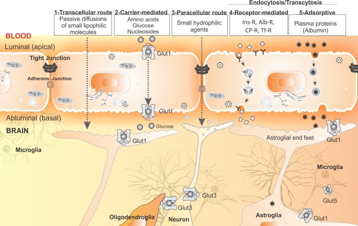
A number of parameters (physicochemical properties such as molecular weight (MW), lipophilicity, pKa, hydrogen bonding and biological factors) may influence trafficking of a particular substance to cross the BBB and enter the CNS. In general, transportation across BBB is classified into: 1) passive diffusion, which is largely dependent upon the physicochemical properties (in particular lipophilicity) of a compound, 2) paracellular trafficking of small hydrophilic compounds, 3) facilitated transport through carrier-mediated transport through transporters such as glucose transporter (Glut1) and large neutral transporter (LAT1), 4) receptor-mediated endocytosis/transcytosis, and 5) fluid-phase (adsorptive) endocytosis.
While various compounds (e.g., antitumor chemotherapies) exploit the transcellular path, the facilitated route is mostly used for macromolecules and hydrophilic compounds. To facilitate the inward and outward transportation, the carrier-mediated transporters are classified into two classes of transporters, namely outward (efflux) and inward (influx) transporters.
Fig. 4 represents the schematic illustration of BBB transport machineries.
Fig. 4 .
Schematic representation of transport machineries at the BBB site for shuttling of endogenous and/or exogenous substrates. 1) Lipid-soluble small substrates (<500 Da) are able to diffuse across the membrane – they may be effluxed back into the blood circulation through efflux transporters (e.g., P-gp, MRP4). 2) Carrier-mediated transport machineries (e.g., Glut1, Lat1) are responsible for small endogenous molecules (e.g., amino acids, nucleosides, and glucose). 3) Some small hydrophilic molecules can be transported via paracellular route. 4) Larger molecules (e.g., Ins-R=Insulin receptor; Alb-R=Albumin receptor; CP-R=Ceruloplasmin receptor; Tf-R=Transferrin receptor) are transported through receptor-mediated endocytosis/transcytosis using vesicular trafficking towards the brain parenchyma. 5) Some large proteins (e.g., albumin) are transported across the BBB by adsorptive-mediated endocytosis/transcytosis. Of the carrier-mediated transporters, glucose transporters (Gluts) are responsible for the traverse of glucose from blood to brain and between different cells within the brain parenchyma. Adherens junctions provide a path for the cell-to-cell intercommunication within the endothelial cells of BBB.
Passive diffusion and permeation
The central role of BBB is to protect the brain from entering of toxic compounds, thus many compounds (in particular the lipophilic drugs) can passively defuse through transcellular route, where nutrients are actively transported into the brain and possibly toxic compounds are expelled via active efflux pumps. This clearly means that the BBB permeation is a multifactorial and intricate process. And accordingly, the theoretical modeling of this process requires advanced computational methods. Currently, two major approaches are exploited for computational models, i.e. passive and active approaches. The passive diffusion-controlled permeability is dependent upon the inherent physicochemical characteristics (e.g., logP, solubility and surface area) of compounds, and basically molecular descriptor based methods are used to generate predictive models.
The ligand-receptor (i.e., influx/efflux transporters, or receptor-mediated endocytosis) active/facilitated transport can be considered for carrier/receptor mediated trafficking. In short, predictive in-silico models suitable for both the lead identification and the lead optimization processes should include both categories. The most commonly used type of data appear to be the logBB values that are described as the ratio of the steady-state concentration of a designated compound in the brain to that of the blood, i.e. LogBB=Log([CBrain]/[CBlood]). The most commonly used in vitro model is the Transwell™ system based on BCECs, which consists of a porous membrane support submerged in the culture media. As shown in Fig. 4, this system is normally characterized by the two-direction diffusion, i.e. apical to basal (A to B) or basal to apical (B to A). Given existence of large number of drug-like compounds (e.g., ChemNavigator), a very small number of molecules have been drawn to carefully monitor the main permeation driving/limiting force (i.e. passive diffusion, active influx or active efflux), however the data available mostly represent both passive and active transport phenomena. For detailed information, reader is directed to see (Wolburg 2006). In short, in a simplistic view, the absorption of drug molecules across a BBB depends upon: 1) the rate of drug dosing which takes into account the administered dose (mass) and the dosing interval (τ; time), 2) the interactions of drug molecules with circulating biomolecules blood (protein binding), 3) drug biostability and clearance, 3) the apparent absorption rate constant for the drug (Ka; time- 1 ). Clearly, the stability of drug during the absorption process and importantly the intrinsic permeability of BBB to the drug are critical factors in determining logBB.
The passive diffusion involves the movement of drug molecules down a concentration or electrochemical gradient without the expenditure of energy and the overall flux (J) of a drug in one dimension (i.e. the net mass of drug that diffuses through a unit area per unit time) which can be described by Equation (1).

Where; J is the flux of drug; D is the diffusion coefficient of drug across the cellular barrier; Kp is a global partition coefficient (cell membrane/aqueous fluid); A is the surface area of barrier available for absorption; x is the thickness of absorption barrier, and (dC/dx)t is the concentration gradient of drug across the absorption barrier.
Passive diffusion/permeation route is the main route for the entry of many anticancer chemotherapy agents even though they can mostly be pumped out by ABC efflux pumps.
Various drugs are substrate to the carrier-mediated transport machineries of BBB. Fig. 5 demonstrates the schematic representation of efflux and influx transport machineries of BCECs.
Fig. 5 .
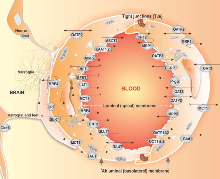
Carrier-mediated transport machineries (efflux and the influx transporters) at the BBB sites of the BBB. Astrocytes, neurons and microglial cells intercommunicate with brain capillary endothelial cells. The transport machineries of BBB are asymmetrically distributed at the luminal and abluminal sites. P-gp: P-glycoprotein. MRP1: multidrug resistance associated protein 1. MRP4: multidrug resistance associated protein 4. MRP5: multidrug resistance associated protein 5. BCRP: breast cancer resistance protein. Glut1: glucose transporter 1. MCT1: monocarboxylate transporter 1. LAT1: large neutral amino acids transporter 1. ASCT2: neutral amino acid transporter 2. EAAT: amino acid transporters. CNT2: concentrative nucleoside transport 2. SERT: serotonin transporter. NET: norepinephrine transporter. CRT: creatine transporter. TAUT: taurine transporter. OATP2: organic anion-transporting polypeptide. OAT3: organic anion transporter 3. OATP1A2: organic anion-transporting polypeptide. OAT2: organic anion transporter 2. OCT 1, 2: organic cation transporter 1, 2. ENT2: equilibrative nucleoside transport 2.
Efflux transporters
A key physiological function of ABC transporters is the protection of cells from many toxic insults from either endogenous or exogenous molecules that can enter the cell by diffusion or active uptake (Fletcher et al 2010). However, the ABC efflux pumps at the BBB site also represent the major blockade control on the entrance of several compounds by pumping them out of the CNS. Of these efflux pumps, P-glycoprotein (P-gp; MDR1/ABCB1 with 12 transmembrane segments) is an ATP-binding cassette (ABC) drug transport protein that is predominantly found in the apical membranes of a number of epithelial cell types in the body as well as the brain microvessel endothelial cells. It has been shown that mouse mdr1a and the human MDR1 P-gpactively transport the ivermectin, dexamethasone, digoxin, and cyclosporine and, to a lesser extent, morphine across a polarized kidney epithelial cell layer in vitro. The investigator reported that the injection of radiolabeled substrates of P-gp in mdr1a knockout and wild-type mice resulted in 20- to 50-fold higher levels of radioactivity in the mdr1a knockout mice brain for digoxin and cyclosporine (Schinkel et al 1995b). These researchers generated mice with a genetic disruption of the drug-transporting mdr1a P-gp and showed that the P-gp knockout mice were overall healthy but they accumulated much higher levels of substrate drugs in the brain with markedly slower elimination. For drugs (e.g., anticancer agents) that are P-gp substrates, this can lead to dramatically increased toxicity (Schinkel et al 1995a).
Overexpression of P-gp in cancer cells in response to the chemotherapy agents (multidrug resistance) along with other chemoresistance mechanisms make brain tumor therapy a challenging hurdle (Robey et al 2010, Cascorbi and Haenisch 2010). The MDR1 gene detection has been reported in all grade brain tumors and in endothelial cells of newly formed capillaries, thus impairing drug access at the tumor cell level (Fattori et al 2007).
It should be stated that most of anticancer agents are substrates ABC. Of these, the MDR1/ABCB1 can pump out a wide range of compounds (e.g., acebutolol, actinomycin D, amprenavir, azidopine, betamethasone, calcein-AM, cepharanthin, cerivastatin, chloroquine, cimetidine, clarithromycin, colchicine, cortisol, cyclosporine, daunorubicin, dexamethasone, digitoxin, digoxin, dipyridamole, docetaxel, domperidone, doxorubicin, eletriptan, emetine, epinastine, erythromycin, estradiol-17b-D-glucuronide, estrone, ethynylestradiol, etoposide, fexofenadine, grepafloxacin, imatinib, indinavir, irinotecan, ivermectin, lansoprazole, levofloxacin, loperamide, losartan, lovastatin, methylprednisolone, mitoxantrone, morphine, neostigmine, omeprazole, pantoprazole, prazosin, prednisolone, puromycin, quinidine2, ramosetron, ranitidine, reserpine, ritonavir, saquinavir, somatostain, sparfloxacin, talinolol, paclitaxel, terfenadine, trimethoprim, vecuronium, verapamil, vinblastine, vincristine), while the MRP4/ABCC4 is able to efflux a narrower spectrum (e.g., cAMP, cGMP, Dehydroepiandrosterone-3-sulfate, Estradiol-17b-D-glucuronide, Folate, Methotrexate, Prostaglandin E1, Prostaglandin E2).
Besides, the functional expression of multidrug resistance-associated proteins (MRPs) which actively transport a broad range of anionic compounds out of the cell were reported in the BCECs (Zhang et al 2000, Begley 2004). The breast cancer resistance protein BCRP/ABCG2 has been reported to be expressed as active efflux drug transporters at the BBB that can display efflux functionality similar to that of P-gp and MRP (Leslie et al 2005, Agarwal et al 2011). While many antitumor agents are substrate to P-gp, drugs inhibiting the MDR1 P-gp activity should be co-administered during chemotherapy of the brain tumors. Various compounds were reported as inhibitors for these efflux machineries.
Influx transporters
As described by Ohtsuki and Terasaki (2007), the inward transportation at the BBB site are mediated by several influx transporters that can be divided into several groups. Various compounds are substrate to such influx transport machineries (Ohtsuki and Terasaki 2007).
The energy transport systems include several transporters such as glucose transporter (Glut1) for the transport of glucose and mannose; monocarboxylate transporters (MCTs) for the transport of lactate, short-chain fatty acids, biotin, salicylic acid and valproic acid; and creatine transporter (CRT).
The amino acid transport systems consist of small and large neutral amino acid transporter systems (LAT2/4F2hc and LAT1/4F2hc, respectively) for the transport of neutral amino acids and L-dopa; acidic amino acid transporter for aspartate and glutamate (ASCT2); basic amino acid transporter (BAAT) for arginine and lysine; the b-amino acid transporter for b-alanine and taurine (TAUT); System A (ATA2) for small neutral amino acids; System ASC/system B + .
The organic anion transport systems include oatp2 and oatp14 for digoxin and organic anions, and OCTN2 for the transport of carnitine.
The nucleoside transport systems include CNT2. The peptide transport systems are oligopeptide transporters (PepT1, PepT2), polypeptide transport systems such as OAT3 for PAH, HVA, indoxylsulfate; oatp14 for thyroid hormones. The neurotransmitter transport systems such as GAT2/BGT1, SERT and NET are respectively used for the transport of g-aminobutyric acid, serotonin and norepinephrine. The choline transport system is for the transportation of choline and thiamine at the BBB site; readers are directed to see excellent reviews (Ohtsuki and Terasaki 2007, Terasaki et al 2003).
To enhance the brain uptake of neurotherapeutic agents, some of these transporters have successfully been used in prodrug development. L-dopa and progabide appear to the classical paradigms for such application, while both pyrimidine and purine nucleoside analogs are currently used clinically as anti-metabolite drugs. Nucleoside transporters have been exploited for the design of some anticancer agents (Omidi and Gumbleton 2005). For example, cytarabine is an analog of deoxycytidine (1-b-d-arabinofuranosylcytosine, araC, Cytosar-Us), which is used as combination chemotherapy in the treatment of chronic myelogenous, leukemia, multiple myeloma, Hodgkin’s lymphoma and non-Hodgkin’s lymphomas. Gemcitabine (dFdC, 2',2'-diuorodeoxycytidine, Gemzars) is a broad-spectrum agent that is used for the treatment of a variety of cancers including pancreatic and bladder cancers. Both cytarabine and gemcitabine are substrate to nucleoside transporter 1 (ENT1) (Hubeek et al 2005, Marce et al 2006). Capecitabine (5'-deoxy-5-N-[(pentoxy) carbonyl]-cytidine, Xelodas) is a prodrug, which is employed in the treatment of metastatic colorectal cancer (Mata et al 2001). Nucleoside transporters are responsible for the transportation of two purine nucleoside anti-metabolite drugs, fludarabine (9-b-d-arabinofuranosyl-2-.uoroadenine) (Elwi et al 2009, Molina-Arcas et al 2005), and cladribine (2-chloro-2'-deoxyadenosine, CdA, Leustatins) that are used for the treatment of low-grade lymphomas and chronic lymphocytic leukemia.
Endocytic pathway and transport of macromolecular nanostructures
Exogenous and endogenous macromolecules’ trafficking is mediated through cell membranous vesicular machinery domains that comprise numerous components including lipid rafts, caveolae and clathrin-coated pits. It should be evoked that fluid-phase endocytosis or adsorptive-mediated transcytosis (AMT) is also largely involved in the internalization of macromolecules (Herve et al 2008). All these cell membrane machineries seem to involve in the endocytosis, exocytosis and transcytosis of macromolecules.
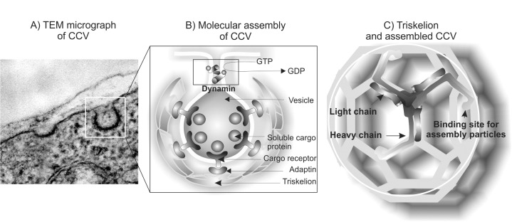
Clathrin-mediated endocytosis is the most widely studied vesicular membrane internalizing system, and the participation of clathrin-coated vesicles has also been investigated in terms of receptor-mediated transport in the BBB. Clathrin forms a non-covalently bound triskelion structure composed of three heavy chains (192 kDa each) and three light chains.
Membranous caveolae domains are flask-shaped invaginations of the plasma membrane coated by a 22 kDa structural protein caveolin-1 that is involved in the travers of small and large molecules (i.e., from small molecules like folate to macromolecules like albumin and lipoproteins). These micro-domains are highly enriched in glycosphingolipids, cholesterol, sphingomyelin, and lipid-anchored membrane proteins. Caveolae have been implicated in a wide range of cellular functions including transcytosis, receptor-mediated uptake, stabilization of lipid rafts and compartmentalization of a number of signaling events at the cell surface. Several studies have also shown that the caveolae-mediated uptake of materials is not limited to macromolecules; in certain cell-types, viruses (e.g. simian virus 40) and even entire bacteria (e.g. specific strains of E. Coli) are engulfed and transferred to intracellular compartments in a caveolae-dependent fashion (Smith and Gumbleton 2006, Omidi and Gumbleton 2005).
Fig. 6 represents schematic illustration of the clathrin-coated vesicles (CCVs) and its main protein “triskelion”.
Fig. 6 .
TEM micrograph (A) and schematic illustration of molecules involved in assembly of the clathrin coated vesicle (B) and its main protein triskelion (C). TEM: transmission electron microscopy; CCV: clathrin coated vesicle.
Endocytosis is the main path of nanoparticles' (NPs) entry into CNS; for detailed information reader is directed to see excellent review by Gumbleton’s group (Smith and Gumbleton 2006). Accordingly, several studies have showed that receptors such as transferrin (Tf) enhance brain delivery of NPs in vivo. For example, to uncover the precise mechanism of such uptake, Chang et al (2009) studied the endocytosis of poly(lactic-co-glycolic acid) (PLGA) NPs (<90 nm) coated with Tf using a BCECs co-culture with astrocytes. They found that, unlike unlabeled NPs, Tf-labeled NPs were profoundly endocytosed through an energy-dependent process and remarked that the Tf-labeled PLGA NPs interact with the cells in a specific manner and enter the cells via the caveolae pathway (Chang et al 2009). However, Tf conjugated biodegradable polymersomes (Tf-PO) with diameter of approximately 100 nm were shown to be uptaken through a clathrin mediated energy-dependent endocytosis in the bEnd.3 cells (Pang et al 2011). Similarly, lactoferrin (Lf)-modified procationic liposomes (Lf-PCLs) were recently evaluated in the primary BCECs and the uptake was found to be mediated by both clathrin-dependent (receptor-mediated) and absorption-mediated transcytosis (Chen et al 2010a).
Further, BBB is able to restrict the transport of IgG from the blood to the brain, while IgG undergoes efflux from the brain parenchyma via reverse transcytosis across the BBB mediated by FcRn. Such fast elimination of therapeutic antibodies from the brain through endocytic pathway is deemed to limit their therapeutic potency (Caram-Salas et al 2011). Besides, the adsorptive-mediated transcytosis (AMT) is deemed to provide a means for the brain delivery of medicines across the BBB, in particular using cationic NPs tagged with cell-penetrating peptides (CPPs). Two classes of CPPs (Tat-derived peptides and Syn-B vectors) have extensively been exploited for the endocytosis of neurotherapeutics (Herve et al 2008), while liposomal nanostructures may be used for the delivery of CNS drugs (Orthmann et al 2010). Clathrin coated pits were shown to play a key role in the transportation of poly(methoxypolyethyleneglycol cyanoacrylate-co-hexadecylcyanoacrylate) (PEG-PHDCA) NPs, in which an energy-dependent endocytosis as well as low-density lipoprotein receptors were shown to be involved (Kim et al 2007).
Despite the profound toxicity of polyethyleneimine (PEI) (Kafil and Omidi 2011), the magnetic nanoparticles modified with PEI (GPEI) havebeen used as a potential vascular drug/gene carrier to brain tumors with results that intra-carotid administration in conjunction with magnetic targeting significantly increased in the tumor entrapment of GPEI as compared to that of intravenous administration (Chertok et al 2010). It was also shown that the magnetic accumulation of cationic GPEI (zeta-potential = + 37.2 mV) in tumor lesions was 5.2-fold higher than that achieved with slightly anionic G100 (zeta-potential = -12 mV) following intra-carotid administration (Chertok et al 2010).
Jallouli et al studied theBBB uptake and transcytosis of 60 nm porous NPs differing in their surface charge and inner composition. Having used maltodextrins with/without a cationic ligand, they showed that the cationic NPs were accumulated mainly around the paracellular area, while neutral NPs were mainly on the cell surface and the dipalmitoylphosphatidyl glycerol (DPPG) NPs were at both paracellular areas and on the surface of cells. It was shown that the filipin can increase the binding and uptake, while the transcytosis of neutral NPs was inhibited by filipin. They concluded that the neutral NPs, like LDL, exploit the caveolae pathway and suggested the neutral and cationic 60 nm porous NPs as potential candidates for drug delivery to the brain (Jallouli et al 2007).
Having used a cross-reacting material 197 (CRM197) which is a non-toxic mutant of diphtheria toxin, Wang et al reported that the apical-to-basal transcytosis of CRM197 can involve the caveolae-mediated pathway in the hCMEC/D3 endothelial cells as the caveolin-1 mRNA and protein expression levels were significantly increased by CRM197. These researchers speculated that the upregulation of caveolin-1 may be mediated via a PI3K/Akt dependent pathway and reduction of the phospho-FOXO1A (forkhead box O) transcription factor. Based upon such findings, it was proposed that carrier protein CRM197-mediated delivery across the BBB is involved in the induction of FOXO1A transcriptional activity and the upregulation of caveolin-1 expression (Wang et al 2010). Similarly, CRM197-grafted polybutylcyanoacrylate NPs have been used for the delivery of zidovudine across human brain-microvascular endothelial cells (Kuo and Chung 2012).
In short, to internalize the exogenous materials, it seems that the BCECs exploit a variety of endocytic pathways (i.e., clathrin-mediated endocytosis, caveolar endocytosis, fluid phase endocytosis and macropinocytosis). Perhaps, the physicochemical characteristics of drug delivery nanocarriers and their interactions with cell surface elements dictate the internalization mechanisms to take place. Likewise, using fixed-size NPs, it has been shown that the surface modifications of nanoparticles (e.g., charge and protein ligands) can affect their mode of internalization by BCECs and thereby the subcellular fate (Georgieva et al 2011).
Targeted therapy of brain tumours
The integrity of BBB in metastatic cancerous tumors appears to be different from the normal ones, thus most promising antitumor drugs effective against cancers outside the brain have failed to provide clinical benefits against brain tumors, in part because of poor penetration of the antitumor drugs into the brain parenchyma (Ningaraj et al 2007). Thus, various cancer antigens have been targeted through conjugated immunotherapies.
Immunotoxins
Targeted immunotoxins are the bioconjugates of cancer specific marker targeting agent (usually monoclonal antibody) with a cytotoxic toxin, which have been developed for the targeted therapy of solid tumor such as GBM (Li and Hall 2010).
Of these, Interleukin-4 (IL-4) is a pleiotropic cytokine which is primarily produced by Th2-type T lymphocytes, mast cells, and basophils. Given that human malignant glioma and astrocytictumor overexpress high-affinity IL-4 receptors, IL-4 was reported to inhibit cell proliferation through theinduction of JAK/STAT pathway. Interestingly, IL-4 conjugation with pseudomonas exotoxin (IL4-PE; NBI-3001) was reported as highly and specifically cytotoxic-targetedbioconjugate against GBM, but with less cytotoxic to hematopoietic and normal brain cells (Shimamura et al 2006, Weber et al 2003). Interleukin-13 (IL-13) secreted by activated type 2 T lymphocytes and mast cells, is a pleiotropic lymphokine. IL-13 receptors (IL-13Rs) were shown to be overexpressed in various solid tumors cells including brain tumor, thus they are considered as tumor-specific markers. This has justified the development of an IL-13 based immunotoxin (e.g., recombinant fusion cytotoxin IL13-PE38QQR or cintredekinbesudotox) for targeted therapy of brain tumor (Shimamura et al 2006).
The epidermal growth factor receptor (EGFR), a 170 kDatransmembrane protein with extracellular receptor domain, is overexpressed in many solid tumors such as GBM. To control EGFR dimerization, immunotoxin TP-38 have been developed. TP-38 is a 43.5 kDa recombinant protein fusing pseudomonas exotoxin (PE-38) with TGF-α which can specifically target the EGFR (Sampson et al 2003).
ONTAK (DAB389IL-2) is a ligand fusion toxin consisting of the full-length sequence of IL-2 gene fused to the enzymatically active and translocating domains of diphtheria toxin (DT) that has been approved by the US Food and Drug Administration (FDA) for cutaneous T-cell lymphoma in 1999 (Frankel et al 2002).
Tf-CRM107 is conjugate protein of DT with a point mutation (CRM107) linked by a thioester bond to human Tf based on the ground that Tf receptor (a transmembrane glycoprotein) is significantly upregulated in dividing cells (Laske et al 1997).
Given the roles of the serine protease urokinase-type plasminogen activator (uPA) and its receptor (uPAR) in glioma-cell invasion and neovascularization, the recombinant fusion protein DTAT (encoding DT, a linker, and the downstream 135-amino terminal fragment portion of human urokinase plasminogen activator) was developed (Vallera et al 2002). It targets uPAR and delivers the potent catalytic portion of DT to the uPAR presenting cells simultaneously targeting both overexpressed uPAR on GBM cells and on tumor neovasculature. A bi-specific immunotoxin DTAT13 has also been developed to target concurrently uPAR and IL-13 receptor expressing GBM cells (Rustamzadeh et al 2006).
However, the main pitfall for these targeted toxins is their limited access to brain due to the excellent barrier presence of BBB, in which the diffusion rate of these immunotoxins into brain tumors is tightly controlled. Besides, the conjugated toxins are foreign proteins and cancer patients often develop neutralizing antibodies that impairs the retreatment strategies in case of recurrence.
Other strategies
Since cell membrane ion channels, as essential biomachineries, play a pivotal role in cell proliferation as well as cancer cell development and progression, the calcium-dependent potassium (KCa) channels have been targeted in brain tumor using various strategies (Ningaraj et al 2002, Hu et al 2007). Further, recent investigations have proven that the kinase inhibitors are thepromising new class of therapeutics that may control gliomas, in which the specificity of receptor tyrosine kinase inhibitors (RTKIs) on various solid tumors have been shown (Ningaraj et al 2007).
Angiogenesis also plays a central role in malignant primary brain tumor growth, in which the vascular endothelial cell growth factor (VEGF) and the basic Fibroblast Growth Factor (bFGF) were shown to bind to their receptors to promote glioma growth. While the VEGFR is expressed in human high grade glioma (but not in normal cells), it has been targeted using mAbs and macromolecular nanosystems (Jensen 2009).
In short, to enhance the dose of therapeutic agent to a brain tumor, a number of strategies have been exploited including: 1) increasing drug plasma concentration (e.g., intra-arterial infusion), 2) chemical modification to increase drug permeability, 3) design of inactive drug precursors (the so-called prodrugs) that could more easily cross the blood–brain barrier before conversion to a drug with active formulation, and 4) osmotic disruption of the blood–brain barrier using osmotic-disruptive agents such as mannitol (Provenzale et al 2005). In fact, the most significant challenges facing brain tumor drug delivery is development and advancement of effective brain targeting technology with BBB crossing potential. Recent advances in nanotechnology appear to provide promising solutions to this challenge. Several nanocarriers (polymer and lipid based NPs, dendrimers, nanogels, nanoemulsions and nanosuspensions) appear to provide promising drug delivery platform (Wong et al 2011).
Multimodal nanomedicines and theranostics
Endeavors to engineer multimodal nanomedicines and theranostics have resulted in various nanosystems showing great promising in vivo corollaries (Roger et al 2011). Of these, liposomal nanoformulations have widely been studied because they are deemed to the most biocompatible nano-scaled delivery system that can passively (through enhanced permeation and retention (EPR) effects) and/or actively (by conjugation of homing devices) target tumors (Micheli et al 2012). Various specific features (e.g., vesicle size, chemical affinity, and thermal/pH sensitiveness) of liposomes can be tuned affecting the targeting potential of liposomes. It has been shown that the BBB may be circumvented by NPs with a size dimension smaller than 50 nm or through lipid-mediated transport or receptor-mediated and PEG-assisted processes (Veiseh et al 2005).
Cross-linked iron oxide nanoparticles
Given that the precise delineation of tumor margins is a crucial matter for successful surgical resection of brain tumors, the development of multimodal imaging and therapy nanosystems are essential for intraoperatively visualizing tumor boundaries.
Cross-linked iron oxide (CLIO) NPs conjugated to Near-infrared (NIR, at a range of 700-900 nm) fluorescence detection avoids the background fluorescence interference of natural biomolecules. A CLIO-C5.5, which is detectable NP by both magnetic resonance imaging and fluorescence, has been recently developed (Veiseh et al, 2005). The probe was fabricated by coating iron oxide nanoparticles (NPs) with covalently bound bi-functional poly(ethylene glycol) (PEG) polymers that were subsequently functionalized with chlorotoxin (Cltx), a gliomatumor-targeting molecule, and the NIR fluorescing molecule Cy5.5. These researchers showed a significantly higher degree of internalization of NPC-Cy5.5 conjugates with high stability and prolonged retention (at least 24 h) within targeted glioma cells. Such NIR CLIO NPs havesuccessfully been used for the accuracy of tumor margin determination as similarly reported for orthotopictumors implanted in hosts with differing immune responses to the tumor (Trehin et al 2006).
In 2009, Veiseh et al reported the development of an iron oxide nanoparticle coated with polyethylene glycol-grafted chitosan which was able to cross the BBB and target brain tumors in a genetically engineered mouse model. The nanoprobe was conjugated to a tumor-targeting agent, Cltx, and a NIR fluorophore. Using in vivo magnetic resonance, biophotonic imaging, and histologic and biodistribution analyses, they showed an innocuous toxicity profile induced by the nanoprobe, while it showed a sustained retention in tumors and suggested its application for the diagnosis and treatment of a variety of tumor types in brain (Veiseh et al 2009).
To develop seamless nanosystems, a NS (polymer coated MNP core conjugated with green fluorescent protein (GFP) encoding DNA and Cltx) was engineered and their accumulation in the tumor site and specifically enhanced uptake of NPs into cancer cells were shown (Kievit et al 2010).
MNPs coated with dextran and functionalized with an anti-insulin-like-growth-factor binding protein 7 (anti-IGFBP7) single domain antibody was engineered and conjugated with Cy5.5. The developed anti-IGFBP7 armed MNP-Cy5.5 was able to selectively bind to abnormal vessels within a glioblastoma, while the MRI, NIR imaging, and fluorescent microscopy studies showed corresponding spatial and temporal changes (Tomanek et al 2012).
Fig. 7 represents the transmission electron microscopy and the laser scanning confocal microscopy images of incorporated multifunctional nanoprobe (PEG-Cltx-Cy5.5 magnetic nanoparticles) with target glioma cells.
Fig. 7 .
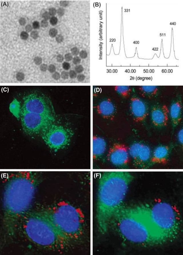
Transmission electron microscopy (A), X-ray diffraction (B), and confocal fluorescent images (C-F) of multifunctional PEG-Cltx-Cy5.5 magnetic nanoparticles (NPC-Cy5.5) used as ultrasound-MRI responsive agents. C) Rat cardiomyocytes (rCM). D) 9L glioma. E) Top section of the NPC-Cy5.5 treated 9L cells. F) Middle section of the NPC-Cy5.5 treated 9L cells. Data were adapted with permission from (Veiseh et al 2005).
Other theranostic systems
While use of chemotherapy and immunotherapy has been limited due to poor blood-brain barrier penetration, stimuli responsive multimodal nanomedicines may provide promising clinical outcomes.
NIR ultrasound (US) can transiently permeabilize the BBB and thus increase passive diffusion of therapies, while subsequent implementation of an external magnetic field can actively enhance the localization of a chemotherapeutic agent immobilized on a novel magnetic nanoparticle. This notion has successfully been examined and resulted in the significantly improved delivery of 1,3-bis(2-chloroethyl)-1-nitrosourea in rodent gliomas (Chen et al, 2010b). Besides, the MNPs conjugated with NIR fluorescing dyes (e.g., Cyanine dyes such as Cy5.5 or indocynine green) and a therapeutic agent can be used for simultaneous imaging and therapy of brain tumor as previously reported (Veiseh et al 2010, Veiseh et al 2009).
Fig. 8 represents the combined use of focused ultrasound (FUS) and magnetic targeting for the synergistic delivery of therapeutic MNPs across the BBB.
Fig. 8 .
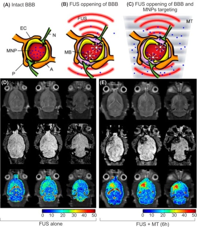
MRIgFUS drug delivery across BBB using polymer coated MNPs conjugated with Epirubicin. A) Intact CNS capillaries. B) Disruption of the BBB through activated of MBs by FUS, enhancing passive influx of therapeutic MNPs. C) Active delivery of the therapeutic MNPs to the brain through the use of combined magnetic targeting MT with FUS. D) In vivo imaging of MNP distribution in the brain using FUS alone. E) In vivo imaging of MNPs distribution in the brain using FUS and MT after 6 h post treatment. MRIgFUS: MRI-guided focused ultrasound. FUS: focused ultrasound. BBB: blood-brain barrier. MNPs: magnetic nanoparticles. MB: microbubbles. MT: magnetic targetingA: astrocyte. EC: endothelial cell. N: neuron. P: pericyte. Data were adapted with permission from (Liu et al 2010).
Liu et al showed that neither FUS alone nor magnetic targeting (MT) alonecaused significant MNP accumulation in the experimental brainsite. However, when FUS and MT were combined, MNP accumulation increased dramatically in brainsite. This study is a proof of technology that clarifies the effectiveness of the integrated nanomedicine platform to enhance and monitor the delivery of multifunctional nanoparticles to the brain (Liu et al 2012). Using MRI-guided focused ultrasound (MRIgFUS), macromolecules and even stem cells can be noninvasive delivered to the specific brain regionsfrom the blood (Burgess et al 2011). While MRIgFUS methodology shows great promising clinical results (Fig. 8), its combination with other imaging systems may advance its sensitivity and also responsiveness to exogenous stimuli. Epirubicin (a cytotoxic agent similar to doxorubicin) conjugated MNPs were profoundly able to alter the spin–spin relaxation rate (R2) and thus be used as anindicator of the MRI contrast agent (Liu et al 2010).
Also, quantum dots (QDs) armed antibody targeting brain tumor marker molecules may provide an imaging and therapy theranostics, however these NSs need to be further furnished with moieties to cross the BBB. They also need to be conjugated with chemotherapy agent to exert antitumor impacts in the target cells. Since the NIR pulsed laser can be used to stimulate the photoacoustic (PA) properties, conjugation of chemotherapies with an enhancing contrast agent may result in simultaneous detection and therapy using NIR US and fluorescence as non-invasive techniques, which may provide a more effective and tolerable means of tumor detection and treatment.
In 2010, Bellavance et al reported thedevelopment of a novel cationic liposome formulation composed of DPPC: DC-Chol:DOPE:DHPE Oregon Green, which was shown to possess efficient internalization and intracellular delivery to F98 and U-118 glioblastoma (GBM) cells in pH-sensitive manner. At which point, they suggested such liposomal formulation as a novel potent and efficient cytosolic delivery of intracellular therapeutics such as chemotherapy agents to the glioblastoma (Bellavance et al 2010).
Of the liposomal nanoformulations, the selectivity of PEGylated immunoliposomes based on monoclonal antibodies against the glial fibrillary acidic protein (GFAP) and the E2 extracellular loop of connexin 43 (MAbE2Cx43) with respect to the focus of a glioma was studied in experiments on animals with intracranial C6 glioma (Chekhonin et al 2012). The PEGylated stealth immunoliposomes labeled with a fluorescent (Dil C18) or a paramagnetic gadolinium-diethylenetriaminepentaacetic acid (Gd-DTPA) were injected to the tumor-bearing rats and fluorescent-labeled liposomal nanocontainers were detected at the periphery of the glioma 48 h after injection. The rats injected with paramagnetic immunoliposomes carrying MAbE2Cx43 showed distinct accumulation of the paramagnetic contrast agent at the periphery of the glioma, indicating usefulness of these immunoliposomal nano-containers for the targeted delivery of diagnostic and therapeutic drugs to the peritumoral invasion zone of high-grade gliomas (Chekhonin et al 2012).
Using novel quaternary ammonium beta-cyclodextrin (QAbetaCD) NPs (with 65-88 nm diameter and controllable cationic properties), Gil et al reported successful delivery of doxorubicin (DOX) across the BBB. They showed that QAbetaCD NPs are not toxic to bovine brain microvessel endothelial cells (BBMVECs) at concentrations up to 500 mg/mL. They also showed that the DOX/QAbetaCD complexes can kill U87 cells as effectively as DOX alone, while the QAbetaCD NPs completely protect BBMVECs from the cytotoxicity of DOX. And as a result, it was suggested that the QAbetaCD NPs acts as safe and effective delivery system for anticancer agents such as DOX for brain tumors (Gil et al 2009).
Complete resection in low-grade gliomas that show no MRI-enhanced images appear to be a challenging therapy which demands a robust and specific probe for detection and possibly on demand therapy. To tackle this, fluorescently photostable QDs armed with antibody targeting the epidermal growth factor receptor (EGFR) have been used and tumor cells were visualized with contrast ratios as high as 1000: 1 compared to normal brain tissue (Kantelhardt et al 2010).
In 2011, a dual-targeting drug carrier (PAMAM-PEG-WGA-Tf) was developed based on the PEGylated fourth generation PAMAM dendrimer with transferrin (Tf) and wheat germ agglutinin (WGA) on the periphery and doxorubicin (DOX) loaded in the interior. Having possessed nano-scale size (~ 20 nm), the PAMAM-PEG-WGA-Tf efficiently inhibited the growth rate of C6 glioma cells, while it reduced the cytotoxicity of DOX to the normal cells. These researchers reported significantly the increase and accumulation of DOX in the tumor site (due to the targeting effects of both Tf and WGA) and suggested it to be used as a BBB penetrating agent with tumor targeting properties (He et al 2011).
Hollow gold nanospheres (HAuNS) can generate profound photoacoustic signals and also induce efficient photothermal ablation (PTA) potential that can be used for simultaneous imaging and PTA of the target cancer cells. HAuNS targeted to integrins (overexpressed in glioma) were injected to tumor bearing mice, whose treatment with near-infrared laser resulted in an image-guided local tumor PTA therapy with photoacoustic molecular imaging (Lu et al 2011).
Concluding remarks
Despite the implementation of current therapeutic modalities for brain tumor therapy (i.e., surgical, radiological, and chemotherapeutic interventions), the malignant glioma as a severe primary brain tumor show high recurrence rate and an extremely high mortality rate within 2 years of diagnosis. While patients with primary brain tumors and brain metastases have a very poor prognosis, they show poor responsiveness to chemotherapy modalities, mainly because of excellent function of the BBB that selectively controls traverse of the administered chemotherapies. This clearly highlights essentiality for the advancement of brain tumor therapies towards smarter pharmaceutical. Nanomedicines appear to represent great promise in the therapy of brain tumors as they protect therapeutic agent, cross the BBB and allow the sustained liberation of encapsulated drugs (Orringer et al 2009). Once armed with homing and imaging devices, these nanomedicines they can undoubtedly act as seamless smart multimodal nanomedicines that can be used for the simultaneous targeting of tumor specific marker(s) and therapy. Such potentials of multimodal nanomedicines can provide monitoring means and on-demand therapy for brain tumor patients. For the design and engineering of such intelligent multimodal nanomedicines and theranostics, thus, the transportation mechanisms of BBB should be fully understood.
Ethical Issues
No ethical issues to be promulgated.
Conflict of interests
No conflict of interests to be declared.
Acknowledgments
This work was supported by Research Center for Pharmaceutical Nanotechnology (RCPN) and The Research and Technology Affairs at Tabriz University of Medical Sciences. Authors are thankful to Dr Mark Gumbleton, Welsh School of Pharmacy at Cardiff University. Authors are also grateful to Miss. R. Ilghami and Mr. F. Shokraneh (BioImpacts’ Editorial Team) for their editorial assistances.
References
- Abbott NJ . 2005 Dynamics of CNS barriers: evolution, differentiation, and modulation. Cell Mol Neurobiol, 25(1), 5-23 [DOI] [PMC free article] [PubMed] [Google Scholar]
- Abbott NJ, Hughes CC, Revest PA and Greenwood J . 1992 Development and characterisation of a rat brain capillary endothelial culture: towards an in vitro blood-brain barrier. J Cell Sci, 103(Pt 1), 103(Pt 1) 23-37 [DOI] [PubMed] [Google Scholar]
- Abbott NJ, Ronnback L and Hansson E . 2006 Astrocyte-endothelial interactions at the blood-brain barrier. Nat Rev Neurosci, 7(1), 41-53 [DOI] [PubMed] [Google Scholar]
- Afonso PV, Ozden S, Prevost MC, Schmitt C, Seilhean D, Weksler B, et al. 2007 Human blood-brain barrier disruption by retroviral-infected lymphocytes: role of myosin light chain kinase in endothelial tight-junction disorganization. J Immunol, 179(4), 2576-83 [DOI] [PubMed] [Google Scholar]
- Agarwal S, Hartz AM, Elmquist WF and Bauer B . 2011 Breast cancer resistance protein and P-glycoprotein in brain cancer: two gatekeepers team up. Curr Pharm Des, 17(26), 2793-802 [DOI] [PMC free article] [PubMed] [Google Scholar]
- Armulik A, Genove G, Mae M, Nisancioglu MH, Wallgard E, Niaudet C, et al. 2010 Pericytes regulate the blood-brain barrier. Nature, 468(7323), 557-61 [DOI] [PubMed] [Google Scholar]
- Balabanov R and Dore-Duffy P . 1998 Role of the CNS microvascular pericyte in the blood-brain barrier. J Neurosci Res, 53(6), 637-44 [DOI] [PubMed] [Google Scholar]
- Balbuena P, Li W and Ehrich M . 2011 Assessments of tight junction proteins occludin, claudin 5 and scaffold proteins ZO1 and ZO2 in endothelial cells of the rat blood-brain barrier: cellular responses to neurotoxicants malathion and lead acetate. Neurotoxicology, 32(1), 58-67 [DOI] [PubMed] [Google Scholar]
- Banks WA . 2010 Blood-brain barrier as a regulatory interface. Forum Nutr, 63, 102-10 [DOI] [PubMed] [Google Scholar]
- Barar J and Omidi Y . 2008 Bioelectrical and permeability properties of brain microvasculature endothelial cells: Effects of tight junction modulators. J Biol Sci, 8(3), 556-562 [Google Scholar]
- Begley DJ . 2004 ABC transporters and the blood-brain barrier. Curr Pharm Des, 10(12), 1295-312 [DOI] [PubMed] [Google Scholar]
- Bellavance MA, Poirier MB and Fortin D . 2010 Uptake and intracellular release kinetics of liposome formulations in glioma cells. Int J Pharm, 395(1-2), 251-9 [DOI] [PubMed] [Google Scholar]
- Betz AL, Firth JA and Goldstein GW . 1980 Polarity of the blood-brain barrier: distribution of enzymes between the luminal and antiluminal membranes of brain capillary endothelial cells. Brain Res, 192(1), 17-28 [DOI] [PubMed] [Google Scholar]
- Burgess A, Ayala-Grosso CA, Ganguly M, Jordao JF, Aubert I and Hynynen K . 2011 Targeted delivery of neural stem cells to the brain using MRI-guided focused ultrasound to disrupt the blood-brain barrier. PLoS One, 6(11), e27877 [DOI] [PMC free article] [PubMed] [Google Scholar]
- Caram-Salas N, Boileau E, Farrington GK, Garber E, Brunette E, Abulrob A, et al. 2011 In vitro and in vivo methods for assessing FcRn-mediated reverse transcytosis across the blood-brain barrier. Methods Mol Biol, 763, 383-401 [DOI] [PubMed] [Google Scholar]
- Cascorbi I and Haenisch S . 2010 Pharmacogenetics of ATP-binding cassette transporters and clinical implications. Methods Mol Biol, 596, 95-121 [DOI] [PubMed] [Google Scholar]
- Chang J, Jallouli Y, Kroubi M, Yuan XB, Feng W, Kang CS, et al. 2009 Characterization of endocytosis of transferrin-coated PLGA nanoparticles by the blood-brain barrier. Int J Pharm, 379(2), 285-92 [DOI] [PubMed] [Google Scholar]
- Chekhonin VP, Baklaushev VP, Yusubalieva GM, Belorusova AE, Gulyaev MV, Tsitrin EB, et al. 2012 Targeted delivery of liposomal nanocontainers to the peritumoral zone of glioma by means of monoclonal antibodies against GFAP and the extracellular loop of Cx43. Nanomedicine, 8(1), 63-70 [DOI] [PubMed] [Google Scholar]
- Chen H, Tang L, Qin Y, Yin Y, Tang J, Tang W, et al. 2010. a Lactoferrin-modified procationic liposomes as a novel drug carrier for brain delivery. Eur J Pharm Sci, 40(2), 94-102 [DOI] [PubMed] [Google Scholar]
- Chen PY, Liu HL, Hua MY, Yang HW, Huang CY, Chu PC, et al. 2010. b Novel magnetic/ultrasound focusing system enhances nanoparticle drug delivery for glioma treatment. Neuro Oncol, 12(10), 1050-60 [DOI] [PMC free article] [PubMed] [Google Scholar]
- Chertok B, David AE and Yang VC . 2010 Polyethyleneimine-modified iron oxide nanoparticles for brain tumor drug delivery using magnetic targeting and intra-carotid administration. Biomaterials, 31(24), 6317-24 [DOI] [PMC free article] [PubMed] [Google Scholar]
- Cohen-Kashi Malina K, Cooper I and Teichberg VI . 2009 Closing the gap between the in-vivo and in-vitro blood-brain barrier tightness. Brain Res, 1284, 12-21 [DOI] [PubMed] [Google Scholar]
- Correale J and Villa A . 2009 Cellular elements of the blood-brain barrier. Neurochem Res, 34(12), 2067-77 [DOI] [PubMed] [Google Scholar]
- Elwi AN, Damaraju VL, Kuzma ML, Baldwin SA, Young JD, Sawyer MB, et al. 2009 Human concentrative nucleoside transporter 3 is a determinant of fludarabine transportability and cytotoxicity in human renal proximal tubule cell cultures. Cancer Chemother Pharmacol, 63(2), 289-301 [DOI] [PubMed] [Google Scholar]
- Fattori S, Becherini F, Cianfriglia M, Parenti G, Romanini A and Castagna M . 2007 Human brain tumors: multidrug-resistance P-glycoprotein expression in tumor cells and intratumoral capillary endothelial cells. Virchows Arch, 451(1), 81-7 [DOI] [PubMed] [Google Scholar]
- Fletcher JI, Haber M, Henderson MJ and Norris MD . 2010 ABC transporters in cancer: more than just drug efflux pumps. Nat Rev Cancer, 10(2), 147-56 [DOI] [PubMed] [Google Scholar]
- Frankel AE, Powell BL and Lilly MB . 2002 Diphtheria toxin conjugate therapy of cancer. Cancer Chemother Biol Response Modif, 20, 301-13 [PubMed] [Google Scholar]
- Georgieva JV, Kalicharan D, Couraud PO, Romero IA, Weksler B, Hoekstra D, et al. 2011 Surface characteristics of nanoparticles determine their intracellular fate in and processing by human blood-brain barrier endothelial cells in vitro. Mol Ther, 19(2), 318-25 [DOI] [PMC free article] [PubMed] [Google Scholar]
- Gil ES, Li J, Xiao H and Lowe TL . 2009 Quaternary ammonium beta-cyclodextrin nanoparticles for enhancing doxorubicin permeability across the in vitro blood-brain barrier. Biomacromolecules, 10(3), 505-16 [DOI] [PubMed] [Google Scholar]
- Gumbleton M and Audus KL . 2001 Progress and limitations in the use of in vitro cell cultures to serve as a permeability screen for the blood-brain barrier. J Pharm Sci, 90(11), 1681-98 [DOI] [PubMed] [Google Scholar]
- Hawkins BT and Davis TP . 2005 The blood-brain barrier/neurovascular unit in health and disease. Pharmacol Rev, 57(2), 173-85 [DOI] [PubMed] [Google Scholar]
- He H, Li Y, Jia XR, Du J, Ying X, Lu WL, et al. 2011 PEGylated Poly(amidoamine) dendrimer-based dual-targeting carrier for treating brain tumors. Biomaterials, 32(2), 478-87 [DOI] [PubMed] [Google Scholar]
- Herve F, Ghinea N and Scherrmann JM . 2008 CNS delivery via adsorptive transcytosis. The AAPS journal, 10(3), 455-72 [DOI] [PMC free article] [PubMed] [Google Scholar]
- Hu J, Yuan X, Ko MK, Yin D, Sacapano MR, Wang X, et al. 2007 Calcium-activated potassium channels mediated blood-brain tumor barrier opening in a rat metastatic brain tumor model. Mol Cancer, 6, 22 [DOI] [PMC free article] [PubMed] [Google Scholar]
- Hubeek I, Stam RW, Peters GJ, Broekhuizen R, Meijerink JP, Cohen-Kashi MalinaVan Wering ER, et al. 2005 The human equilibrative nucleoside transporter 1 mediates in vitro cytarabine sensitivity in childhood acute myeloid leukaemia. Br J Cancer, 93(12), 1388-94 [DOI] [PMC free article] [PubMed] [Google Scholar]
- Jallouli Y, Paillard A, Chang J, Sevin E and Betbeder D . 2007 Influence of surface charge and inner composition of porous nanoparticles to cross blood-brain barrier in vitro. Int J Pharm, 344(1-2), 103-9 [DOI] [PubMed] [Google Scholar]
- Jensen RL . 2009 Brain tumor hypoxia: tumorigenesis, angiogenesis, imaging, pseudoprogression, and as a therapeutic target. J Neurooncol, 92(3), 317-35 [DOI] [PubMed] [Google Scholar]
- Jinga VV, Gafencu A, Antohe F, Constantinescu E, Heltianu C, Raicu M, et al. 2000 Establishment of a pure vascular endothelial cell line from human placenta. Placenta, 21(4), 325-36 [DOI] [PubMed] [Google Scholar]
- Kafil V and Omidi Y . 2011 Cytotoxic Impacts of Linear and Branched Polyethylenimine Nanostructures in A431 Cells. BioImpacts, 1(1), 23-30 [DOI] [PMC free article] [PubMed] [Google Scholar]
- Kantelhardt SR, Caarls W, Cohen-Kashi MalinaVan WeringDe Vries AH, Hagen GM, Jovin TM, Schulz-Schaeffer W, et al. 2010 Specific visualization of glioma cells in living low-grade tumor tissue. PLoS One, 5(6), e11323 [DOI] [PMC free article] [PubMed] [Google Scholar]
- Kievit FM, Veiseh O, Fang C, Bhattarai N, Lee D, Ellenbogen RG, et al. 2010 Chlorotoxin labeled magnetic nanovectors for targeted gene delivery to glioma. ACS Nano, 4(8), 4587-94 [DOI] [PMC free article] [PubMed] [Google Scholar]
- Kim HR, Gil S, Andrieux K, Nicolas V, Appel M, Chacun H, et al. 2007 Low-density lipoprotein receptor-mediated endocytosis of PEGylated nanoparticles in rat brain endothelial cells. Cell Mol Life Sci, 64(3), 356-64 [DOI] [PMC free article] [PubMed] [Google Scholar]
- Kniesel U and Wolburg H . 2000 Tight junctions of the blood-brain barrier. Cell Mol Neurobiol, 20(1), 57-76 [DOI] [PMC free article] [PubMed] [Google Scholar]
- Krause D, Kunz J and Dermietzel R . 1993 Cerebral pericytes--a second line of defense in controlling blood-brain barrier peptide metabolism. Adv Exp Med Biol, 331, 149-52 [DOI] [PubMed] [Google Scholar]
- Krizbai IA and Deli MA . 2003 Signalling pathways regulating the tight junction permeability in the blood-brain barrier. Cell Mol Biol (Noisy-le-grand), 49(1), 23-31 [PubMed] [Google Scholar]
- Kuo YC and Chung CY . 2012 Transcytosis of CRM197-grafted polybutylcyanoacrylate nanoparticles for delivering zidovudine across human brain-microvascular endothelial cells. Colloids Surf B Biointerfaces, 91, 242-9 [DOI] [PubMed] [Google Scholar]
- Laske DW, Youle RJ and Oldfield EH . 1997 Tumor regression with regional distribution of the targeted toxin TF-CRM107 in patients with malignant brain tumors. Nat Med, 3(12), 1362-8 [DOI] [PubMed] [Google Scholar]
- Leslie EM, Deeley RG and Cole SP . 2005 Multidrug resistance proteins: role of P-glycoprotein, MRP1, MRP2, and BCRP (ABCG2) in tissue defense. Toxicol Appl Pharmacol, 204(3), 216-37 [DOI] [PubMed] [Google Scholar]
- Li YM and Hall WA . 2010 Targeted toxins in brain tumor therapy. Toxins, 2(11), 2645-62 [DOI] [PMC free article] [PubMed] [Google Scholar]
- Liebner S, Czupalla CJ and Wolburg H . 2011 Current concepts of blood-brain barrier development. Int J Dev Biol, 55(4-5), 467-76 [DOI] [PubMed] [Google Scholar]
- Liu HL, Hua MY, Yang HW, Huang CY, Chu PC, Wu JS, et al. 2010 Magnetic resonance monitoring of focused ultrasound/magnetic nanoparticle targeting delivery of therapeutic agents to the brain. Proc Natl Acad Sci U S A, 107(34), 15205-10 [DOI] [PMC free article] [PubMed] [Google Scholar]
- Liu HL, Yang HW, Hua MY and Wei KC . 2012 Enhanced therapeutic agent delivery through magnetic resonance imaging-monitored focused ultrasound blood-brain barrier disruption for brain tumor treatment: an overview of the current preclinical status. Neurosurg Focus, 32(1), E4 [DOI] [PubMed] [Google Scholar]
- Lu W, Melancon MP, Xiong C, Huang Q, Elliott A, Song S, et al. 2011 Effects of photoacoustic imaging and photothermal ablation therapy mediated by targeted hollow gold nanospheres in an orthotopic mouse xenograft model of glioma. Cancer Res, 71(19), 6116-21 [DOI] [PMC free article] [PubMed] [Google Scholar]
- Marce S, Molina-Arcas M, Villamor N, Casado FJ, Campo E, Pastor-Anglada M, et al. 2006 Expression of human equilibrative nucleoside transporter 1 (hENT1) and its correlation with gemcitabine uptake and cytotoxicity in mantle cell lymphoma. Haematologica, 91(7), 895-902 [PubMed] [Google Scholar]
- Mata JF, Garcia-Manteiga JM, Lostao MP, Fernandez-Veledo S, Guillen-Gomez E, Larrayoz IM, et al. 2001 Role of the human concentrative nucleoside transporter (hCNT1) in the cytotoxic action of 5[Prime]-deoxy-5-fluorouridine, an active intermediate metabolite of capecitabine, a novel oral anticancer drug. Mol Pharmacol, 59(6), 1542-8 [DOI] [PubMed] [Google Scholar]
- Micheli MR, Bova R, Magini A, Polidoro M and Emiliani C . 2012 Lipid-Based Nanocarriers for CNS-Targeted Drug Delivery. Recent Pat CNS Drug Discov, 7(1), 71-86 [DOI] [PubMed] [Google Scholar]
- Molina-Arcas M, Marce S, Villamor N, Huber-Ruano I, Casado FJ, Bellosillo B, et al. 2005 Equilibrative nucleoside transporter-2 (hENT2) protein expression correlates with ex vivo sensitivity to fludarabine in chronic lymphocytic leukemia (CLL) cells. Leukemia, 19(1), 64-8 [DOI] [PubMed] [Google Scholar]
- Nakhlband A and Omidi Y . 2011 Barrier Functionality of Porcine and Bovine Brain Capillary Endothelial Cells. BioImpacts, 1(3), 153-159 [DOI] [PMC free article] [PubMed] [Google Scholar]
- Nico B and Ribatti D. 2012. Morphofunctional aspects of the blood-brain barrier. Curr Drug Metab. [DOI] [PubMed]
- Ningaraj NS, Rao M, Hashizume K, Asotra K and Black KL . 2002 Regulation of blood-brain tumor barrier permeability by calcium-activated potassium channels. J Pharmacol Exp Ther, 301(3), 838-51 [DOI] [PubMed] [Google Scholar]
- Ningaraj NS, Salimath BP, Sankpal UT, Perera R and Vats T . 2007 Targeted brain tumor treatment-current perspectives. Drug Target Insights, 2, 197-207 [PMC free article] [PubMed] [Google Scholar]
- Ohtsuki S and Terasaki T . 2007 Contribution of carrier-mediated transport systems to the blood-brain barrier as a supporting and protecting interface for the brain; importance for CNS drug discovery and development. Pharm Res, 24(9), 1745-58 [DOI] [PubMed] [Google Scholar]
- Omidi Y, Barar J, Ahmadian S, Heidari HR and Gumbleton M . 2008 Characterization and astrocytic modulation of system L transporters in brain microvasculature endothelial cells. Cell Biochem Funct, 26(3), 381-91 [DOI] [PubMed] [Google Scholar]
- Omidi Y, Campbell L, Barar J, Connell D, Akhtar S and Gumbleton M . 2003 Evaluation of the immortalised mouse brain capillary endothelial cell line, b .End3, as an in vitro blood-brain barrier model for drug uptake and transport studies. Brain Res, 990(1-2), 95-112 [DOI] [PubMed] [Google Scholar]
- Omidi Y and Gumbleton M. 2005. Biological Membranes and Barriers, In: Biomaterials for delivery and targeting of proteins and nucleic acids. Mahato R (Ed.) New York, CRC Press, 220-263.
- Orlowski M, Sessa G and Green JP . 1974 Gamma-glutamyl transpeptidase in brain capillaries: possible site of a blood-brain barrier for amino acids. Science, 184(4132), 66-8 [DOI] [PubMed] [Google Scholar]
- Orringer DA, Koo YE, Chen T, Kopelman R, Sagher O and Philbert MA . 2009 Small solutions for big problems: the application of nanoparticles to brain tumor diagnosis and therapy. Clin Pharmacol Ther, 85(5), 531-4 [DOI] [PMC free article] [PubMed] [Google Scholar]
- Orthmann A, Zeisig R, Koklic T, Sentjurc M, Wiesner B, Lemm M, et al. 2010 Impact of membrane properties on uptake and transcytosis of colloidal nanocarriers across an epithelial cell barrier model. J Pharm Sci, 99(5), 2423-33 [DOI] [PubMed] [Google Scholar]
- Pang Z, Gao H, Yu Y, Chen J, Guo L, Ren J, et al. 2011 Brain delivery and cellular internalization mechanisms for transferrin conjugated biodegradable polymersomes. Int J Pharm, 415(1-2), 284-92 [DOI] [PubMed] [Google Scholar]
- Powell DW . 1981 Barrier function of epithelia. Am J Physiol, 241(4), G275-88 [DOI] [PubMed] [Google Scholar]
- Provenzale JM, Mukundan S and Dewhirst M . 2005 The role of blood-brain barrier permeability in brain tumor imaging and therapeutics. AJR Am J Roentgenol, 185(3), 763-7 [DOI] [PubMed] [Google Scholar]
- Ramsauer M, Kunz J, Krause D and Dermietzel R . 1998 Regulation of a blood-brain barrier-specific enzyme expressed by cerebral pericytes (pericytic aminopeptidase N/pAPN) under cell culture conditions. J Cereb Blood Flow Metab, 18(11), 1270-81 [DOI] [PubMed] [Google Scholar]
- Reese TS and Karnovsky MJ . 1967 Fine structural localization of a blood-brain barrier to exogenous peroxidase. J Cell Biol, 34(1), 207-17 [DOI] [PMC free article] [PubMed] [Google Scholar]
- Robert AM and Robert L . 1998 Extracellular matrix and blood-brain barrier function. Pathol Biol (Paris), 46(7), 535-42 [PubMed] [Google Scholar]
- Robey RW, Massey PR, Amiri-Kordestani L and Bates SE . 2010 ABC transporters: unvalidated therapeutic targets in cancer and the CNS. Anticancer Agents Med Chem, 10(8), 625-33 [DOI] [PMC free article] [PubMed] [Google Scholar]
- Roger M, Clavreul A, Venier-Julienne MC, Passirani C, Montero-Menei C and Menei P . 2011 The potential of combinations of drug-loaded nanoparticle systems and adult stem cells for glioma therapy. Biomaterials, 32(8), 2106-16 [DOI] [PubMed] [Google Scholar]
- Rubin LL and Staddon JM . 1999 The cell biology of the blood-brain barrier. Annu Rev Neurosci, 22, 11-28 [DOI] [PubMed] [Google Scholar]
- Rustamzadeh E, Vallera DA, Todhunter DA, Low WC, Panoskaltsis-Mortari A and Hall WA . 2006 Immunotoxin pharmacokinetics: a comparison of the anti-glioblastoma bi-specific fusion protein (DTAT13) to DTAT and DTIL13. J Neurooncol, 77(3), 257-66 [DOI] [PubMed] [Google Scholar]
- Sampson JH, Akabani G, Archer GE, Bigner DD, Berger MS, Friedman AH, et al. 2003 Progress report of a Phase I study of the intracerebral microinfusion of a recombinant chimeric protein composed of transforming growth factor (TGF)-alpha and a mutated form of the Pseudomonas exotoxin termed PE-38 (TP-38) for the treatment of malignant brain tumors. J Neurooncol, 65(1), 27-35 [DOI] [PubMed] [Google Scholar]
- Schinkel AH, Mol CA, Wagenaar E, Cohen-Kashi MalinaVan WeringDe VriesVan Deemter L, Smit JJ and Borst P . 1995. a Multidrug resistance and the role of P-glycoprotein knockout mice. Eur J Cancer, 31A(7-8), 1295-8 [DOI] [PubMed] [Google Scholar]
- Schinkel AH, Wagenaar E, Cohen-Kashi MalinaVan WeringDe VriesVan DeemterVan Deemter L, Mol CA and Borst P . 1995. b Absence of the mdr1a P-Glycoprotein in mice affects tissue distribution and pharmacokinetics of dexamethasone, digoxin, and cyclosporin A. J Clin Invest, 96(4), 1698-705 [DOI] [PMC free article] [PubMed] [Google Scholar]
- Shi F and Audus KL . 1994 Biochemical characteristics of primary and passaged cultures of primate brain microvessel endothelial cells. Neurochem Res, 19(4), 427-33 [DOI] [PubMed] [Google Scholar]
- Shimamura T, Husain SR and Puri RK . 2006 The IL-4 and IL-13 pseudomonas exotoxins: new hope for brain tumor therapy. Neurosurg Focus, 20(4), E11 [DOI] [PubMed] [Google Scholar]
- Smith M, Omidi Y and Gumbleton M . 2007 Primary porcine brain microvascular endothelial cells: biochemical and functional characterisation as a model for drug transport and targeting. J Drug Target, 15(4), 253-68 [DOI] [PubMed] [Google Scholar]
- Smith MW and Gumbleton M . 2006 Endocytosis at the blood-brain barrier: from basic understanding to drug delivery strategies. J Drug Target, 14(4), 191-214 [DOI] [PubMed] [Google Scholar]
- Stamatovic SM, Keep RF, Wang MM, Jankovic I and Andjelkovic AV . 2009 Caveolae-mediated internalization of occludin and claudin-5 during CCL2-induced tight junction remodeling in brain endothelial cells. J Biol Chem, 284(28), 19053-66 [DOI] [PMC free article] [PubMed] [Google Scholar]
- Tatsuta T, Naito M, Oh-Hara T, Sugawara I and Tsuruo T . 1992 Functional involvement of P-glycoprotein in blood-brain barrier. J Biol Chem, 267(28), 20383-91 [PubMed] [Google Scholar]
- Terasaki T, Ohtsuki S, Hori S, Takanaga H, Nakashima E and Hosoya K . 2003 New approaches to in vitro models of blood-brain barrier drug transport. Drug Discov Today, 8(20), 944-54 [DOI] [PubMed] [Google Scholar]
- Tilling T, Korte D, Hoheisel D and Galla HJ . 1998 Basement membrane proteins influence brain capillary endothelial barrier function in vitro. J Neurochem, 71(3), 1151-7 [DOI] [PubMed] [Google Scholar]
- Tomanek B, Iqbal U, Blasiak B, Abulrob A, Albaghdadi H, Matyas JR, et al. 2012 Evaluation of brain tumor vessels specific contrast agents for glioblastoma imaging. Neuro Oncol, 14(1), 53-63 [DOI] [PMC free article] [PubMed] [Google Scholar]
- Trehin R, Figueiredo JL, Pittet MJ, Weissleder R, Josephson L and Mahmood U . 2006 Fluorescent nanoparticle uptake for brain tumor visualization. Neoplasia, 8(4), 302-11 [DOI] [PMC free article] [PubMed] [Google Scholar]
- Vallera DA, Li C, Jin N, Panoskaltsis-Mortari A and Hall WA . 2002 Targeting urokinase-type plasminogen activator receptor on human glioblastoma tumors with diphtheria toxin fusion protein DTAT. J Natl Cancer Inst, 94(8), 597-606 [DOI] [PubMed] [Google Scholar]
- Veiseh O, Gunn JW and Zhang M . 2010 Design and fabrication of magnetic nanoparticles for targeted drug delivery and imaging. Adv Drug Deliv Rev, 62(3), 284-304 [DOI] [PMC free article] [PubMed] [Google Scholar]
- Veiseh O, Sun C, Fang C, Bhattarai N, Gunn J, Kievit F, et al. 2009 Specific targeting of brain tumors with an optical/magnetic resonance imaging nanoprobe across the blood-brain barrier. Cancer Res, 69(15), 6200-7 [DOI] [PMC free article] [PubMed] [Google Scholar]
- Veiseh O, Sun C, Gunn J, Kohler N, Gabikian P, Lee D, et al. 2005 Optical and MRI multifunctional nanoprobe for targeting gliomas. Nano Lett, 5(6), 1003-8 [DOI] [PubMed] [Google Scholar]
- Wakui S, Furusato M, Hasumura M, Hori M, Takahashi H, Kano Y, et al. 1989 Two- and three-dimensional ultrastructure of endothelium and pericyte interdigitations in capillary of human granulation tissue. J Electron Microsc (Tokyo), 38(2), 136-42 [PubMed] [Google Scholar]
- Wang P, Xue Y, Shang X and Liu Y . 2010 Diphtheria toxin mutant CRM197-mediated transcytosis across blood-brain barrier in vitro. Cell Mol Neurobiol, 30(5), 717-25 [DOI] [PMC free article] [PubMed] [Google Scholar]
- Weber F, Asher A, Bucholz R, Berger M, Prados M, Chang S, et al. 2003 Safety, tolerability, and tumor response of IL4-Pseudomonas exotoxin (NBI-3001) in patients with recurrent malignant glioma. J Neurooncol, 64(1-2), 125-37 [DOI] [PubMed] [Google Scholar]
- Wolburg H. 2006. The Endothelial Frontier, In: Boold Brain Barriers. Dermietzel R, Spray DC and Nedergaard M (Ed.), Weinheim, WILEY-VCH Verlag GmbH & Co. KGaA.
- Wong HL, Wu XY and Bendayan R. 2011. Nanotechnological advances for the delivery of CNS therapeutics. Adv Drug Deliv Rev, (in press). [DOI] [PubMed]
- Zhang Y, Han H, Elmquist WF and Miller DW . 2000 Expression of various multidrug resistance-associated protein (MRP) homologues in brain microvessel endothelial cells. Brain Res, 876(1-2), 148-53 [DOI] [PubMed] [Google Scholar]


