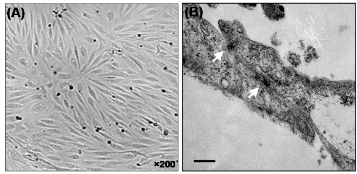
Fig. 1.

Light microscopy (A) and transmission electron micrographs (B) of the porcine brain capillary endothelial (PBCE) cells. Squamous morphology of the confluent cells (A) and two elongated primary PBCE cells cultured onto the Transwell™ polycarbonate membranes display typical morphology of the endothelial cells. The white arrows (B) represent the tight junctional elements between two flattened primary PBCE cells. Bar equals to 200 nm.
