Abstract
Sialolithasis is the most common salivary gland disease. A case of an unusually large sialolith arising in the submandibular gland is presented, along with a review of the management of giant salivary gland calculi.
INTRODUCTION
Sialolithiasis is the most common disease affecting adult salivary glands, accounting for more than 50% of all salivary gland conditions. Twelve per 1000 adult population are reported to suffer from the condition each year, with males affected more than females (1). Salivary stones most commonly occur in the submandibular glands (up to 90% of cases) and parotid glands (5 to 20%). The sublingual gland and minor salivary glands are rarely affected. The right and left sides are affected equally, with bilaterally arising stones being rare, accounting for less than 3% of cases (2). 88% of salivary calculi are reported to be less than 10mm in size (3) with review of the literature showing the occurrence of abnormally large (>15mm) salivary calculi to be rare. Salivary gland stones can occur at any age, yet occur most commonly between the third and sixth decades of life. This pattern is the same for giant calculi. Sialolithiasis of any size is deemed rare in the paediatric population.
The typical symptoms of salivary lithiasis are pain and swelling due to obstruction of the salivary ducts, classically at mealtimes. Giant salivary calculi have however been reported to remain asymptomatic for many months prior to presentation (4).
CASE REPORT
A 58-year-old male patient was referred from his general dental practitioner with a weeks history of pain in the right floor of mouth and submandibular region, exacerbated by swallowing. The patient gave a past medical history of oesophagitis and osteoarthritis, and took Esomeprazole and Diclofenac regularly. The patient had no known allergies and was a non-smoker.
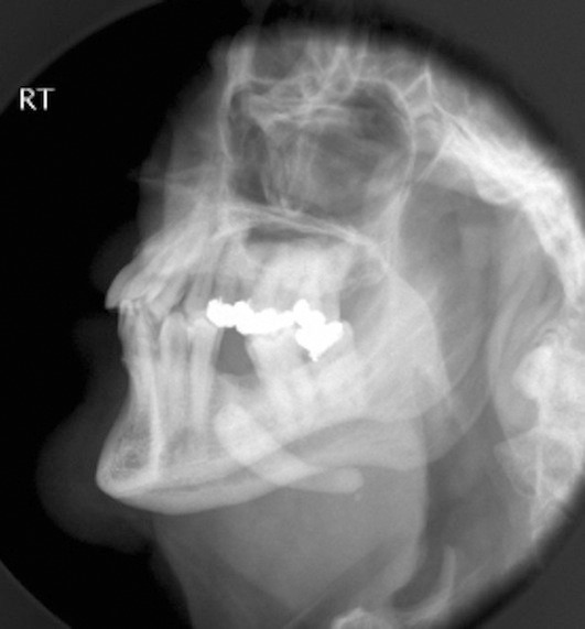
Clinical examination revealed right submandibular swelling and tenderness, with a large salivary calculus palpable in the right floor of mouth. The presence of the sialolith was confirmed on plain radiograph (Fig. 1) with a large opaque mass being evident in the right submandibular region.
Fig. 1.

Plain radiograph demonstrating large radiopaque submandibular mass
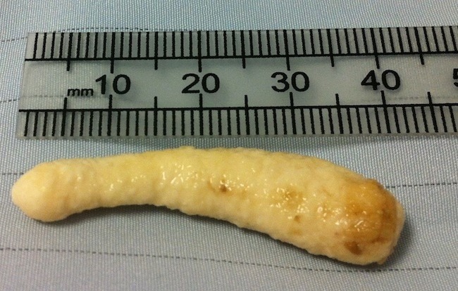
The patient underwent excision of the right submandibular gland and stone via a standard extra-oral approach, without complication. Examination of the stone (Fig. 2) showed a hard, elongated calculus weighing 3.0g and measuring 41mm. Histopathological examination of the gland showed features of chronic sialedenitis.
Fig. 2.

Irregular submandibular sialolith, measuring 40mm
DISCUSSION
Many different aetiological theories have been proposed for salivary gland formation. These include inflammatory, infective, mechanical, neurogenic and chemical. Stone formation is currently thought to be multifactorial, leading to the precipitation of amorphous tricalcic phosphate around an organic matrix of salivary mucin, desquamated epithelial cells and bacteria. Crystallisation occurs and this structure becomes the initial hydroxyapatite focus. This initial focus acts as a catalyst that attracts and supports the deposition of different substances. Giant salivary calculi are thought to form in salivary ducts, which allow expansion and permit salivary flow around the stone. Stones may slowly increase in size, remaining asymptomatic for a more substantial period of time (2). Subsequently, most giant salivary calculi adopt an oval or elongated shape. Giant calculi are described as being hard in texture, yellow in colour and with a porous aspect (5). The stone in our case was classical in appearance for a giant salivary calculi developing within the submandibular duct and gland hilum.
Several factors predispose the submandibular gland to stone disease. These include the length and calibre of its duct, as well as the direction of flow and salivary content. Wharton’s ducts are longer and of larger calibre than parotid (Stenson’s) ducts. These dimensions, along with the need for saliva to flow against gravity, are thought to result in slower salivary flow rates. Saliva produced in the submandibular gland is also more alkaline than that produced in the parotid glands, with a higher calcium and mucin concentration (6). The predisposition to calculi, and ability to tolerate expansion, lead to a higher incidence of giant calculi associated with this gland (4).
Diagnosis of giant salivary lithiasis is often straightforward from a thorough history and examination. Special investigations can be used to confirm diagnosis and plan treatment.
Plain radiography will detect opaque stones (80 to 95% of sialoliths), with intra-oral occlusal radiographs particularly useful. Computerised tomography (CT) scanning is more expensive, yet has been described as the most accurate non-invasive technique for defining the location of stones (5,7). Sialography allows the whole duct system to be visualised, demonstrating calculi of all sizes and also glandular damage from chronic obstruction.
Ultrasound provides an excellent, non-invasive method of detecting sialoliths. Stones that are greater than 1.5mm and of high mineral content are reported to be identifiable on ultrasound with an accuracy of 99% (8). In cases of clinically evident giant sialoliths, ultrasound imaging may aid treatment planning by the detection of further small stones. It is also described as the best method of demonstrating salivary flow post-stone removal (8).
The location and size of calculi are important factors when planning intervention for large calculi. The goal of treatment for giant calculi, as for standard size stones, is restoration of normal salivary secretion. Although chronic sialedenitis secondary to persistent obstruction from a giant calculus leads to a fibrotic and poorly functioning gland, symptoms apparently resolve after calculi removal (9). The stone should be removed by the least invasive method available, with the risk of complications minimised. Sialodochotomy is a well-reported technique for the intra-oral removal of ductal stones, including giant calculi. Possible complications include duct stenosis and lingual nerve damage. Sialendoscopy is now an established intervention for stone removal, and has been described for use in giant salivary calculi (10). The incorporation of extracorporeal short-wave lithotripsy to endoscopic removal has also been shown to be an effective modality and an alternative to conventional excision.
Submandibular gland excision is recommended in cases of substantial intra-glandular caliculli, which are inaccessible via a trans-oral approach. Also, when multiple small stones are present in the vertical and comma portions of Wharton’s duct, sialadenectomy is recommended (10). Excision of the gland is reported to carry a risk of up to 8% for temporary or permanent marginal mandibular nerve palsy. There was no reported damage to the nerve in the reported case.
Giant salivary calculi are rare, yet most cases present with the classical picture of salivary colic. Although modern methods of stone investigation and intervention have been reported for the treatement of giant calculi, transoral sialolithotomy with sialodochoplasty or sialodenectomy remain the mainstay of treatment.
REFERENCES
- 1.Leung AK, Choi MC, Wagner GA. Multiple sialoliths and a sialolith of unusual size in the submandibular duct: a case report. Oral Surg Oral Med Oral Pathol Oral Radiol Endod 1999; 87: 331. [DOI] [PubMed] [Google Scholar]
- 2.McKenna JP, Bostock DJ, McMenamin PG. Sialolithiasis. Am Fam Physician 1987; 36: 119–25 [PubMed] [Google Scholar]
- 3.Lustman J, Regev E, Melamed Y. Sialothiasis: a survey on 245 patients and review of the literature. Int J Oral Maxillofac Surg 1990; 19: 135–8 [DOI] [PubMed] [Google Scholar]
- 4.Manjunath R, Burman R. Giant submandibular sialolith of remarkable size in the comma area of Wharton’s Duct: a case report. J Oral Maxillofac Surg 2009; 67:1329–32 [DOI] [PubMed] [Google Scholar]
- 5.Oteri G, Procopio RM, Cicciu M. Giant Salivary Gland Calculi (GSGC): Report of two cases. The Open Dentistry Journal 2011; 5:90–95 [DOI] [PMC free article] [PubMed] [Google Scholar]
- 6.Raksin SZ, Gould SM, William AC. Submandibular gland sialolith of unusual shape and size. J Oral Surg 1975; 33:142–45 [PubMed] [Google Scholar]
- 7.Weissman JL. Imaging of the salivary gland. Semin Ultrasound CT MR 1995; 16: 546–68 [DOI] [PubMed] [Google Scholar]
- 8.Yoshimura Y, Inoue Y, Odagawa T. Sonographic examination of sialolithiasis. J Oral Maxillofac Surg 1989; 47:907–12 [DOI] [PubMed] [Google Scholar]
- 9.Yoshimura Y, Morishita T, Sugihara T. Salivary gland function after sialolithiasis: Scintographic examination of the submandibular glands with 99m Tc-pertechnate. J Oral Maxillofac Surg 1989; 47:704–10 [DOI] [PubMed] [Google Scholar]
- 10.Wallace E, Tauzin M, Hagan I, et al. Management of giant sialoliths: Review of the literature and preliminary experience with interventional sialendoscopy. Laryngoscope. 2010; 120:1974–8 [DOI] [PubMed] [Google Scholar]


