Abstract
Intraneural Hemangioma of the digital nerve is rare and so far three cases have been reported in the literature. We present a case of 12- year- old boy with painless soft tissue mass in the right hand and numbness on the radial aspect of the index finger. Magnetic Resonance Imaging showed an isointense subcutaneous lesion without discrete borders in the first web space classically of hemangioma with the radial digital nerve extension. On exploration, the intraneural extension of the hemangioma was confirmed and total resection, microsurgical primary digital nerve repair was done. The patient became better and at 6 months follow up the index finger sensation improved. The patient had no reccurence and he is still under follow up.
Keywords: Digital artery, Intraneural, Hemangioma
Introduction
Intraneural hemangioma of digital nerve is an extremely rare benign lesion with mesodermic origin. The first case was reported by Sommer in 1922. Treatment requires microneural dissection and excisional nerve grafting. [1–5]. We report an interesting case of intra-neural hemangioma originating from the princeps pollicis artery to the right index finger involving the radial digital nerve and discuss the management.
Case Report
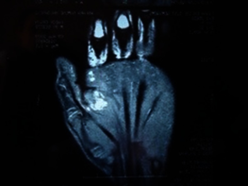
A 12- year- old boy presented with a painless swelling in the right hand of 3 years duration; He also had tingling and numbness in index finger. There was no history of trauma and relevant medical condition. Physical examination revealed a soft, painful mass, approximately 1.5 cm in diameter on the volar aspect of the first web space in the right hand with all clinical features of hemangioma. The patient had hypoesthesia on the radial aspect of the index finger. There was no motor disturbance in the hand and fingers. Passive and active ranges of motion were unaffected. MRI imaging revealed a subcutaneous lesion that was isointense with muscle on T1- weighted images.(Figure 1). The lesion demonstrated hyperintesity without discrete borders in the first web space on T2-weighted images with fat saturation and radial digital nerve involvement. (Figure 2)
Fig. 1.
Sagittal T1-weighted image; reveals a small subcutaneous lesion on the volar aspect of the hand
Fig. 2.
Transverse T2- weighted image demonstrated a hyper intense lesion with radial digital nerve involvement
The operation was performed under general anesthesia using a pneumatic tourniquet and operating microscope. Exploration revealed a dark blue hemangioma of approximately 2 × 1.5 × 1.5 cm, originating from the volar surface of Princeps pollicis artery with radial digital nerve extension. (Figure 3). This hemangioma was then resected off the surronding tissue and the radial digital nerve dissection showed segmental interfascicular extension of hemangioma into the digital nerve. Hence resection of the nerve segment (20 mm) was done carefully. The digital nerve ends were adequately mobilised and coapted using 8/0 nylon suture without tension. Histopathologic examination revealed thin-walled dilated capillaries embedded in fibroadipous stroma with peripheral nerve fibers, consistent with the hemangioma and the intraneural extension.
Fig. 3.

The cavarneuos hemangioma originated from princeps pollicis artery and the radial digital nerve appears expanded, segmentally distorted and discoloured secondary to the intraneural extension
Sensation on the radial side of the index finger was evaluated and two point discrimination test was performed. Sensory deficit was reported with two point discrimination of 10 mm. The sensation on the index finger improved progressively, and the two point discrimination was 6 mm at the end of 6 months follow up. There was no reccurence of the lesion observed during follow up.
Discussion
Peripheral nerve tumors comprise less than 5% of all tumors of the hand [6]
Intraneural hemangioma is a rare lesion, especially in a peripheral nerve. Sato is the first to report on the peripheral nerve hemangiomas [7]. They present either as a tumor of variable size or with spontaneous localized pain while motor or sensory deficit is rare. Treatment may require microneural dissection and excision of the lesion or segmental nerve resection and grafting[1].
Surgical excision is the only feasible method for treatment of intraneural hemangioma, and careful microsurgical dissection is mandatory to prevent recurrence and minimize iatrogenic injury to the surrounding nerve fascicles. [8]. It is not always possible to excise intraneural lesions totally, and an incomplete resection carries a high risk of recurrence [9] .Large segmental nerve lesions require interfascicualr nerve grafting.
We report a case of hemangioma with radial digital nerve extension in a young boy for whom careful microsurigcal excision and primary nerve repair gave a good result with sensory recovery and no recurrence. Pre-operatvive planning using MRI is essential in delinating the lesion and histopathaology is the gold standard tool for diagnosis. The surgery should be total excision of the lesion and if possible reconstruct with primary nerve grafting. One should always look for the recurrence in the follow up.
References
- 1.Prosser AJ, Burke FD. Hemangioma of the median nerve associated with Reynaud’s phenomenon. J Hand Surg (Br) 1987;12:227–228. doi: 10.1016/0266-7681_87_90019-2. [DOI] [PubMed] [Google Scholar]
- 2.Sommer R. Angioma in peripheral nervous system. Deutsche Ztschr Chir. 1922;173:65–77. doi: 10.1007/BF02815674. [DOI] [Google Scholar]
- 3.Kon M, Vuursteen P. An intraneural hemangioma of a digital nerve: case report. J Hand Surg (Am) 1981;6:357–358. doi: 10.1016/s0363-5023(81)80041-x. [DOI] [PubMed] [Google Scholar]
- 4.Nagay L, Mc Cabe SJ, Wolff TW. Hemangioma of the digital nerve: a case report. J Hand Surg (Br) 1990;15:487–488. doi: 10.1016/0266-7681(90)90098-o. [DOI] [PubMed] [Google Scholar]
- 5.Kerimoglu U, Uzumcugil A, Yilmaz G, Ayvaz M. Intraneural hemangioma of digital nerve diagnosed with MR imaging. Skeletal Radiol. 2007;36:157–160. doi: 10.1007/s00256-006-0094-4. [DOI] [PubMed] [Google Scholar]
- 6.Strickland JW, Steichen JB. Nerve tumors of the hand and forearm. J Hand Surg [Am] 1997;2:285–291. doi: 10.1016/s0363-5023(77)80128-7. [DOI] [PubMed] [Google Scholar]
- 7.Sato S. Ueber das cavernose Angiome am peripherischen Nerve systems. Arch Klin Chir. 1913;100:553–574. [Google Scholar]
- 8.Chatillon CE, Guitot MC, Jacgues L. Lipomatous, vasculer, and chondromatous benign tumors of the peripheral nerves: representative cases and review of the literature. Neurosurg Focus. 2007;22:E18. doi: 10.3171/foc.2007.22.6.19. [DOI] [PubMed] [Google Scholar]
- 9.Mestdagh H, Lecomte-Houcke M, Reyford H (1990) Intraneural hemangioma of the posterior tibial nerve. J Bone Joint Surg Br [DOI] [PubMed]


