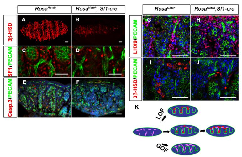
Fig. 4. Constitutively active Notch signaling results in a loss of Leydig cells.
The coelomic domain of the gonad is upwards, anterior is leftwards and posterior is rightwards. (A–D) Compared with wild type (A,C), RosaNotch; Sf1-cre mouse gonads (B,D) showed a loss of Leydig cells at 13.5 dpc based on immunofluorescent staining for 3β-HSD (red, A,B) or SF1 (red, C,D). PECAM1 (green) labels vasculature and germ cells in C and D. (E,F) In view of the low levels of activated caspase 3 (red), this loss was not due to cell death (PECAM1, green; Syto13 stains DNA, blue; n=3 gonads; P=0.7813). (G–J) LHX9 (red), which is expressed in interstitial somatic cells (G), shows an increase in RosaNotch; Sf1-cre gonads (H; n=3 gonads; P<0.01), whereas Leydig cell numbers are decreased based on 3β-HSD staining (I,J). Images shown are representative staining of at least three sections from three independent gonads. (K) A summary diagram of Notch/Hes1 results: loss of Notch function (LOF) leads to Leydig cell differentiation (red, oval cells), whereas gain of Notch function (GOF) maintains interstitial mesenchymal progenitor cells (purple, spindle shaped), and restricts their differentiation into Leydig cells. Scale bars: 50 μm.

