Abstract
We would like to present a rare case of alobar holoprosencephaly (HPE) in a fetus diagnosed by routine sonography in the second trimester. Structural sonography demonstrated multiple facial anomalies including absent nasal bone, flat facial profile, hypotelorism, fusion of the orbits and proboscis. After counseling, termination of pregnancy was performed by vaginally administered misoprostol. Karyotyping of amniotic fluid cells revealed an isochromosome 18q, resulting in a trisomy 18q and monosomy 18p. A stillborn female of 390 g with several congenital anomalies was born. Postmortem examination demonstrated several anomalies including the HPE, cyclopia, double fused eye, absence of the nose, and the presence of a proboscis. In the literature only a few cases have been published.
Keywords: Alobar holoprosencephaly, cyclopia, proboscis, prenatal diagnosis, isochromosome 18q, sonography
We present a case with alobar holoprosencephaly (HPE) and proboscis diagnosed at 21+1 weeks of gestation during routine sonographic scanning. Chromosome analysis demonstrated an abnormal karyotype 46,XX,i(18)(q10). Isochromosome 18q is a rare cytogenetic abnormality. The phenotypical features of this chromosomal abnormality are variable and overlap with trisomy 18 and monosomy 18p.1 HPE is rarely described in trisomy 18, and occasionally in monosomy 18p.1 Actually, isochromosome 18q associated with alobar HPE was only described eight times before. We give a review of the literature and describe a case of alobar HPE, diagnosed by routine sonography in the second trimester, associated with an isochromosome 18q.
Case Report
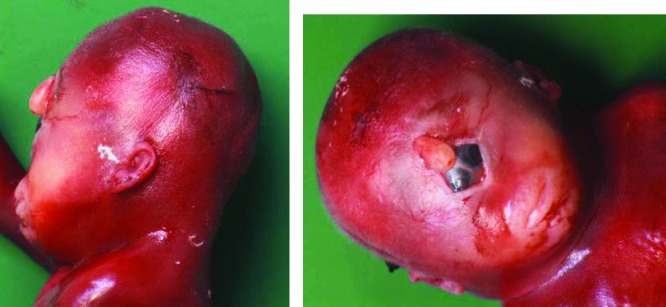
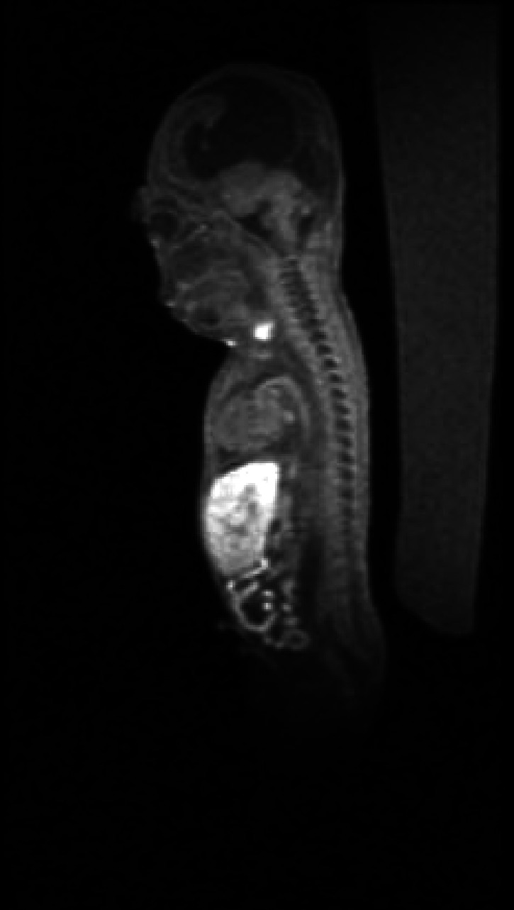
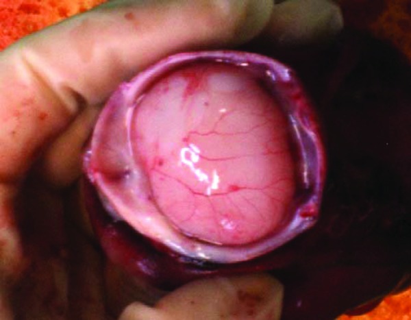
A 37-year-old, gravida 2, para 1 woman was seen for routine sonographic scanning at 20+5 weeks of gestation. Obstetric history revealed a spontaneous birth of a male fetus of 3080 g at 40+3 weeks of gestation. The parents were nonconsanguineous and without dysmorphic features or congenital anomalies. The family history of the mother mentioned a sister who died at the age of 17 because of an intracranial bleeding from an aneurysm. There was no history of infection or drug abuse, and serological screening for HIV, hepatitis B, and syphilis was negative. Until then, the pregnancy had been uneventful. The patient had declined first-trimester aneuploidy screening. At routine sonography, an abnormal image of the fetal brain and facial structures was seen. The patient was referred to our hospital for detailed ultrasound examination. An alobar HPE with facial anomalies including absent nasal bone, flat facial profile, hypotelorism, fusion of the orbits and proboscis were noted. Other anomalies seen were a single umbilical artery, abnormal four-chamber view of the heart, especially abnormal shape of the right atrium, and cystic kidneys. Amniocentesis was performed at 21+1 weeks of gestation and an abnormal karyotype 46,XX,i(18)(q10) was diagnosed. The fetus therefore had a trisomy of the long arm and a monosomy of the short arm of chromosome 18. The parents decided to terminate the pregnancy on the basis of the ultrasound abnormalities. Eight hours after inducing labor with vaginally administered misoprostol, a stillborn female fetus was delivered at 21+3 weeks of gestation. Birth weight was 390 g (normal weight at 21 weeks: 360 g). Several congenital anomalies were confirmed at postmortem examination including a cyclopia with a double fused eye, the absence of the nose, and the presence of a proboscis (Fig. 1). Postmortem magnetic resonance imaging scan was performed. The coronal slides gave a definite view of the monoventricular cavity and the proboscis (Fig. 2). Autopsy demonstrated further the alobar HPE (Fig. 3), absence of the corpus callosum, perimembranous ventricular septum defect, bicuspid pulmonal artery valves, malrotation of the small bowel, bilateral hydronephrosis, right megaureter, and uterus bicornis.
Figure 1.

Postnatal image of proboscis and cyclopia with a double fused eye.
Figure 2.

Postmortem magnetic resonance imaging coronal slide demonstrating the monoventricle of the brain of the fetus.
Figure 3.

Postmortem image at autopsy demonstrating the monoventricle of the brain.
Discussion
In our case, the fetal karyotyping showed an isochromosome 18q, resulting in a monosomy 18p and trisomy 18q. This chromosome aberration occurred de novo because both parents had a normal karyotype. The HPE4 gene, TGIF, is located on the distal part of chromosome 18, namely 18p11.31.2 Hemizygosity of HPE4 does not automatically result in the phenotype of HPE, suggesting that multiple genetic and environmental factors are involved in the development of the HPE phenotypes. For the de novo case, the recurrence risk for siblings is not significantly increased above that of the general population.3
There have only been seven cases4,5,6,7,8,9,10 previously reported of isochromosome 18q in combination with HPE (Table 1). Of interest, Levy-Mozziconacci et al9 described a case similar to ours, with a proboscis and a bicornuate uterus, related to i(18)(q10). The karyotype abnormality in that particular case, however, was a dic(18)(p11.3), which means the fetus had three copies of the q-arm and three copies of a small part of the p-arm, excluding the locus where HPE4 is located, therefore making their case different from ours.
Table 1. Cases in the Literature of Isochromosome 18q in Combination with Holoprosencephaly.
| Author (Year) | Karyotype | Phenotypes Apart from Holoprosencephaly | Gravida, Parity | Maternal Age (y) |
|---|---|---|---|---|
| Wurster-Hill et al (1991)4 | 46XYi(18q) | Microcephaly, hypotelorism, hypoplastic nose, short neck, hypoplasia of radius and ulna, aplasia of first metacarpals, radial deviation of the hands, “rocker bottom” feet | G1, P0 | 31 |
| Spinner et al (1991)5 | 46XXi(18q) | Bicornuate uterus, proboscis | G1, P0 | 25 |
| Van Essen et al (1993)6 | 46XXi(18q) | DiGeorge anomaly, streak ovaries | G1, P0 | 26 |
| Graf et al (2002)7 | 46XX,i(18)(q10) | Omphalocele, bilateral hypoplastic forearm with radial deviation of hands | G1, P0 | 21 |
| De Pater et al (1997)8 | Mosaic 46XX,inv | Hypotelorism, spina bifida | G7, P4 | 27 |
| Levy-Mozziconacci et al (1996)9 | 46XX,-18, + dic(18)(p11,3) | Cyclopia, proboscis, radial deviation of left hand, agenesis of left thumb, aplasia of right thumb, bilateral clubfeet, multicystic kidney | G1, P0 | 27 |
| Froster-Iskenius et al (1984)10* | 46XX,i(18q) | Hypoplastic olfactory bulbs | G5, P2 | 37 |
| Present case (2010) | 46, XX,i(18)(q10) | Single umbilical artery, cyclopia with double fused eyes, proboscis, absence of corpus callosum, perimembranous ventricular septum defect, bicuspid pulmonal artery valves, malrotation of small bowel, bilateral hydronephrosis, right megaureter, bicornuate uterus | G2, P1 | 37 |
Froster-Iskenius et al reported a case of isochromosome 18q exhibiting olfactory bulbs as a minor manifestation of holoprosencephaly.10
Abnormalities in the forearms and hand positioning were described in another case11 with an isochromosome 18q without HPE, but not in ours. Although the mother in our case is 37 years old, reviewing the other reported cases, it is unlikely that there is an association between isochromosome 18q and increased maternal age.1 This case stresses the importance of standard sonography for all pregnant women between 18 and 21 weeks to detect any congenital anomalies of the fetus.
Acknowledgments
The authors thank Dr. D. de Jong, Department of Radiology, for providing the magnetic resonance image.
References
- 1.Pal S, Siti M I, Ankathil R, Zilfalil B A. Two cases of isochromosome 18q syndrome. Singapore Med J. 2007;48:e146–e150. [PubMed] [Google Scholar]
- 2.Portnoï M F, Gruchy N, Marlin S. et al. Midline defects in deletion 18p syndrome: clinical and molecular characterization of three patients. Clin Dysmorphol. 2007;16:247–252. doi: 10.1097/MCD.0b013e328235a572. [DOI] [PubMed] [Google Scholar]
- 3.Lim A S, Lim T H, Kee S K. et al. Holoprosencephaly: an antenatally-diagnosed case series and subject review. Ann Acad Med Singapore. 2008;37:594–597. [PubMed] [Google Scholar]
- 4.Wurster-Hill D H, Marin-Padilla J M, Moeschler J B, Park J P, McDermet M. Trisomy 18 and 18p- features in a case of isochromosome 18q [46,XY,i(18q)]: prenatal diagnosis and autopsy report. Clin Genet. 1991;39:142–148. doi: 10.1111/j.1399-0004.1991.tb03001.x. [DOI] [PubMed] [Google Scholar]
- 5.Spinner N B, Eunpu D L, Austria J R, Mamunes P. Holoprosencephaly in a newborn girl with 46,XX,i(18q) Am J Med Genet. 1991;39:11–12. doi: 10.1002/ajmg.1320390104. [DOI] [PubMed] [Google Scholar]
- 6.Essen A J van, Schoots C J, Lingen R A van, Mourits M J, Tuerlings J H, Leegte B. Isochromosome 18q in a girl with holoprosencephaly, DiGeorge anomaly, and streak ovaries. Am J Med Genet. 1993;47:85–88. doi: 10.1002/ajmg.1320470117. [DOI] [PubMed] [Google Scholar]
- 7.Graf M D, Gill P, Krew M, Schwartz S. Prenatal detection of structural abnormalities of chromosome 18: associations with interphase fluorescence in situ hybridization (FISH) and maternal serum screening. Prenat Diagn. 2002;22:645–648. doi: 10.1002/pd.354. [DOI] [PubMed] [Google Scholar]
- 8.de Pater J M, Scheres J M, Brons J. Abnormal chromosome 18 in prenatal diagnosis with holoprosencephaly. Prenat Diagn. 1997;17:887–888. [PubMed] [Google Scholar]
- 9.Levy-Mozziconacci A, Piquet C, Scheiner C. et al. i(18q) in amniotic and fetal cells with a normal karyotype in direct chorionic villus sampling: cytogenetics and pathology. Prenat Diagn. 1996;16:1156–1159. doi: 10.1002/(SICI)1097-0223(199612)16:12<1156::AID-PD13>3.0.CO;2-K. [DOI] [PubMed] [Google Scholar]
- 10.Froster-Iskenius U, Coerdt W, Rehder H, Schwinger E. Isochromosome 18q with karyotype 46,XX,i(18q). Cytogenetics and pathology. Clin Genet. 1984;26:549–554. doi: 10.1111/j.1399-0004.1984.tb01102.x. [DOI] [PubMed] [Google Scholar]
- 11.Sahoo T, Naeem R, Pham K. et al. A patient with isochromosome 18q, radial-thumb aplasia, thrombocytopenia, and an unbalanced 10;18 chromosome translocation. Am J Med Genet A. 2005;133A:93–98. doi: 10.1002/ajmg.a.30535. [DOI] [PubMed] [Google Scholar]


