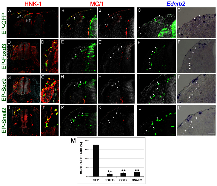
Fig. 2.
Foxd3, Sox9 and Snail2 inhibit melanocyte specification and dorsolateral migration. (A-L′) Late plasmid EP (35 ss) performed before melanoblast emigration in the flank. Thirty hours later, control labeled cells were present in the dorsolateral pathway, and expressed MC/1 and Ednrb2 (A-C′). By contrast, Foxd3-, Sox9- or Snail2-transfected cells were found in the ventral pathway, negative for MC/1 and Ednrb2 (arrowheads, D-L′). (M) Quantification of the percentage of MC/1+/GFP+ cells (**P=0.01, P<0.01, P=0.01 for Foxd3, Sox9 and Snail2, respectively). Scale bars: 100 μm in A; 120 μm in D,G,J; 60 μm in E,D′,G′,J′; 75 μm in B-C′,H,H′,K. DM, dermomyotome.

