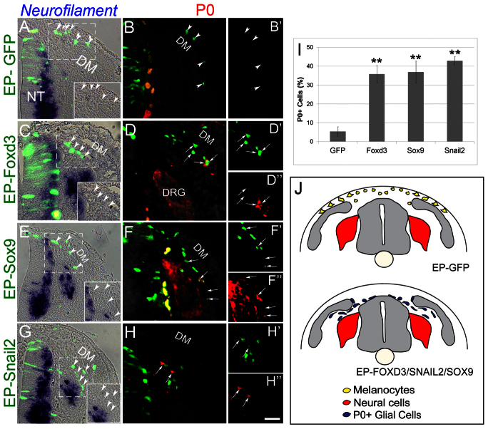
Fig. 3.
Foxd3, Sox9 and Snail2 upregulate P0 expression in prospective melanocytes. (A-B′) Late-emigrating NC progenitors that received control GFP at 35 ss, flank level, migrate dorsolaterally and are negative for neurofilament or P0 (arrowheads). (C-H″) EP with Foxd3, Sox9 or Snail2 upregulates P0 (arrows) but not neurofilament (arrowheads) and cells are diverted ventrally (see also Fig. 2). (I) Quantification of the percentage of P0-expressing cells in all treatments (**P<0.01). (J) Schematic representation of the results suggesting that putative melanoblasts lose neurogenic ability but keep the potential to develop into glia under these experimental conditions. Orange and yellow cells in B,D,F represent autofluorescence of blood cells. Scale bars: 70 μm in A,B,B′,E,G; 60 μm in F-F″; 55 μm in C; 50 μm in D-D″,H-H″. DM, dermomyotome.

