Abstract
T-box genes are potent regulators of mesoderm development in many metazoans. In chordate embryos, the T-box transcription factor Brachyury (Bra) is required for specification and differentiation of the notochord. In some chordates, including the ascidian Ciona, members of the Tbx2 subfamily of T-box genes are also expressed in this tissue; however, their regulatory relationships with Bra and their contributions to the development of the notochord remain uncharacterized. We determined that the notochord expression of Ciona Tbx2/3 (Ci-Tbx2/3) requires Ci-Bra, and identified a Ci-Tbx2/3 notochord CRM that necessitates multiple Ci-Bra binding sites for its activity. Expression of mutant forms of Ci-Tbx2/3 in the developing notochord revealed a role for this transcription factor primarily in convergent extension. Through microarray screens, we uncovered numerous Ci-Tbx2/3 targets, some of which overlap with known Ci-Bra-downstream notochord genes. Among the Ci-Tbx2/3 notochord targets are evolutionarily conserved genes, including caspases, lineage-specific genes, such as Noto4, and newly identified genes, such as MLKL. This work sheds light on a large section of the notochord regulatory circuitry controlled by T-box factors, and reveals new components of the complement of genes required for the proper formation of this structure.
Keywords: Ciona, Brachyury, Notochord
INTRODUCTION
T-box (or Tbx) genes comprise an evolutionarily conserved class of transcription factors defined by the presence of a distinctive DNA-binding domain. Members of the Tbx family are crucial regulators of embryogenesis (Naiche et al., 2005), and mutations in T-box genes are linked to several human genetic diseases (Packham and Brook, 2003), further underscoring the importance of Tbx transcription factors.
The discovery and functional characterization of Brachyury (Bra), the founding Tbx family member, revealed its involvement in the specification and development of the notochord (Herrmann et al., 1990). The notochord, the defining feature of chordates, provides structural support for developing embryos throughout this phylum (Jiang and Smith, 2007); a basement membrane and a peri-notochordal sheath consisting of extracellular matrix (ECM) proteins contribute to its rigidity (Stemple, 2005). Elongation of the chordate body plan is in part accomplished through convergent extension (CE) of notochord cells via mediolateral intercalation (Keller et al., 2000), which requires cells to become motile and polarized, to remodel cell-adhesive contacts and to interact with ECM components. Following intercalation, pressure exerted on the notochordal sheath by the formation and expansion of vacuoles provides the tail with further rigidity (Stemple, 2005). Studies in all chordates examined thus far have shown that Bra is indispensible for notochord fate commitment (Showell et al., 2004). Conversely, few studies have addressed the composition and regulation of the battery of genes required for later notochord morphogenetic events.
In addition to the group containing Bra, phylogenetic analyses have categorized vertebrate T-box genes into four further subfamilies, including that of Tbx2, which in mammals has four members, Tbx2, Tbx3, Tbx4 and Tbx5 (Naiche et al., 2005). The paralogs Tbx2 and Tbx3 are important regulators of heart formation (Harrelson et al., 2004), brain morphogenesis (Manning et al., 2006) and cell migration (Fong et al., 2005). In humans, altered levels of these genes have deleterious results: haploinsufficiency caused by mutations in TBX3 plays a role in ulnar-mammary syndrome (UMS) (Packham and Brook, 2003), and recently TBX2 and TBX3 have been found to be amplified or upregulated in different cancers (Lu et al., 2010). These transcriptional regulators can contribute to tumorigenesis through inhibition of apoptosis and by increasing invasiveness through control of cell adhesion genes (Lu et al., 2010). A thorough understanding of the contributions of Tbx2/3 genes to normal development will thus inform studies of these transcription factors in pathological processes.
Previous studies have suggested that Tbx2 subfamily members could also contribute to notochord formation. Xenopus and zebrafish notochords both express tbx3, and the zebrafish tbx2b gene is also found in the notochord (Dheen et al., 1999; Takabatake et al., 2000); however, their individual functions and regulatory interactions in this tissue remain incompletely characterized. Impaired Tbx2b activity results in defects in notochord specification, which precludes the study of its involvement in later stages of notochord morphogenesis (Dheen et al., 1999; Fong et al., 2005). Hence, the roles and targets of tbx2 and tbx3 genes in the development of this structure have yet to be investigated in any chordate.
We have addressed this point using the ascidian Ciona intestinalis, a powerful model organism for studies of gene regulation and function during notochord morphogenesis. The ascidian notochord is tractable, consisting of only 40 post-mitotic cells where Ciona Brachyury (Ci-Bra) is exclusively expressed (Corbo et al., 1997; Passamaneck and Di Gregorio, 2005). Ciona notochord cells intercalate through cellular and molecular mechanisms similar to those used by vertebrates (Jiang et al., 2005; Munro and Odell, 2002b), and require an intact extracellular matrix (Veeman et al., 2008). Formation of intercellular lumens similar to vacuoles also contributes to notochord elongation (Dong et al., 2009; Miyamoto and Crowther, 1985).
Ascidians diverged from the main chordate lineage before the genome duplications associated with the emergence of vertebrates; accordingly, and due to the loss of the ancestral Tbx4/5 ortholog (Horton et al., 2008), the Ciona genome contains only a single Tbx2 subfamily member, Ci-Tbx2/3 (Takatori et al., 2004). Among other territories, Ci-Tbx2/3 is expressed in the notochord after its fate specification, making Ci-Tbx2/3 both an ideal candidate regulator of notochord differentiation and the only other Tbx gene besides Ci-Bra with detectable expression in this tissue. As previous work in Ciona has uncovered numerous Ci-Bra downstream genes (e.g. Kugler et al., 2008; Takahashi et al., 1999), the identification of shared Ci-Bra and Ci-Tbx2/3 targets, and the elucidation of the notochord gene regulatory circuitry powered by these transcription factors, are greatly facilitated.
In this study, we focused on the transcriptional regulation and developmental role of Ci-Tbx2/3 in the Ciona notochord. We have analyzed the relationship between Ci-Tbx2/3 and Ci-Bra, and studied the effects of alterations in Ci-Tbx2/3 function on different stages of notochord development, including mediolateral intercalation and lumen formation. Finally, expression profiling identified several Ci-Tbx2/3-dependent notochord genes with various functions, including genes previously reported as Ci-Bra targets. These results shed light on an extensive, crucial branch of the notochord gene regulatory network, components of which appear to be shared between ascidians and higher chordates.
MATERIALS AND METHODS
Embryo culture, electroporation and morpholino injections
Adult Ciona intestinalis were purchased from M-REP (Carlsbad, CA). Culturing, electroporations and X-gal staining were carried out as previously described (Oda-Ishii and Di Gregorio, 2007). Ci-Bra mutant embryos were kindly provided by Drs Shota Chiba and William Smith (UC Santa Barbara, CA, USA).
The Ci-Tbx2/3 morpholino (30 fmol), 5′-CGCTATCGGTCAAACACATTCAGAG-3′ (Gene Tools), was co-injected into unfertilized eggs with ~1 μg Ci-Bra>lacZ (Corbo et al., 1997). The Ci-Tbx2/3 shRNA plasmid was constructed as described previously (Nishiyama and Fujiwara, 2008) using the oligonucleotides listed in supplementary material Table S2.
Whole-mount in situ hybridization
Whole-mount in situ hybridizations using BCIP/NBT detection were carried out as previously described (José-Edwards et al., 2011). The Ci-Tbx2/3 cDNA was constructed by PCR amplifying two overlapping halves of its coding region from cDNA with the primers listed in supplementary material Table S2, then digested and ligated into the pCR4-TOPO vector (Invitrogen). EST clones used for whole-mount in situ hybridization were prepared as described previously (José-Edwards et al., 2011); other select cDNAs (supplementary material Table S3) were cloned into the pGEM-T (Promega) vector.
Double-fluorescent whole-mount in situ hybridization and immunostaining experiments were performed as described previously (Wagner and Levine, 2012). Anti-DIG-POD (Roche) and rabbit anti-GFP (Invitrogen) antibodies were used at 1:500 and 1:1000 dilutions, respectively. Antisense DIG-labeled probes were visualized through incubation with rhodamine-tyramide working solution (Perkin Elmer) for 5 minutes to 1 hour at room temperature. Embryos were then blocked for 1 hour in TNBS [100 mM Tris (pH 7.5), 150 mM NaCl, 0.5% Roche blocking reagent, 2% normal goat serum] and incubated overnight at 4°C with 1:500 goat anti-rabbit Alexa Fluor 488 (Invitrogen) secondary antibody.
Plasmid construction
Regions of the Ci-Tbx2/3 locus were amplified from Ciona intestinalis genomic DNA. Subsequent deletions and mutations were made using unique restriction sites or through PCR with the oligonucleotides provided in the supplementary material Table S2. These fragments were cloned into the lacZ-containing plasmid pFBΔSP6 (Oda-Ishii and Di Gregorio, 2007).
The Ci-Tbx2/3 DBD was amplified from cDNA employing the primers: 5′-AACCATGGATAACATGGAACTGTGGGAGC-3′ and 5′-CTACTAGTTCCGCTCCCTGAGTCTCG-3′, and used to replace the Ci-FoxN2/3DBD fragment within the Ci-Bra>en::Ci-FoxN2/3DBD::GFP plasmid (kindly supplied by Dr Yale Passamaneck, Kewalo Marine Laboratory, Hawaii, USA) to create Ci-Bra>en::Ci-Tbx2/3DBD::GFP. Excision of engrailed from Ci-Bra>Ci-Tbx2/3DBD::GFP generated Ci-Bra>Ci-Tbx2/3DBD::GFP, and Ci-Bra>Ci-Tbx2/3DBD::VP16::GFP was subsequently constructed by amplifying two copies of the VP16 domain from a 4X-VP16 construct using the primers 5′-TTACTAGTGGATCCGCCCCCCCG-3′ and 5′-TTACTAGTCGGCAACCCACCGTACTC-3′, and inserting the product into the Ci-Bra >Ci-Tbx2/3DBD::GFP plasmid.
Immunostaining, microscopy and quantitative analysis
For either direct visualization of fluorescence or immunostaining, embryos were fixed as for whole-mount in situ hybridization at room temperature for 30 minutes to 1 hour. Double immunofluorescence experiments were performed using 1:1000 rabbit anti-GFP and mouse anti-β-Gal (Promega) primary antibodies and 1:1000 goat anti-rabbit Alexa Fluor 488 and goat anti-mouse Alexa Fluor 555 (Invitrogen) secondary antibodies. Select embryos were counterstained with 1 U rhodamine-phalloidin (Invitrogen) in 1× PBS/0.2% Triton X-100 for 3 hours at room temperature. All fluorescent samples were mounted in VECTASHIELD with DAPI (Vector Laboratories). Images were obtained with a Leica DMR microscope with the exception of confocal images, which were acquired at the Weill Cornell Medical College Optical Microscopy Core Facility using a Zeiss LSM 510 microscope. Data presented in graphs represent average values with error bars indicating the s.d. garnered from at least three independent experiments. Statistical significance for the phenotypic classes of Ci-Tbx2/3 mutants, when compared with Ci-Bra >GFP samples, was determined using a χ2 test.
Microarray screens
Approximately 100-300 fluorescent embryos expressing the Ci-Bra>Tbx2/3DBD::GFP, Ci-Bra>Tbx2/3VP16::GFP or Ci-Bra>GFP transgenes were manually selected at ~7 hpf and ~10.5 hpf (Hotta et al., 2007). RNA was extracted using the RNeasy micro kit (Qiagen) from three biological replicates for each construct and used to create labeled probes for hybridization to a Ciona intestinalis Affymetrix GeneChip (Christiaen et al., 2008) by the Weill Cornell Genomics Resources Core Facility. Results were RMA summarized from raw data (CEL files) and quantile normalized before proceeding with ANOVA to statistically analyze the data (Weill Cornell Epigenomics Core Facility). Probe sets with P≤0.05 with an absolute fold change cut-off of 2 were considered further. The microarray dataset has been deposited into the NCBI Gene Expression Omnibus (accession number GSE42267).
RESULTS
Ci-Tbx2/3 notochord expression depends upon Ci-Bra
In Ciona, transcripts for Ci-Tbx2/3 are found in a number of spatial domains, including the notochord (Fig. 1A) (Imai et al., 2004; Takatori et al., 2004). Unlike in other chordates, such as zebrafish (Dheen et al., 1999; Martin and Kimelman, 2008), Ci-Bra and Ci-Tbx2/3 are the only Tbx genes present in the Ciona notochord. This fact both simplified the analysis of the contribution of the T-box gene family to notochord development and provided an ideal system for studying the regulatory interactions between these factors.
Fig. 1.

Ci-Tbx2/3 notochord expression is controlled by Ci-Bra. (A-C) Whole-mount in situ hybridization for Ci-Tbx2/3 on mid-tailbud wild-type embryos (A) and the offspring of Ci-Bra heterozygous animals (B,C). The percentage of embryos exhibiting each phenotype is provided in the upper right corner of each panel, whereas the total number of embryos scored is in the lower left corner. Notochord expression is highlighted by a red arrowhead, while a white arrowhead denotes lack of expression in this domain. Colored arrowheads indicate other expression domains as follows: blue, CNS; green, epidermis. Scale bar: 40 μm.
Expression of Ci-Bra begins at the 64-cell stage, concomitant with notochord fate restriction (Corbo et al., 1997), and precedes expression of Ci-Tbx2/3, suggesting that the notochord expression of Ci-Tbx2/3 could be mediated by Ci-Bra. To assess this, we analyzed the expression pattern of Ci-Tbx2/3 in Ci-Bra−/− mutant embryos (Chiba et al., 2009). We performed whole-mount in situ hybridization for Ci-Tbx2/3 on the offspring of Ci-Bra+/− animals as homozygotes do not survive to adulthood (Chiba et al., 2009). We determined that, compared with controls and wild-type siblings (Fig. 1A,B), Ci-Tbx2/3 transcripts were not detectable in the notochord of Ci-Bra−/− embryos (Fig. 1C), signifying that Ci-Bra is indispensable for the expression of Ci-Tbx2/3 in this domain. In the absence of notochord-specific Ci-Bra, Ci-Tbx2/3 expression in the epidermis and CNS is detected at normal levels, emphasizing that other tissue-specific factors likely activate Ci-Tbx2/3 in its additional expression domains. Thus, Ci-Tbx2/3 lies downstream of Ci-Bra in the notochord gene regulatory network.
Characterization of a Ci-Tbx2/3 notochord cis-regulatory module (CRM)
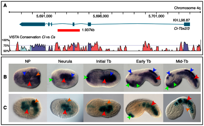
The previous results prompted us to investigate in detail the cis-regulatory mechanisms mediating the hierarchical interaction between Ci-Bra and Ci-Tbx2/3 in the notochord. Whole-genome VISTA alignments between Ciona intestinalis and its sister species Ciona savignyi (Frazer et al., 2004) highlighted several stretches of non-coding sequence conservation across the ~15 kb Ci-Tbx2/3 locus (Fig. 2A). As such areas are often seen as hallmarks of functional cis-regulatory DNA, we tested the ability of six genomic fragments encompassing conserved regions within the Ci-Tbx2/3 locus to direct tissue-specific expression of the lacZ reporter in vivo. Constructs containing genomic fragments located 5′ of the Ci-Tbx2/3 transcription start site and within the first intron exhibited staining only in Ci-Tbx2/3 expression domains outside of the notochord (supplementary material Fig. S1), indicating that the Ci-Tbx2/3 locus harbors multiple tissue-specific CRMs. Conversely, upon electroporation of a 1.9 kb region spanning part of the second and third introns (Fig. 2A), the resulting embryos displayed robust notochord activity (Fig. 2C), recapitulating the onset and pattern of the endogenous gene in this domain (Fig. 2B). Genome-wide ChIP studies, published after this CRM was identified, showed the highest Ci-Bra binding peaks within the first intron in early (110-cell stage) embryos (Kubo et al., 2010); however, we found that these areas lacked notochord staining (supplementary material Fig. S1). Nonetheless, another peak was contained in the 1.9 kb region with notochord activity, consistent with our results (Kubo et al., 2010).
Fig. 2.
Identification of a CRM that recapitulates Ci-Tbx2/3 notochord expression. (A) Top: map of the Ci-Tbx2/3 locus. Exons and introns are denoted by teal rectangles and lines, respectively. A red box indicates the notochord CRM. Bottom: VISTA alignment illustrating the sequence conservation across the Ci-Tbx2/3 locus between Ciona intestinalis (Ci) and Ciona savignyi (Cs), obtained using the following parameters: calculation window, 100 bp; minimum conservation width, 100 bp; conservation identity, 50%. Purple peaks indicate conserved coding regions; light blue peaks indicate conserved 5′- or 3′-UTR; pink peaks indicate conserved non-coding regions. (B) Ci-Tbx2/3 whole-mount in situ hybridization. (C) X-Gal staining of C. intestinalis embryos carrying the Ci-Tbx2/3 notochord CRM in A. (B,C) Expression territories are highlighted with arrowheads: red, notochord; orange, muscle; blue, CNS; green, epidermis. In most panels, dorsal is upwards and anterior is towards the left. NP, neural plate; Tb, tailbud.
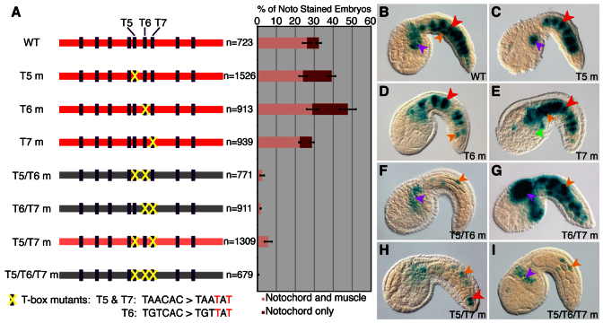
In order to identify the minimal cis-regulatory sequences necessary for the activity of this notochord CRM, we tested the ability of truncations of the 1.9 kb region to retain notochord expression (supplementary material Fig. S2). A 294 bp region drove consistent lacZ expression in the notochord and was enriched for T-box binding sites, containing nine sequence blocks (T1-T9) matching the generic TNNCAC Ci-Bra consensus binding site (e.g. Dunn and Di Gregorio, 2009) (Fig. 3A,B), supporting a role for Ci-Bra in the activation of this notochord CRM.
Fig. 3.
Activity of the Ci-Tbx2/3 notochord CRM requires T-box binding sites. (A) Left: schematic representation of the 294 bp Ci-Tbx2/3 notochord CRM. Red boxes represent constructs with notochord activity; configurations exhibiting weak or lack of notochord activity are depicted by light-red and gray boxes, respectively. Dark purple rectangles depict T-box binding sites. Right: percentage of embryos exhibiting notochord staining when electroporated with the wild-type (WT) or T-box (T) mutant plasmids to the left of each bar. (B-I) Microphotographs showing X-Gal stained embryos expressing the constructs in A. Arrowheads are colored as follows: red, notochord; orange, muscle; purple, mesenchyme. m, mutant.
To determine the functional requirements of such sites for notochord activity, we analyzed the 294 bp CRM further. Two overlapping halves of this sequence, 172 bp (T1-T6) and 151 bp (T6-T9), directed weakened notochord expression; however, notochord expression of the 151 bp fragment was lost upon removal of sites T6 and T7 (supplementary material Fig. S2). Given this result, we first mutagenized T6 as it was common to both partially active halves of the CRM. Because mutation of T6 alone did not abolish notochord activity (Fig. 3A,D), we examined the contribution of T5 and T7, the T-box sites adjacent to T6. However, as with T6, we found that obliterating either of those sites individually did not result in a decrease in notochord activity (Fig. 3A,C,E). Rather, we observed an apparent increase in the number of embryos exhibiting notochord staining upon mutation of either T5 or T6. In the wild-type CRM, we observed that notochord staining is often accompanied by ectopic muscle staining (light pink bars in the chart in Fig. 3A). This is likely due to the T-box sites being bound by the muscle-specific T-box factor(s) Tbx6b and/or Tbx6c when the CRM is isolated from its genomic context. Consistently, a peak of Tbx6b occupancy was found in the region corresponding to the 294 bp CRM in early embryos (supplementary material Fig. S1) (Kubo et al., 2010). Muscle cells are large and located more superficially than notochord cells, such that strong muscle staining can mask the notochord activity. When T5 and T6 were mutated, more embryos exhibited notochord-specific expression (maroon bars in the chart in Fig. 3A) because the loss of muscle staining increased the visibility of the notochord.
Because disruptions of single T-box sites did not eliminate lacZ notochord expression, we created constructs carrying compound mutations and found that disruption of T6 in combination with either T5 or T7 resulted in a drastic abrogation of notochord activity (Fig. 3A,F,G). Simultaneous disruption of T5 and T7 was less effective, with 6% of embryos still exhibiting notochord staining (Fig. 3A,H), suggesting that the individual T-box sites are mostly, although not completely, redundant. Any residual notochord activity was lost upon simultaneous mutation of T5, T6 and T7 (Fig. 3A,I).
The Ciona savignyi region corresponding to the 294 bp notochord CRM also harbors a number of T-box sites, and the notochord activity of the Ci-Tbx2/3 294 bp CRM in C. savignyi embryos was similar to that in C. intestinalis (supplementary material Fig. S3). These results strongly suggest that the cis-regulatory strategy that ensures notochord expression of Ci-Tbx2/3 is evolutionarily conserved between these two species.
Roles of Ci-Tbx2/3 in notochord formation
In prior studies, notochord expression of Ci-Tbx2/3 was detected starting at neurulation (Imai et al., 2004; Takatori et al., 2004) when intercalation first begins (Munro and Odell, 2002b). However, we observed Ci-Tbx2/3 notochord expression from the neural plate through tailbud stages (Fig. 2B), suggesting that Ci-Tbx2/3 might contribute to various steps of notochord differentiation, including CE.
To determine whether Ci-Tbx2/3 participates in notochord formation, we disrupted its function using several approaches. Morpholino oligonucleotide (MO) knockdown of Ci-Tbx2/3 resulted in extensive defects in body axis length and abnormally shaped heads; the shortened, curled tails of morphants (supplementary material Fig. S4A-A″) suggested that the notochord fails to extend and acquire rigidity upon Ci-Tbx2/3 knockdown. The severity of the MO-induced phenotypes, owing to the expression of Ci-Tbx2/3 in multiple territories (Fig. 1A), prevented further characterization of notochord-specific defects. To circumvent this limitation, we expressed mutant forms of Ci-Tbx2/3 in the notochord using the Ci-Bra promoter region (Corbo et al., 1997). Because the domain structure of Ci-Tbx2/3 is uncharacterized except for the evolutionarily conserved DNA-binding domain (DBD), we built three versions of Ci-Tbx2/3. We first created a dominant-negative by cloning the DBD downstream of the Ci-Bra promoter (Ci-Tbx2/3DBD); the resulting protein would be expected to simply bind target genes and neither activate nor repress transcription. We also engineered heterologous fusions of the DBD with the VP16 activation (Ci-Tbx2/3VP16) or Engrailed repression (Ci-Tbx2/3En) domains (Kugler et al., 2008; Sadowski et al., 1988) to create constitutive activator or repressor forms, respectively. All constructs also encoded GFP to allow for the detection of incorporation of the transgenes.
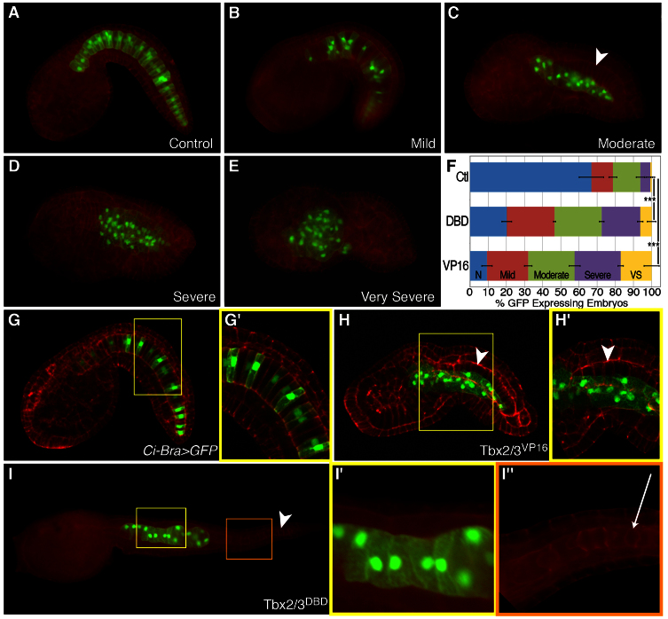
CE transforms the Ciona notochord from a monolayer of 40 notochord cells into a cylindrical rod a single cell in diameter (Munro and Odell, 2002b) (Fig. 4A). When Ciona zygotes were electroporated with either the Ci-Tbx2/3DBD or the Ci-Tbx2/3VP16 plasmid, the resulting embryos showed defective intercalation and impaired tail elongation (Fig. 4). Taking into account mosaic incorporation of the transgenes (Di Gregorio and Levine, 2002), which results in variable severity of the phenotype and produces a mixture of transgenic and non-transgenic notochord cells, we grouped the observed phenotypes into four classes: mild (mostly normal, but displaying incomplete intercalation along some points of the tail, Fig. 4B), moderate (two rows of notochord cells, Fig. 4C), severe (more than two rows of notochord cells, Fig. 4D) and very severe (transgenic cells mislocalized to the trunk, Fig. 4E). The incidence of abnormal notochord development was significantly increased in Ci-Tbx2/3DBD and Ci-Tbx2/3VP16 embryos (79.8% and 90.7%, respectively) compared with control embryos expressing the neutral Ci-Bra>GFP transgene (Corbo et al., 1997) (~33%, Fig. 4F). Despite their abnormalities, Ci-Tbx2/3DBD and Ci-Tbx2/3VP16 embryos still contained 40 notochord cells, even though Ci-Tbx2/3 is expressed prior to the last cell division of notochord precursors (Jiang and Smith, 2007); therefore, Ci-Tbx2/3 appeared not to affect notochord cell mitosis. Ci-Tbx2/3 shRNA constructs and the Ci-Tbx2/3En fusion also produced similar notochord phenotypes (supplementary material Fig. S4); however, the low fluorescence of embryos electroporated with the latter construct prevented its use in further experiments.
Fig. 4.
Perturbation of Ci-Tbx2/3 function causes defects in notochord morphogenesis. (A) Ci-Bra>GFP control embryo. (B-E) Classification of notochord intercalation defects observed in embryos electroporated with either Ci-Bra>Tbx2/3DBD::GFP or Ci-Bra>Tbx2/3VP16::GFP. (F) Quantification of phenotypic categories displayed in A-E. n=1581 for control (Ctl), n=586 for DBD and n=309 for VP16; N, normal; VS, very severe. ***P<0.001 compared with controls. (G-H′) Confocal z-stacks of Ci-Bra>GFP controls (G,G′) and Ci-Bra>Tbx2/3VP16::GFP (H,H′) stage-matched embryos. (I-I″) Larva (~17.5 hpf) expressing Ci-Bra>Tbx2/3DBD::GFP. (G′,H′,I′,I″) Higher magnification views of the areas boxed in yellow or orange in G, H and I, respectively. White arrowheads indicate non-transgenic notochord cells, whereas a white arrow (I″) indicates a lumen between non-transgenic cells. All embryos were counterstained with rhodamine-phalloidin (red).
As described above, electroporated embryos contained different proportions of transgenic and wild-type notochord cells (Fig. 4G-H′). In controls, notochord cells appeared in single file along the length of the tail, intercalated cells had aligned nuclei, and both GFP-positive and GFP-negative cells were disk-shaped (Fig. 4G,G′; supplementary material Movie 1), characteristic of proper notochord formation. By contrast, no such uniformity was seen in embryos expressing either of the altered forms of Ci-Tbx2/3: the transgenic cells in these animals varied in shape and size, appeared in multiple planes and their nuclei were variably located (Fig. 4H,H′; supplementary material Movie 2). In moderately affected Ci-Tbx2/3DBD and Ci-Tbx2/3VP16 embryos, transgenic and non-transgenic notochord cells often appeared spatially separated. For example, in Fig. 4H, fluorescent cells harboring Ci-Tbx2/3VP16 are located ventral-laterally of the non-transgenic notochord cells. This segregation is surprising as cells from both sides of the midline mix randomly during notochord intercalation (Miyamoto and Crowther, 1985; Munro and Odell, 2002b) in Ci-Bra>GFP control embryos (Fig. 4G,G′). Taken together, this phenomenon, along with the different phenotypic variations observed, suggest that these intercalation defects could be due to dose-dependent deficiencies in the motility of the transgenic notochord cells.
At the final stages of notochord differentiation, the notochord normally begins to turn into a hollow tube through the formation of extracellular lumens (Fig. 4I-I″) (Dong et al., 2009). These structures were absent in Ci-Tbx2/3VP16- or Ci-Tbx2/3DBD-expressing cells (Fig. 4I′). Overall, therefore, Ci-Tbx2/3 contributes to different stages of notochord differentiation.
We also monitored the development of tissues adjacent to the notochord in transgenic embryos. The CNS activity of the marker CRM Ci-ETR (Satou et al., 2001) was unaffected by expression of Ci-Tbx2/3VP16 (supplementary material Fig. S5A,B); therefore, the phenotype induced by notochord-specific expression of Ci-Tbx2/3VP16 appeared cell-autonomous. Muscle cells also appeared to be properly specified; however, they failed to elongate along the anterior-posterior axis (supplementary material Fig. S5C,D), consistent with previous observations correlating the elongation of the notochord with the shape of muscle cells (Di Gregorio et al., 2002; Munro and Odell, 2002a).
Differential regulation of target genes by Ci-Tbx2/3
In order to identify candidate Ci-Tbx2/3 target genes, we performed genome-wide microarray analyses on Ci-Tbx2/3DBD- and Ci-Tbx2/3VP16-expressing embryos. Total RNA was isolated from transgenic embryos and stage-matched Ci-Bra>GFP controls at the mid-late neurula and mid-tailbud I/II stages (Hotta et al., 2007), with the aim of capturing targets expressed throughout and following intercalation. These screens yielded 94 probe sets with significant differential expression of at least twofold, corresponding to 81 genes. The datasets obtained from the two stages were mostly non-overlapping, highlighting the dynamic nature of the notochord transcriptome regulated by Ci-Tbx2/3. Furthermore, Ci-Tbx2/3DBD and Ci-Tbx2/3VP16 appear predominantly to regulate distinct sets of targets, although the phenotypes induced by these transgenes are similar; this is consistent with a related screen that found that the VP16 domain influenced TBX2 target selection (Butz et al., 2004). Notably, our results also indicate that Ci-Tbx2/3VP16 may act as an activator in only a few instances (e.g. for Nicastrin); instead, in the majority of cases, this chimera appears to operate as a dominant-negative form because, surprisingly, several genes that emerged from the Ci-Tbx2/3VP16 screen were downregulated (e.g. Noto4, Table 1).
Table 1.
Notochord gene targets from microarray screen of embryos expressing Tbx2/3DBD or Tbx2/3VP16
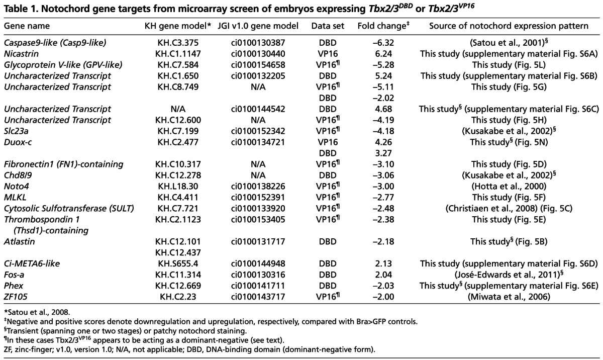
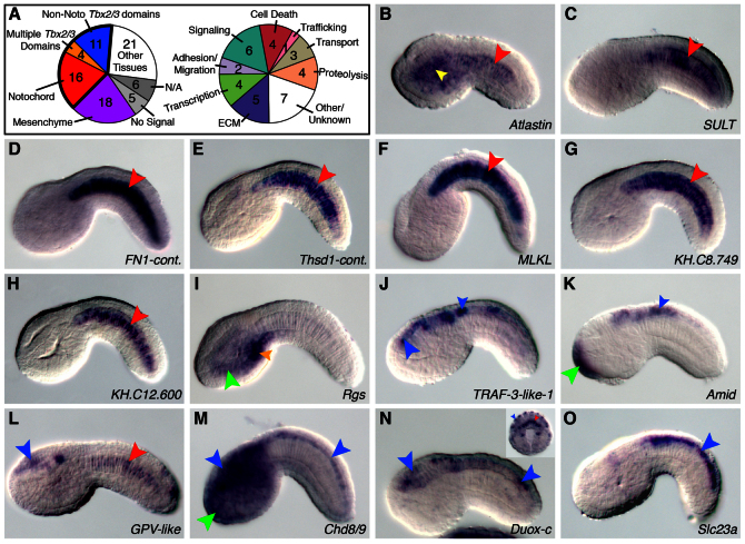
To validate the microarray results, we performed whole-mount in situ hybridization for the 81 candidate targets and observed a detectable signal for 70 genes (Fig. 5A). As expected, the screen identified several notochord genes (20/70, ~29%) (Fig. 5A-H; Table 1; supplementary material Fig. S6), 13 of which are novel, including fibronectin 1-containing (FN1-cont.), thrombospondin 1-containing (Thsd1-cont.) and mixed linage kinase domain-like (MLKL). The microarray data suggested that Ci-Tbx2/3 serves to activate the notochord expression of these genes, as the majority were downregulated when the function of this transcription factor was impaired (Table 1).
Fig. 5.
Expression patterns of candidate Ci-Tbx2/3 targets identified via microarray screen. (A) Left: pie chart summarizing the expression patterns of 81 potential Ci-Tbx2/3 target genes. The ‘multiple Tbx2/3 domains’ category represents those genes found in the notochord and at least one non-notochord Ci-Tbx2/3 expression territory. Right: pie chart depicting putative functions for the 31 targets expressed in Ci-Tbx2/3 expression domains (outlined in bold on the left). Some genes are present in multiple categories. (B-O) Whole-mount in situ hybridization on early to late tailbud embryos for the genes listed in the lower right corner of each panel. The inset in N shows a 110-cell stage embryo probed for Duox-c expression. Stained territories are denoted by colored arrowheads as follows: red, notochord; orange, muscle; blue, CNS; green, epidermis; yellow, endoderm. Larger arrowheads signal expression in domains matching that of Ci-Tbx2/3, whereas smaller arrowheads indicate expression in other areas. Cont., containing; N/A, not assayed.
Interestingly, despite the fact that Ci-Tbx2/3DBD and Ci-Tbx2/3VP16 were expressed almost exclusively in the notochord, we identified a number of targets (11/70, ~16%) in the CNS and epidermis, other Ci-Tbx2/3 expression domains (Fig. 5A,I-K; supplementary material Table S1). We observed that these non-notochord targets, including Rgs and claudin gene family member 3, tend to be upregulated (supplementary material Table S1), implying that they are normally repressed by Ci-Tbx2/3 in the notochord, but activated in the CNS and/or epidermis. In vertebrates, Tbx2 and Tbx3 are thought to act as both activators and repressors (Washkowitz et al., 2012), and the direction of regulation of their targets can vary in a context-dependent manner, such that a gene may be upregulated in one circumstance, but downregulated in another (Butz et al., 2004; Chen et al., 2001; Paxton et al., 2002). The above results, and the fact that we obtained similar phenotypes upon expression of Ci-Tbx2/3DBD, Ci-Tbx2/3VP16 and Ci-Tbx2/3En (Fig. 4; supplementary material Fig. S4), suggested that Ci-Tbx2/3 could both activate and repress transcription, similar to its vertebrate counterparts. However, we cannot rule out that Ci-Tbx2/3 may indirectly regulate some of these downstream genes.
A third category of Ci-Tbx2/3 targets was expressed in both the notochord and other Ci-Tbx2/3 expression domains (Fig. 5A,L-O). Duox-c was transiently expressed in the notochord in early embryos (110-cell stage), but became restricted to various regions of the CNS at later stages (Fig. 5N). Downregulation of Duox-c in the notochord occurs following the neural plate stage (data not shown) when Ci-Tbx2/3 expression commences in this domain (Fig. 2B), consistent with the hypothesis that Ci-Tbx2/3 might repress transcription. This suggests that Ci-Tbx2/3 may also contribute to the temporal control of notochord expression. Furthermore, Slc23a is expressed in the posterior nerve cord at tailbud stages (Fig. 5O), like Ci-Tbx2/3, but switches to the notochord in larvae (Kusakabe et al., 2002). This temporal and spatial variability indicates that the function of Ci-Tbx2/3 is likely modified locally by tissue-specific regulators.
Ci-Tbx2/3 controls genes involved in a wide variety of cellular processes
Fig. 5A categorizes the 31 bona fide targets detected in Ci-Tbx2/3 expression territories according to their predicted functions, e.g. cell death, ECM, transcription, adhesion, signaling and others. This suggests that Ci-Tbx2/3 controls a wide array of cellular processes required to develop the notochord, CNS and epidermis properly, and explains the severe developmental defects observed when the function of this transcriptional regulator is manipulated.
The most extensively characterized notochord gene controlled by Ci-Tbx2/3 is Noto4 (Table 1), a gene originally identified as a transcriptional target of Ci-Bra (Takahashi et al., 1999). Noto4 has been shown to be required for notochord intercalation (Yamada et al., 2011); Noto4 morphants display intercalation defects evocative of the phenotypes observed in embryos expressing the Ci-Tbx2/3DBD and Ci-Tbx2/3VP16 transgenes, strongly suggesting that Noto4 is one of the main effectors of Ci-Tbx2/3. In addition to Noto4, cytosolic sulfotransferase (SULT) (Fig. 5C) had been previously identified as a positive mediator of cell migration in Ciona heart precursors (Christiaen et al., 2008). The downregulation of this gene in Ci-Tbx2/3VP16 embryos could contribute to the impairment of notochord cell motility that we observed (Fig. 4).
Among the developmental regulators controlled by Ci-Tbx2/3 is Fos-a, a notochord transcription factor that we previously positioned downstream of Ci-Bra (José-Edwards et al., 2011). Fos-a appeared among the Ci-Tbx2/3 targets from the microarray analysis of mid-tailbud embryos. Interestingly, Fos-a is normally not expressed at the mid-tailbud stage (José-Edwards et al., 2011) and is upregulated in Ci-Tbx2/3DBD transgenics (Table 1), suggesting that it may be repressed by Ci-Tbx2/3. Preliminary in situ and qPCR results confirm this result (data not shown). Together, these observations hint at the existence of a transcriptional circuit presided over by Ci-Bra that activates both Fos-a and Ci-Tbx2/3 and is later counterbalanced by Ci-Tbx2/3.
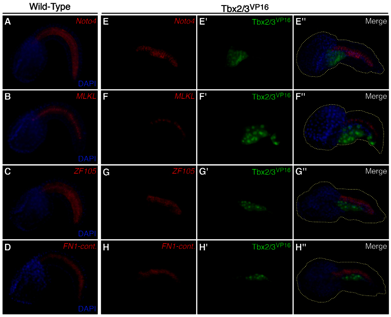
As outlined above, despite the presence of the VP16 activation domain, Ci-Tbx2/3VP16 acts also as a dominant-negative form. To verify this point, and to further validate the results of the screens in vivo, we looked at the expression of a subset of notochord targets downregulated in embryos expressing Ci-Tbx2/3VP16 (Fig. 6). Taking advantage of the mosaic incorporation of the transgene, we expected that candidate targets of Ci-Tbx2/3VP16-mediated repression would be absent in notochord cells containing the transgene, but expressed normally in non-transgenic cells. Indeed, we found that Noto4, MLKL, ZF105 and FN1-cont., which are normally expressed throughout the notochord in control wild-type embryos (Fig. 6A-D), are detected only in non-transgenic cells in Ci-Tbx2/3VP16 embryos (Fig. 6E-H). Accordingly, transgenic notochord cells do not express these genes at detectable levels (Fig. 6E′-H″). These results confirm that, in different contexts, Ci-Tbx2/3VP16 can either activate or prevent transcription.
Fig. 6.
Ci-Tbx2/3 is required for the expression of notochord genes. (A-H″) Fluorescent whole-mount in situ hybridization of wild-type (A-D) or Ci-Tbx2/3VP16::GFP-expressing (green) embryos (E-H′) with probes (red) for Noto4 (A,E-E″), MLKL (B,F-F″), ZF105 (C,G-G″) and FN1-cont. (D,H-H″), and nuclei counterstained with DAPI (blue). Cont., containing.
DISCUSSION
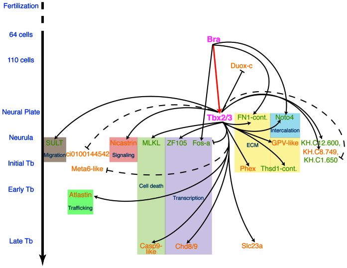
A complete understanding of how the developing body plan is shaped requires the reconstruction of the gene circuitry and cis-regulatory relationships responsible for cell differentiation in a particular lineage. Through this study, we have identified Ci-Tbx2/3 as both an effector and a regulator of a wide branch of the gene network downstream of Ci-Bra (Fig. 7). The elucidation of numerous Ci-Tbx2/3 targets has allowed us to gain insights into the mechanisms of transcriptional control underlying notochord morphogenesis.
Fig. 7.
The notochord gene network downstream of Ci-Bra and Ci-Tbx2/3. The onset of notochord expression of Ci-Bra, Ci-Tbx2/3 and some of the newly identified Ci-Tbx2/3 target genes are plotted against the timeline of Ciona developmental stages on the left. Arrows indicate activation of gene expression, whereas bars signify negative regulation. Broken lines are used for those genes that retain weak notochord expression, possibly owing to incomplete repression by Ci-Tbx2/3. Colored boxes encompass genes with similar putative functions. Gene names in green indicate Ci-Tbx2/3-regulated targets that have also been identified as targets of Ci-Bra in previous studies. Names in orange are used for genes that are Ci-Tbx2/3-specific targets. Cont., containing; ECM, extracellular matrix.
Regulation of Ci-Tbx2/3 expression in the notochord
We have provided evidence that Ci-Bra is required for the notochord expression of another T-box gene, Ci-Tbx2/3. We gathered support for this regulatory relationship from multiple lines of evidence, including the identification of nine T-box binding sites within the Ci-Tbx2/3 notochord CRM. Detailed examination of the minimal sequences required for the notochord activity of the CRM revealed that the T-box sites function redundantly. The Ciona intestinalis genome is highly polymorphic (Dehal et al., 2002), which can lead to faster than usual transcription factor binding site turnover (Satou et al., 2012). The distinctive architecture of the Ci-Tbx2/3 CRM ensures the precise expression of this transcription factor: the high number and redundancy of T-box sites could counteract the effects of inactivating mutations within its sequence. The ability of the 294 bp construct to direct a similar level of notochord expression in the sister ascidian species Ciona savignyi supports this hypothesis. In particular, although the C. savignyi sequence is also enriched in T-box sites, these sites often have differing sequences, orientations and spacing than their counterparts in C. intestinalis, yet C. savignyi embryos retain the ability to properly interpret the cis-regulatory information contained in the C. intestinalis CRM. This finding hints at a considerable degree of flexibility in the functional requirements of the CRM, which may enable it to sustain changes in its sequence.
Role of Ci-Tbx2/3 in notochord formation and identification of both novel and conserved targets
We showed that upon disruption of Ci-Tbx2/3, CE is greatly impaired by the clustering of transgenic notochord cells that fail to interdigitate into a single row. Consistent with Ci-Tbx2/3 being a downstream target of Ci-Bra, lack of intercalation was also observed in Ci-Bra mutant embryos (Chiba et al., 2009). In other systems, Tbx2 subfamily genes are expressed in tissues undergoing similar morphogenetic changes. For example, in zebrafish embryos, Tbx2b-deficient ectodermal cells failed to migrate to the neural plate (Fong et al., 2005). Therefore, the ability to coordinate complex cellular movements seems to be an evolutionarily conserved function for these factors.
Through microarray analysis, we uncovered 31 bona fide Ci-Tbx2/3 targets found in its various expression territories, most importantly the notochord. Although leaky activity of the Ci-Bra promoter in the mesenchyme (Corbo et al., 1997) could account for some of the genes found in this domain, the transient expression of Ci-Tbx2/3 in the mesenchyme (Takatori et al., 2004) argues that some of these 18 genes may also represent Ci-Tbx2/3 targets. Nevertheless, among the 31 Ci-Tbx2/3 targets are genes with various putative functions and also genes that are presumably lineage specific, such as Noto4. Although lineage specific, Noto4 contains a conserved phosphotyrosine binding domain (PTB) (Yamada et al., 2011). Proteins containing this domain, such as integrins, can bind ECM-contacting proteins (Uhlik et al., 2005). Furthermore, deletion of regions of Noto4 outside of the PTB caused notochord cells to take on a spherical, as opposed to their normally columnar, shape (Yamada et al., 2011), suggesting that Noto4 could also interact with the cytoskeleton and thus affect notochord intercalation through different mechanisms.
A large fraction of Ci-Tbx2/3 targets seem to be involved in the construction and remodeling of the ECM, such as FN1-cont. In both the Xenopus mesoderm (Davidson et al., 2006) and Ciona notochord (Veeman et al., 2008) loss of the ECM components fibronectin and laminin, respectively, results in a lack of CE. Thrombospondin-type 1 domain containing (Thsd1-cont.) proteins mediate cell-cell and cell-matrix interactions in the ECM surrounding the tissues in which they are expressed (Adams, 2001), including the notochord (Urry et al., 1998; Wu et al., 2009). Additionally, extracellular leucine-rich repeat-containing proteins, such as GPV-like, have also been shown to contribute to ECM organization in various animals (Mancuso et al., 2012). The presence of these genes within our list of Ci-Tbx2/3 targets indicates that an analogous loss of ECM integrity could have caused the deficiency of intercalation seen in transgenic embryos. Intercalation is also accomplished through the non-canonical Wnt/planar cell polarity (PCP) pathway. Of note, our results suggested that Ci-Tbx2/3 represses a gene encoding a receptor similar to the LRP canonical Wnt co-receptors (LRP-like); it seems conceivable that this could indirectly allow participation of Dsh, a mediator of both non-canonical and canonical/β-catenin Wnt signaling, in PCP at the expense of canonical Wnt signaling to promote notochord cell intercalation.
In vertebrates, the roles of Tbx2/Tbx3 in cancer parallel their functions during embryogenesis. For example, these proteins were recently shown to contribute to epithelial-mesenchymal transition (EMT) (Humtsoe et al., 2012; Wang et al., 2012). Ectopic expression of TBX2 promoted EMT-like invasiveness of mammary epithelial cells, causing the formation of lamellipodial protrusions and overexpression of ECM remodelers; both E-cadherin and the tight junction protein ZO1 were also downregulated (Wang et al., 2012). Claudins are another component of tight junctions that help maintain stable contacts between cells (Steed et al., 2010). Interestingly, among the upregulated genes in our screen was claudin gene family member 3. Greater notochord cell-cell interactions in embryos where Ci-Tbx2/3 function is impaired could account for the clustering of transgenic notochord cells that we observed. Therefore, overall, cell adhesion and ECM genes appear to be recurrent Tbx2/Tbx3 targets. Notably, although TBX3 was found to be upregulated in EMT-prone squamous cell carcinoma cells, DUOX1 was downregulated in a screen for genes differentially expressed in EMT (Humtsoe et al., 2012). Our finding that Ci-Tbx2/3 repressed Duox-c in the notochord suggests that TBX3 may similarly regulate the expression of DUOX1 during EMT. Exploration of the consequences of this relationship that we identified in Ciona could thus inform studies of related morphological changes in other biological contexts.
Even though not all Ciona genes possess unequivocal vertebrate counterparts, we found parallels with targets identified in previous screens in NIH3T3, ROS17/2.8 (Chen et al., 2001) and HEK293 (Butz et al., 2004) cells overexpressing TBX2. Similarities include targets belonging to the same gene ontology families, such as cytosolic sulfotransferase (SULT), cathepsin and Rgs proteins, and genes involved in ECM formation, cell adhesion and apoptosis. Tail regression of Ciona larvae prior to metamorphosis involves apoptosis of various tissues, including those of the notochord, an event involving caspases (Chambon et al., 2002; Nishiyama and Fujiwara, 2008). Although we did not see evidence of apoptosis during tailbud stages, we found that tail resorption in Ci-Tbx2/3VP16-expressing larvae was delayed and juveniles retained more tail remnants than wild-type controls (data not shown). Among the notochord genes regulated by Ci-Tbx2/3 was Caspase9-like (Fig. 7). Caspases are conserved targets for Tbx2 subfamily members: overexpression of TBX2 in SW13 adenocarcinoma led to reductions in the activation of caspases 8 and 9 (Ismail and Bateman, 2009). Another Ci-Tbx2/3 notochord target, MLKL, was very recently implicated in a mechanism of cell death known as necroptosis (Sun et al., 2012; Zhao et al., 2012); its role in embryonic development remains unclear, but its contribution to notochord morphogenesis can now be investigated using Ciona as a model.
Together, these results indicate that Ci-Tbx2/3 has retained ancestral target genes common to the main chordate lineage and has also acquired control over derived, lineage-specific genes.
The T-box family and the notochord gene regulatory network
The interactions between T-box transcription factors in vertebrates can be exceedingly complex given the large number of family members, many of which are co-expressed. The hierarchical relationship that we identified indicates that, in addition to controlling a specific cohort of genes, Ci-Tbx2/3 serves to both reinforce and refine the Ci-Bra-downstream gene regulatory network. The comparison of Ci-Tbx2/3-regulated genes identified here to those previously classified as Ci-Bra targets suggests that Ci-Bra is required for the initial activation of shared targets that have the same onset as Ci-Tbx2/3, such as Noto4 and FN1-cont., whereas Ci-Tbx2/3 is required to maintain the notochord expression of these genes later in development. Shared targets expressed at later stages of notochord formation may be indirectly regulated by Ci-Bra using Ci-Tbx2/3 as an intermediary, or alternatively Ci-Bra and Ci-Tbx2/3 may cooperatively control expression of these target genes (green font, Fig. 7). By contrast, Ci-Tbx2/3 serves to temporally limit the notochord activity of yet another set of Ci-Bra targets, including Fos-a. As noted above, there is also a conspicuous group of Ci-Tbx2/3 targets that do not seem to overlap with those of Ci-Bra (orange font, Fig. 7). These results imply that, despite belonging to the same family and binding similar consensus sequences (Tada and Smith, 2001), T-box transcription factors maintain distinct activities. Consistently, previous work has shown that target specificity is influenced by the DBD itself (Conlon et al., 2001), and it is noteworthy that the T-domains of Ci-Bra and Ci-Tbx2/3 share only 47% identity.
The lack of recognizable activation or repression domains in the Ci-Tbx2/3 protein sequence make its categorization challenging. Nevertheless, the observation that Ci-Tbx2/3 targets are often expressed in a mutually exclusive fashion in the notochord and CNS suggests that Ci-Tbx2/3 works as an activator and is counteracted by localized repressors. However, the similarity of the phenotypes induced by Ci-Tbx2/3DBD, Ci-Tbx2/3VP16 and Ci-Tbx2/3En, and the temporal regulation of some of the notochord targets suggest that Ci-Tbx2/3 can ultimately operate as both an activator and a repressor in a context-dependent fashion. This functional duality could depend upon the tissue-specific availability of distinct Ci-Tbx2/3 interacting partners and provides a powerful mechanism that ensures fine-tuned control of tissue identity in the developing embryo.
Supplementary Material
Acknowledgements
We thank Drs Chiba and Smith (UC Santa Barbara, CA, USA) for providing the Ci-Bra−/− embryos.
Footnotes
Funding
This work was supported by the National Institutes of Health/National Institute of General Medical Sciences (NIH/NIGMS) [GM100466] along with supplemental funding from the American Recovery and Reinvestment Act [HD050704-05S1], by the March of Dimes Birth Defects Foundation [1-FY11-468] and by the Charles A. Frueauff Foundation [to A.D.G.]. D.S.J.-E. was supported in part by an NIH training grant [T32 GM008539]. I.O.-I. was supported in part by a fellowship from the Uehara Memorial Foundation (Japan). Research reported in this publication was supported by the National Institute of General Medicine of the NIH. Deposited in PMC for release after 12 months.
Competing interests statement
The authors declare no competing financial interests.
Supplementary material
Supplementary material available online at http://dev.biologists.org/lookup/suppl/doi:10.1242/dev.094227/-/DC1
References
- Adams J. C. (2001). Thrombospondins: multifunctional regulators of cell interactions. Annu. Rev. Cell Dev. Biol. 17, 25-51 [DOI] [PubMed] [Google Scholar]
- Butz N. V., Campbell C. E., Gronostajski R. M. (2004). Differential target gene activation by TBX2 and TBX2VP16: evidence for activation domain-dependent modulation of gene target specificity. Gene 342, 67-76 [DOI] [PubMed] [Google Scholar]
- Chambon J.-P., Soule J., Pomies P., Fort P., Sahuquet A., Alexandre D., Mangeat P.-H., Baghdiguian S. (2002). Tail regression in Ciona intestinalis (Prochordate) involves a Caspase-dependent apoptosis event associated with ERK activation. Development 129, 3105-3114 [DOI] [PubMed] [Google Scholar]
- Chen J., Zhong Q., Wang J., Cameron R. S., Borke J. L., Isales C. M., Bollag R. J. (2001). Microarray analysis of Tbx2-directed gene expression: a possible role in osteogenesis. Mol. Cell. Endocrinol. 177, 43-54 [DOI] [PubMed] [Google Scholar]
- Chiba S., Jiang D., Satoh N., Smith W. C. (2009). Brachyury null mutant-induced defects in juvenile ascidian endodermal organs. Development 136, 35-39 [DOI] [PMC free article] [PubMed] [Google Scholar]
- Christiaen L., Davidson B., Kawashima T., Powell W., Nolla H., Vranizan K., Levine M. (2008). The transcription/migration interface in heart precursors of Ciona intestinalis. Science 320, 1349-1352 [DOI] [PubMed] [Google Scholar]
- Conlon F. L., Fairclough L., Price B. M., Casey E. S., Smith J. C. (2001). Determinants of T box protein specificity. Development 128, 3749-3758 [DOI] [PubMed] [Google Scholar]
- Corbo J. C., Levine M., Zeller R. W. (1997). Characterization of a notochord-specific enhancer from the Brachyury promoter region of the ascidian, Ciona intestinalis. Development 124, 589-602 [DOI] [PubMed] [Google Scholar]
- Davidson L. A., Marsden M., Keller R., Desimone D. W. (2006). Integrin α5β1 and fibronectin regulate polarized cell protrusions required for Xenopus convergence and extension. Curr. Biol. 16, 833-844 [DOI] [PubMed] [Google Scholar]
- Dehal P., Satou Y., Campbell R. K., Chapman J., Degnan B., De Tomaso A., Davidson B., Di Gregorio A., Gelpke M., Goodstein D. M., et al. (2002). The draft genome of Ciona intestinalis: insights into chordate and vertebrate origins. Science 298, 2157-2167 [DOI] [PubMed] [Google Scholar]
- Dheen T., Sleptsova-Friedrich I., Xu Y., Clark M., Lehrach H., Gong Z., Korzh V. (1999). Zebrafish tbx-c functions during formation of midline structures. Development 126, 2703-2713 [DOI] [PubMed] [Google Scholar]
- Di Gregorio A., Levine M. (2002). Analyzing gene regulation in ascidian embryos: new tools for new perspectives. Differentiation 70, 132-139 [DOI] [PubMed] [Google Scholar]
- Di Gregorio A., Harland R. M., Levine M., Casey E. S. (2002). Tail morphogenesis in the ascidian, Ciona intestinalis, requires cooperation between notochord and muscle. Dev. Biol. 244, 385-395 [DOI] [PubMed] [Google Scholar]
- Dong B., Horie T., Denker E., Kusakabe T., Tsuda M., Smith W. C., Jiang D. (2009). Tube formation by complex cellular processes in Ciona intestinalis notochord. Dev. Biol. 330, 237-249 [DOI] [PMC free article] [PubMed] [Google Scholar]
- Dunn M. P., Di Gregorio A. (2009). The evolutionarily conserved leprecan gene: its regulation by Brachyury and its role in the developing Ciona notochord. Dev. Biol. 328, 561-574 [DOI] [PMC free article] [PubMed] [Google Scholar]
- Fong S. H., Emelyanov A., Teh C., Korzh V. (2005). Wnt signalling mediated by Tbx2b regulates cell migration during formation of the neural plate. Development 132, 3587-3596 [DOI] [PubMed] [Google Scholar]
- Frazer K. A., Pachter L., Poliakov A., Rubin E. M., Dubchak I. (2004). VISTA: computational tools for comparative genomics. Nucleic Acids Res. 32 Web Server issue, W273-W279 [DOI] [PMC free article] [PubMed] [Google Scholar]
- Harrelson Z., Kelly R. G., Goldin S. N., Gibson-Brown J. J., Bollag R. J., Silver L. M., Papaioannou V. E. (2004). Tbx2 is essential for patterning the atrioventricular canal and for morphogenesis of the outflow tract during heart development. Development 131, 5041-5052 [DOI] [PubMed] [Google Scholar]
- Herrmann B. G., Labeit S., Poustka A., King T. R., Lehrach H. (1990). Cloning of the T gene required in mesoderm formation in the mouse. Nature 343, 617-622 [DOI] [PubMed] [Google Scholar]
- Horton A. C., Mahadevan N. R., Minguillon C., Osoegawa K., Rokhsar D. S., Ruvinsky I., de Jong P. J., Logan M. P., Gibson-Brown J. J. (2008). Conservation of linkage and evolution of developmental function within the Tbx2/3/4/5 subfamily of T-box genes: implications for the origin of vertebrate limbs. Dev. Genes Evol. 218, 613-628 [DOI] [PubMed] [Google Scholar]
- Hotta K., Takahashi H., Asakura T., Saitoh B., Takatori N., Satou Y., Satoh N. (2000). Characterization of Brachyury-downstream notochord genes in the Ciona intestinalis embryo. Dev. Biol. 224, 69-80 [DOI] [PubMed] [Google Scholar]
- Hotta K., Mitsuhara K., Takahashi H., Inaba K., Oka K., Gojobori T., Ikeo K. (2007). A web-based interactive developmental table for the ascidian Ciona intestinalis, including 3D real-image embryo reconstructions: I. From fertilized egg to hatching larva. Dev. Dyn. 236, 1790-1805 [DOI] [PubMed] [Google Scholar]
- Humtsoe J. O., Koya E., Pham E., Aramoto T., Zuo J., Ishikawa T., Kramer R. H. (2012). Transcriptional profiling identifies upregulated genes following induction of epithelial-mesenchymal transition in squamous carcinoma cells. Exp. Cell Res. 318, 379-390 [DOI] [PubMed] [Google Scholar]
- Imai K. S., Hino K., Yagi K., Satoh N., Satou Y. (2004). Gene expression profiles of transcription factors and signaling molecules in the ascidian embryo: towards a comprehensive understanding of gene networks. Development 131, 4047-4058 [DOI] [PubMed] [Google Scholar]
- Ismail A., Bateman A. (2009). Expression of TBX2 promotes anchorage-independent growth and survival in the p53-negative SW13 adrenocortical carcinoma. Cancer Lett. 278, 230-240 [DOI] [PubMed] [Google Scholar]
- Jiang D., Smith W. C. (2007). Ascidian notochord morphogenesis. Dev. Dyn. 236, 1748-1757 [DOI] [PMC free article] [PubMed] [Google Scholar]
- Jiang D., Munro E. M., Smith W. C. (2005). Ascidian prickle regulates both mediolateral and anterior-posterior cell polarity of notochord cells. Curr. Biol. 15, 79-85 [DOI] [PubMed] [Google Scholar]
- José-Edwards D. S., Kerner P., Kugler J. E., Deng W., Jiang D., Di Gregorio A. (2011). The identification of transcription factors expressed in the notochord of Ciona intestinalis adds new potential players to the brachyury gene regulatory network. Dev. Dyn. 240, 1793-1805 [DOI] [PMC free article] [PubMed] [Google Scholar]
- Keller R., Davidson L., Edlund A., Elul T., Ezin M., Shook D., Skoglund P. (2000). Mechanisms of convergence and extension by cell intercalation. Philos. Trans. R. Soc. Lond. B Biol. Sci. 355, 897-922 [DOI] [PMC free article] [PubMed] [Google Scholar]
- Kubo A., Suzuki N., Yuan X., Nakai K., Satoh N., Imai K. S., Satou Y. (2010). Genomic cis-regulatory networks in the early Ciona intestinalis embryo. Development 137, 1613-1623 [DOI] [PubMed] [Google Scholar]
- Kugler J. E., Passamaneck Y. J., Feldman T. G., Beh J., Regnier T. W., Di Gregorio A. (2008). Evolutionary conservation of vertebrate notochord genes in the ascidian Ciona intestinalis. Genesis 46, 697-710 [DOI] [PMC free article] [PubMed] [Google Scholar]
- Kusakabe T., Yoshida R., Kawakami I., Kusakabe R., Mochizuki Y., Yamada L., Shin-i T., Kohara Y., Satoh N., Tsuda M., et al. (2002). Gene expression profiles in tadpole larvae of Ciona intestinalis. Dev. Biol. 242, 188-203 [DOI] [PubMed] [Google Scholar]
- Lu J., Li X.-P., Dong Q., Kung H.-F., He M.-L. (2010). TBX2 and TBX3: the special value for anticancer drug targets. Biochim. Biophys. Acta 1806, 268-274 [DOI] [PMC free article] [PubMed] [Google Scholar]
- Mancuso V. P., Parry J. M., Storer L., Poggioli C., Nguyen K. C. Q., Hall D. H., Sundaram M. V. (2012). Extracellular leucine-rich repeat proteins are required to organize the apical extracellular matrix and maintain epithelial junction integrity in C. elegans. Development 139, 979-990 [DOI] [PMC free article] [PubMed] [Google Scholar]
- Manning L., Ohyama K., Saeger B., Hatano O., Wilson S. A., Logan M., Placzek M. (2006). Regional morphogenesis in the hypothalamus: a BMP-Tbx2 pathway coordinates fate and proliferation through Shh downregulation. Dev. Cell 11, 873-885 [DOI] [PubMed] [Google Scholar]
- Martin B. L., Kimelman D. (2008). Regulation of canonical Wnt signaling by Brachyury is essential for posterior mesoderm formation. Dev. Cell 15, 121-133 [DOI] [PMC free article] [PubMed] [Google Scholar]
- Miwata K., Chiba T., Horii R., Yamada L., Kubo A., Miyamura D., Satoh N., Satou Y. (2006). Systematic analysis of embryonic expression profiles of zinc finger genes in Ciona intestinalis. Dev. Biol. 292, 546-554 [DOI] [PubMed] [Google Scholar]
- Miyamoto D. M., Crowther R. J. (1985). Formation of the notochord in living ascidian embryos. J. Embryol. Exp. Morphol. 86, 1-17 [PubMed] [Google Scholar]
- Munro E. M., Odell G. (2002a). Morphogenetic pattern formation during ascidian notochord formation is regulative and highly robust. Development 129, 1-12 [DOI] [PubMed] [Google Scholar]
- Munro E. M., Odell G. M. (2002b). Polarized basolateral cell motility underlies invagination and convergent extension of the ascidian notochord. Development 129, 13-24 [DOI] [PubMed] [Google Scholar]
- Naiche L. A., Harrelson Z., Kelly R. G., Papaioannou V. E. (2005). T-box genes in vertebrate development. Annu. Rev. Genet. 39, 219-239 [DOI] [PubMed] [Google Scholar]
- Nishiyama A., Fujiwara S. (2008). RNA interference by expressing short hairpin RNA in the Ciona intestinalis embryo. Dev. Growth Differ. 50, 521-529 [DOI] [PubMed] [Google Scholar]
- Oda-Ishii I., Di Gregorio A. (2007). Lineage-independent mosaic expression and regulation of the Ciona multidom gene in the ancestral notochord. Dev. Dyn. 236, 1806-1819 [DOI] [PubMed] [Google Scholar]
- Packham E. A., Brook J. D. (2003). T-box genes in human disorders. Hum. Mol. Genet. 12 Suppl. 1, R37-R44 [DOI] [PubMed] [Google Scholar]
- Passamaneck Y. J., Di Gregorio A. (2005). Ciona intestinalis: chordate development made simple. Dev. Dyn. 233, 1-19 [DOI] [PubMed] [Google Scholar]
- Paxton C., Zhao H., Chin Y., Langner K., Reecy J. (2002). Murine Tbx2 contains domains that activate and repress gene transcription. Gene 283, 117-124 [DOI] [PubMed] [Google Scholar]
- Sadowski I., Ma J., Triezenberg S., Ptashne M. (1988). GAL4-VP16 is an unusually potent transcriptional activator. Nature 335, 563-564 [DOI] [PubMed] [Google Scholar]
- Satou Y., Takatori N., Yamada L., Mochizuki Y., Hamaguchi M., Ishikawa H., Chiba S., Imai K., Kano S., Murakami S. D., et al. (2001). Gene expression profiles in Ciona intestinalis tailbud embryos. Development 128, 2893-2904 [DOI] [PubMed] [Google Scholar]
- Satou Y., Mineta K., Ogasawara M., Sasakura Y., Shoguchi E., Ueno K., Yamada L., Matsumoto J., Wasserscheid J., Dewar K., et al. (2008). Improved genome assembly and evidence-based global gene model set for the chordate Ciona intestinalis: new insight into intron and operon populations. Genome Biol. 9, R152 [DOI] [PMC free article] [PubMed] [Google Scholar]
- Satou Y., Shin-i T., Kohara Y., Satoh N., Chiba S. (2012). A genomic overview of short genetic variations in a basal chordate, Ciona intestinalis. BMC Genomics 13, 208 [DOI] [PMC free article] [PubMed] [Google Scholar]
- Showell C., Binder O., Conlon F. L. (2004). T-box genes in early embryogenesis. Dev. Dyn. 229, 201-218 [DOI] [PMC free article] [PubMed] [Google Scholar]
- Steed E., Balda M. S., Matter K. (2010). Dynamics and functions of tight junctions. Trends Cell Biol. 20, 142-149 [DOI] [PubMed] [Google Scholar]
- Stemple D. L. (2005). Structure and function of the notochord: an essential organ for chordate development. Development 132, 2503-2512 [DOI] [PubMed] [Google Scholar]
- Sun L., Wang H., Wang Z., He S., Chen S., Liao D., Wang L., Yan J., Liu W., Lei X., et al. (2012). Mixed lineage kinase domain-like protein mediates necrosis signaling downstream of RIP3 kinase. Cell 148, 213-227 [DOI] [PubMed] [Google Scholar]
- Tada M., Smith J. C. (2001). T-targets: clues to understanding the functions of T-box proteins. Dev. Growth Differ. 43, 1-11 [DOI] [PubMed] [Google Scholar]
- Takabatake Y., Takabatake T., Takeshima K. (2000). Conserved and divergent expression of T-box genes Tbx2-Tbx5 in Xenopus. Mech. Dev. 91, 433-437 [DOI] [PubMed] [Google Scholar]
- Takahashi H., Hotta K., Erives A., Di Gregorio A., Zeller R. W., Levine M., Satoh N. (1999). Brachyury downstream notochord differentiation in the ascidian embryo. Genes Dev. 13, 1519-1523 [DOI] [PMC free article] [PubMed] [Google Scholar]
- Takatori N., Hotta K., Mochizuki Y., Satoh G., Mitani Y., Satoh N., Satou Y., Takahashi H. (2004). T-box genes in the ascidian Ciona intestinalis: characterization of cDNAs and spatial expression. Dev. Dyn. 230, 743-753 [DOI] [PubMed] [Google Scholar]
- Tassy O., Dauga D., Daian F., Sobral D., Robin F., Khoueiry P., Salgado D., Fox V., Caillol D., Schiappa R., et al. (2010). The ANISEED database: digital representation, formalization, and elucidation of a chordate developmental program. Genome Res. 20, 1459-1468 [DOI] [PMC free article] [PubMed] [Google Scholar]
- Uhlik M. T., Temple B., Bencharit S., Kimple A. J., Siderovski D. P., Johnson G. L. (2005). Structural and evolutionary division of phosphotyrosine binding (PTB) domains. J. Mol. Biol. 345, 1-20 [DOI] [PubMed] [Google Scholar]
- Urry L. A., Whittaker C. A., Duquette M., Lawler J., DeSimone D. W. (1998). Thrombospondins in early Xenopus embryos: dynamic patterns of expression suggest diverse roles in nervous system, notochord, and muscle development. Dev. Dyn. 211, 390-407 [DOI] [PubMed] [Google Scholar]
- Veeman M. T., Nakatani Y., Hendrickson C., Ericson V., Lin C., Smith W. C. (2008). Chongmague reveals an essential role for laminin-mediated boundary formation in chordate convergence and extension movements. Development 135, 33-41 [DOI] [PMC free article] [PubMed] [Google Scholar]
- Wagner E., Levine M. (2012). FGF signaling establishes the anterior border of the Ciona neural tube. Development 139, 2351-2359 [DOI] [PMC free article] [PubMed] [Google Scholar]
- Wang B., Lindley L. E., Fernandez-Vega V., Rieger M. E., Sims A. H., Briegel K. J. (2012). The T box transcription factor TBX2 promotes epithelial-mesenchymal transition and invasion of normal and malignant breast epithelial cells. PLoS ONE 7, e41355 [DOI] [PMC free article] [PubMed] [Google Scholar]
- Washkowitz A. J., Gavrilov S., Begum S., Papaioannou V. E. (2012). Diverse functional networks of Tbx3 in development and disease. Wiley Interdiscip. Rev. Syst. Biol. Med. 4, 273-283 [DOI] [PMC free article] [PubMed] [Google Scholar]
- Wu F.-R., Zhou L.-Y., Nagahama Y., Wang D.-S. (2009). Duplication and distinct expression patterns of two thrombospondin-1 isoforms in teleost fishes. Gene Expr. Patterns 9, 436-443 [DOI] [PubMed] [Google Scholar]
- Yamada S., Ueno N., Satoh N., Takahashi H. (2011). Ciona intestinalis Noto4 contains a phosphotyrosine interaction domain and is involved in the midline intercalation of notochord cells. Int. J. Dev. Biol. 55, 11-18 [DOI] [PubMed] [Google Scholar]
- Zhao J., Jitkaew S., Cai Z., Choksi S., Li Q., Luo J., Liu Z.-G. (2012). Mixed lineage kinase domain-like is a key receptor interacting protein 3 downstream component of TNF-induced necrosis. Proc. Natl. Acad. Sci. USA 109, 5322-5327 [DOI] [PMC free article] [PubMed] [Google Scholar]
Associated Data
This section collects any data citations, data availability statements, or supplementary materials included in this article.


