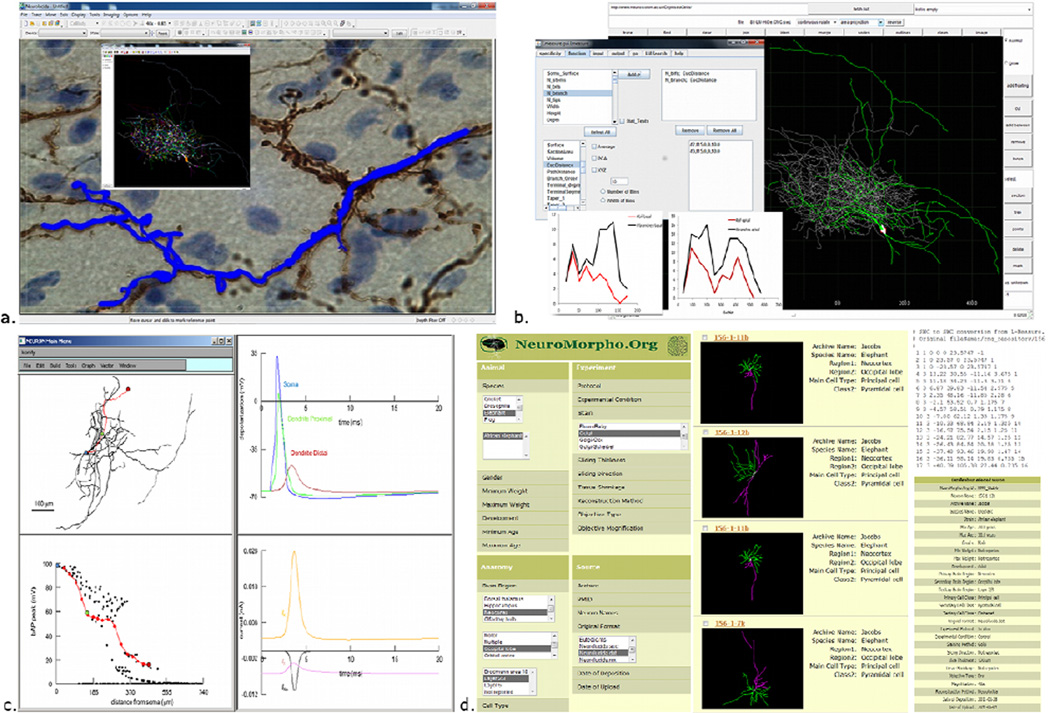
Figure 4. The ecosystem of digital reconstructions: representative resources and tools.
a. Neurolucida allows live or offline tracing and its companion module NeuroExplorer (inset) performs quantitative analyses, b. Digital reconstructions can be viewed and edited using Cvapp, and extensive morphometric analysis can be performed using L-Measure (inset. c. The NEURON simulation environment can distribute biophysical properties on imported neuronal reconstructions (in this example, a hippocampal interneuron; top left) for electrophysiological simulations. Here, the membrane depolarization is recorded (top right) at the soma (blue), proximal dendrite (green) and distal dendrite (red). The peak of back-propagating action potential along the dendrite decreases in amplitude with distance from the soma (bottom left), due to distinct current contributions (bottom right). d. NeuroMorpho.Org hosts digital reconstruction of neuronal morphologies that are published in peer-reviewed journals. Searching across different metadata categories (left) returns a summary result list (middle), which can be individually browsed to download the reconstruction as a standard SWC file (top right) or for additional metadata details (bottom right).

