Abstract
Background/Aim
Excess of fat intake is dramatically increasing in women of childbearing age and results in numerous health complications, including reproductive disorders. Using rabbit does as a biomedical model, the aim of this study was to evaluate onset of puberty, endocrine responses to stimulation and ovarian follicular maturation in females fed a high fat high cholesterol diet (HH diet) from 10 weeks of age (i.e., 2 weeks before normal onset of puberty) or a control diet (C diet).
Methodology/Principal Findings
Three experiments were performed, each including 8 treated (HH group) and 8 control (C group) does. In experiment 1, the endocrine response to Gonadotropin releasing hormone (GnRH) was evaluated at 13, 18 and 22 weeks of age. In experiment 2, the follicular population was counted in ovaries of adult females (18 weeks of age). In experiment 3, the LH response to mating and steroid profiles throughout gestation were evaluated at 18 weeks of age. Fetal growth was monitored by ultrasound and offspring birth weight was recorded. Data showed a significantly higher Luteinizing hormone (LH) response after induction of ovulation at 13 weeks of age in the HH group. There was no difference at 18 weeks, but at 22 weeks, the LH response to GnRH was significantly reduced in the HH group. The number of atretic follicles was significantly increased and the number of antral follicles significantly reduced in HH does vs. controls. During gestation, the HH diet induced intra-uterine growth retardation (IUGR).
Conclusion
The HH diet administered from before puberty onwards affected onset of puberty, follicular growth, hormonal responses to breeding and GnRH stimulation in relation to age and lead to fetal IUGR.
Introduction
Adult lifestyle - generally diet and sedentary habits – as well as environmental chemicals are known factors impacting the fertility of men and women. According to the recent national observational study “Obésité-Epidémiologie” (ObEpi), the prevalence of overweight (defined by a Body Mass Index (BMI) >25 kg/m2) and obesity (BMI >30 kg/m2) among French women is 25% and 15% respectively [1]. The weight of women in childbearing age is dramatically increasing by about 0.5 to 0.7 kg/year. Moreover, in Europe, lipid intake represents more than 32% of food intake with a high proportion of saturated fat [2]. The Normal Weight Obese syndrome, which affects up to 37% of apparently healthy patients, is characterized by a BMI <25 together with a high fat mass (>30%), leading to high inflammatory cytokines and a high level of oxidative stress [3], [4]. Oxidative stress is well known to be detrimental in many tissues, including the female reproductive tract [5]. Compromised oocyte quality, altered pubertal development and hormonal and ovulatory dysfunction have been described.
Early onset of puberty has also been described in animals fed a high fat diet or in models of obese animals. Feng Li et al. [6] showed a dramatic acceleration of the LH pulse frequency concomitant with an early onset of puberty in rat fed a high fat diet. Even the short term (7 days) administration of a fat enriched diet can significantly modify endocrine responses, inducing higher plasma LH concentrations in rats [7]. In pigs, Iberian gilts, which are naturally obese due to leptin resistance subsequent to a mutation in the leptin receptor, have an earlier onset of puberty compared to other breeds [8]. In primates, precocious menarche was reported in rhesus monkeys fed a high calorie diet, in association with high nocturnal plasma LH concentrations [9].
In adults, numerous studies have demonstrated a reduced LH surge in adult obese women. Overweight and obese women were found to have a longer follicular phase and reduced plasma LH concentrations [10], [11], [12]. In a study where 22 fertile, overweight or obese women were compared with 10 fertile, normal-weight women, the overweight group had lower plasma LH concentrations compared with normal weight women and LH concentrations were negatively correlated to BMI and waist circumference [13]. An additional study including 18 premenopausal, eumenorrheic (nonpolycystic syndrome) morbidly obese women and 12 eumenorrheic, normal-weight subjects found a dramatic reduction of both the amplitude and the mean LH concentration in the morbid obesity group [14]. In women, it has been suggested that the pituitary response to endogenous GnRH is attenuated by obesity. Recent work in Assisted Reproductive Technologies (ART) has shown that obese patient require higher doses of gonadotropins and prolonged ovarian stimulation compared to normal weight individuals [15], [16], [17]. Other studies, however, have failed to demonstrate a difference in the ovarian response to stimulation in obese women [18], [19], [20].
Effects of high fat diets were also observed on the ovarian function. In rats, the administration of a cafeteria diet was shown to negatively affect female reproduction by reducing the number of oocytes (median number (interquartile range) of oocytes in cafeteria diet fed females [1(0/6)] vs. chow-fed rats [10(8/12)]) and preantral follicles in the ovary [21]. In obese hyperinsulinemic (fa/fa) adult rats, the ovaries from the obese rat contained more corpora lutea, antral, pre antral and atretic follicles compared to controls, with a positive association between follicular atresia and the expression of the pro apopotic factor of transcription FOXO1 [22]. Similarly, the number of follicles present in the ovary of obese, ob/ob mice, is reduced and granulosa cell apoptosis and follicular atresia are increased [23].
In the same model, excessive lipid storage was shown to induce ovarian function disorders with advanced follicular atresia, apoptosis and defective steroidogenesis [24]. In contrast to what has been found in rodents, fat supplementation in lactating cows did not appear to affect follicular growth, although luteal progesterone was reduced in supplemented cows [25]. In the obese Iberian pig model, the ovarian follicular population, plasma estradiol concentrations and ovulation rates were not different compared to lean pigs [26].
High fat diets appear to induce different effects on reproduction according to the model, the age, the exposure and/or the fat content in the diet. The objective of this work was to explore the effects of a high fat, high cholesterol diet administered from the prepubertal period on the onset of reproductive function, endocrine status and follicular growth, using a previously established rabbit model [27]. The rabbit was chosen as a model because of its decisive advantages over the rat or mouse models for the proposed longitudinal studies [28]. Indeed, it is a preferred model for diet-induced lipid metabolic disorders and insulin resistance [29] and its size allows for repetitive blood sampling and transabdominal ultrasound during pregnancy.
Materials and Methods
Ethical Statement
The experiment was performed in accordance with the International Guiding Principles for Biomedical Research involving Animals as promulgated by the Society for the Study of Reproduction and in accordance with the European Convention on Animal experimentation. The animal studies were approved by the local animal care and use committee (CSU UCEA) and received ethical approval from the local ethics committee (COMETHEA), under protocol number 12/029. Researchers involved in the work with the animals possessed an animal experimentation license (level 1 or 2) delivered by the French veterinary authorities.
Animals and Diet
Forty-eight female New Zealand rabbits (INRA 1077 or PS 19 line) were housed individually with free access to water, under a 8 hours light/16 hours dark photoperiod unless stated below. At 10 weeks of age, they were allocated to one of two groups and fed ad libitum with either a hyperlipidic hypercholesterolemic diet (HH group) (n = 24) or a control diet (C group) (n = 24) containing respectively 7.71% or 1.83% fat and 0.2% or 0% cholesterol as previously described [27].
The fat supplementation consisted of soybean oil, i.e. mainly polyunsaturated fatty acids (N6/N3 = 6.86). The euthanasia of the animals was performed by exsanguination after electronarcosis at the local experimental slaughterhouse, according to the protocol approved by the local ethics committee and the veterinary services.
Experimental Protocol
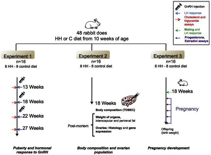
The study was organized in 3 consecutive experiments as summarized in Figure 1.
Figure 1. Schematic representation of the experimental protocol.
Three experiments were performed, each including 8 treated (HH group) and 8 controls does (C group). Experiment 1: Influence of HH diet on puberty and hormonal response. Experiment 2: Influence of HH diet on the ovarian follicular population. Experiment 3: Influence of HH diet on endocrine function during gestation, with fetal growth monitored by ultrasound and recording of offspring birth weight.
Experiment 1 (n = 16)
The first experiment was conducted in does from 10 to 27 weeks of age to evaluate the influence of the HH diet on the onset of puberty and ovulation disorders according to age. Experiments were repeated in the same animals at 13, 18 and 22 weeks of age.
Females were fasted overnight, weighed and blood was collected from the auricular vein into EDTA coated vacutainers for biochemical dosages (total cholesterol and triglycerides, progesterone, leptin and estradiol).
The rabbit being an induced ovulator, puberty can only be assessed by inducing ovulation, either by mating or by chemical stimulation. Here, the onset of puberty was evaluated by injecting 40 µg of a GnRH analogue (Receptal®) IM after 1 week of synchronization with light (16 hours of light/8 hours of dark). Blood was collected for LH assay at 0, 30, 60, 90, 120 and 180 minutes after injection. Blood samples were obtained through a catheter previously placed in the peripheral ear vein into EDTA coated vacutainers which were placed on ice until processing. Samples were centrifuged within an hour of collection and supernatants stored in several aliquots at −20°C until analysis.
Experiment 2 (n = 16)
In the second experiment, ovaries were collected from 18 week old females after slaughter in order to assess follicular populations in the ovaries and ovarian function.
One week before slaughter, 16 does were synchronised by 16 hours of light and 8 hours of dark by day. At 18 weeks of age, the body composition was analysed by Total Body Electrical Conductivity (TOBEC) [30]. After euthanasia, the liver, kidney, ovaries, interscapular and perirenal fat were weighted. All ovaries were flash frozen in liquid nitrogen and then conserved at −80°C for molecular analysis (1 ovary per animal) or fixed in 10% formalin and processed for histological analysis (1 ovary per animal).
For histological analysis, ovaries were embedded in paraffin wax. Four transversal 6 µm sections, each spaced from 100 µm minimum from the others, were stained per ovary using a routine hematoxylin-eosin-saffron (HES). The microscopic observation was performed blindly in two steps: 16 randomly selected fields per ovary were observed with ×10 magnification (4 fields per section, 4 sections per ovary) and 4 randomly selected fields at x5 magnification per ovary (2 fields per section, 2 sections per ovary) using a light microscope combined with a digital camera (DXM 1200, Nikon, Champigny, France). The intermediate magnification (×10) was chosen in order to count all smaller follicles (primordial, primary, secondary) and low magnification (×5) to count larger ones (tertiary, hemorrhagic, luteum corpus). Bodensteiner histological criteria were used for follicle classification [31]. Atretic follicles as assessed by the irregular shape of the pellucid membrane, were also numbered. Repeatability was tested by reproducing measurements 3 times on the same sample by the same experimentator.
The expression of 9 genes involved in ovarian development was studied by RT-qPCR. Total rabbit RNAs were extracted from each sample using Trizol® reagent (Invitrogen Life Technologies, Cergy-Pontoise, France) using the RNeasy Mini kit (QIAGEN SA, Courtaboeuf, France), following the manufacturer’s instructions. RT was performed on each sample using 5 µg Dnase-treated RNA incubated with random hexanucleotide primers with Superscript II (Invitrogen, Cergy-Pontoise, France), according to the manufacturer’s instructions.
Real-time PCR analysis of the different genes was performed using the ABIPrism 7700 HT apparatus (Applied Biosystems). Briefly, PCR was performed in triplicate with the ABsolute blue QPCR SYBR Green ROX mix (Abgene, Les Ulis,France), using 50 ng of cDNA from the RT. Specific primers were used (data supplied as Data S1). Control experiments were performed to ensure that the primers could not amplify any genomic products. Cycle conditions were as follows: one cycle at 50°C for 2 min, followed by 1 cycle at 95°C for 10 min, followed by 45 cycles at 95°c for 15 s and 60°C for 1 min. All expression data were normalized using the mean expression level for each sample of three different genes (H2AFX, CPR2 and YWHAZ). Results were analyzed using Qbase Software (Ghent University, Ghent, Belgium). Each condition (control or HH diet) represents the mean of 5 different animals.
Experiment 3 (n = 16)
The aim of the third experiment was to measure the effects of the HH diet on hormonal response just after mating and during gestation. At 18 weeks of age and after synchronization with light as described in Experiment 2, 8 control and 8 HH rabbit does were mated with 3 different males. The LH response was analysed in samples collected from the peripheral ear vein, through a previously placed catheter, into EDTA coated vacutainers, 0, 30, 60, 90, 120, 180, 240, 300, 360 minutes after mating. Blood samples were collected from the peripheral ear vein for estradiol (E2) and progesterone (P4) assays 3, 6, 13, 20 and 27 days after mating.
Pregnancy was followed by ultrasound scanning at 13, 21 and 27 days of gestation as described previously [27], [32], using a Voluson V8 (General Electrics Healthcare). Does were allowed to deliver naturally and offspring were numbered and weighed at birth.
Total cholesterol and triglycerides were analysed using a colorimetric enzymatic technique (OSR 6116-6187-61118 (OLYMPUS, Hamburg, Germany)).
Hormones were assayed in one single assay to avoid inter-assay variability. LH was measured both after stimulation (experiment 1) or mating (experiment 3) with a multispecies ELISA, with an intra-assay variation of 2.5% for values above 1 ng/ml (LH-DETECT®, Repro-Pharm, France). Leptin was measured in duplicate with a multi-species leptin RIA Kit (LINCO Research, Missouri, USA) (25 µL) with an intra-assay variation below 5% [33]. Estradiol was measured in duplicate by using the I125 E2 Diasorin RIA Kit with an intra-assay of 8% (Sorin diagnostic, Antony, France). Progesterone was measured in duplicate (25 and 40 µL) by direct RIA method (without extraction) using an in-house antibody. Intra-assay variation was 25%.
Statistical Analyses
Non parametric statistical analyses were used. For LH response, areas under the curves (AUCs) were calculated for each animal and data analysed using the Mann and Whitney test. The others variables were analysed by a comparison of means using the Wilcoxon test (PROC NPAR1WAY, SAS version 9.1; SAS Institute, Cary, NC). For progesterone and estradiol values, rabbits were also classified according to the peak value time and a Fisher’s test was used to analyse repartition. All results are expressed as means ± Standard Error of the Mean (SEM) (figures as curves) or as median (quartile1; quartile3) (box plot figures). Significance was defined as P≤0.05.
Results
Experiment 1
Weight and metabolic blood test results
The HH diet did not induce obesity, as no significant difference in body weight was observed at 13, 18, 22 and 27 weeks (data supplied as Data S2).
Plasma cholesterol concentrations were significantly higher in HH does compared to controls, at all times. However, no significant difference was found between HH and Controls for plasma triglyceride concentrations (data supplied as Data S3 and S4).
Hormonal response after induction of ovulation
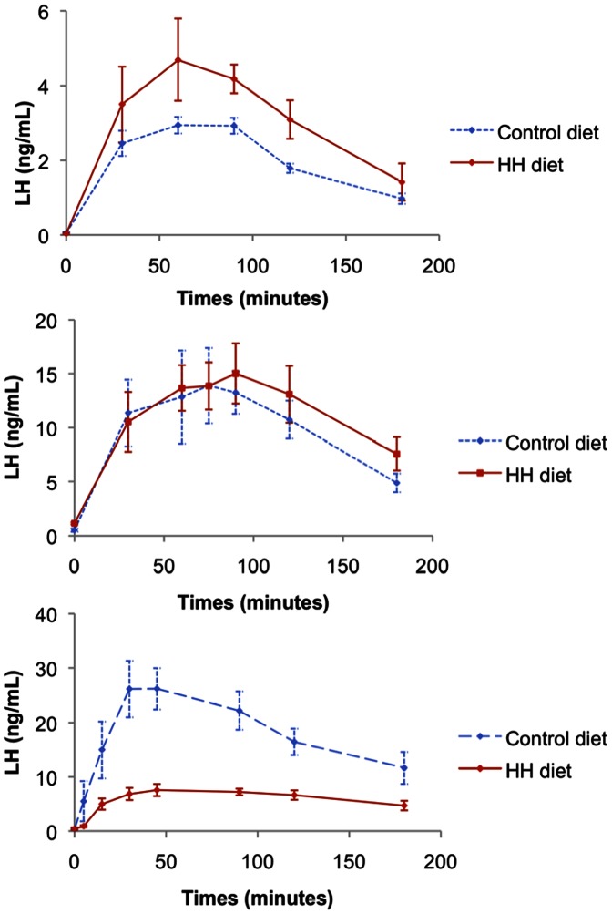
The LH response to GnRH stimulation according to age is shown in Figure 2. At 13 weeks, the peak LH concentrations in response to GnRH was significantly higher in HH does (P<0.02). At 18 weeks, no difference was found between the two groups (P = 0.428). At 22 weeks, the LH response was significantly reduced in HH does (P<0.0001).
Figure 2. Mean (±SEM) LH response after induction of ovulation at 13 (A), 18 (B) and 22 weeks (C) of age (ng/ml).
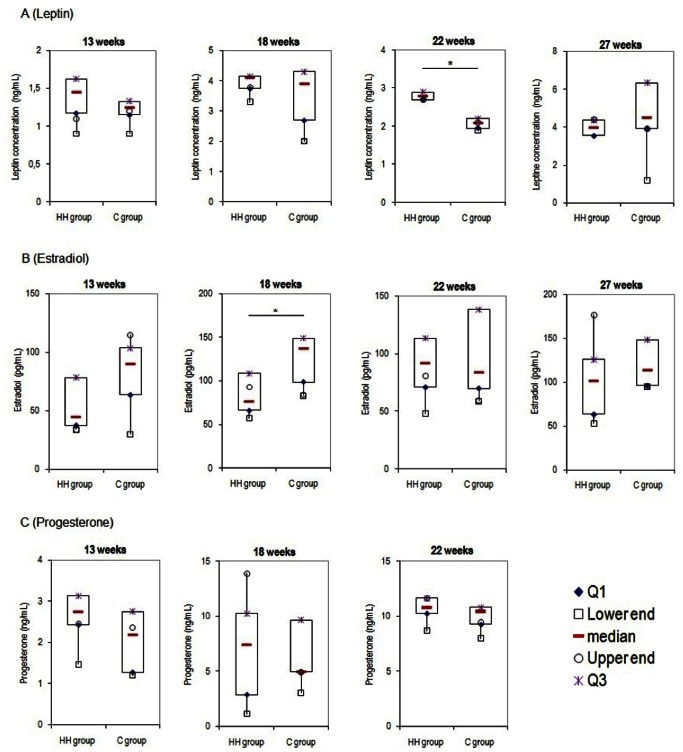
Leptin serum concentrations were significantly higher in HH does compared to controls at 22 weeks of age (2.8 ng/ml (2.7;2.9) vs. 2.1 ng/ml (2.0;2.2), respectively) P<0.05) but not before (Figure 3 (A)). Estradiol concentrations were significantly reduced in HH does at 18 weeks of age (76.0 pg/mL (66.0;108.5) vs. 137.0 pg/mL (98.5;149.0) respectively, P<0.05) (Figure 3 (B)). No difference was found between groups for progesterone concentrations at any time (Figure 3 (C)).
Figure 3. Mean (±SEM) serum leptin, estradiol and progesterone concentrations according to age in HH and Control does.
Each box plot represents the distribution of values in each group at 13, 18, 22 and 27 weeks of age for leptin and estradiol (A and B) and at 13, 18 and 22 weeks of age for progesterone (C). Median values are indicated by the red line within the box. The upper point (in purple) and the lower point (in blue) represent the first and the third quartile respectively. The highest and the lowest values are representing by circle and square respectively. *indicates P<0.05.
Experiment 2
Body composition by TOBEC
At 18 weeks of age, HH does had a significantly higher body lipid content compared with controls (6.11±0.19% vs. 5.67±0.14%, respectively, P = 0.04, other data supplied as Data S5).
Despite a large difference in the means for adipose tissue weight (mean perirenal+interscapular tissue: 101.84 g ±15.41 vs. 82.95 g ±16.90 in HH and Controls, respectively, P = 0.20), there was no significant difference in adipose tissue weight. There was no significant difference in organ weight either (liver, kidney and ovaries), data supplied as Data S5).
Histological results
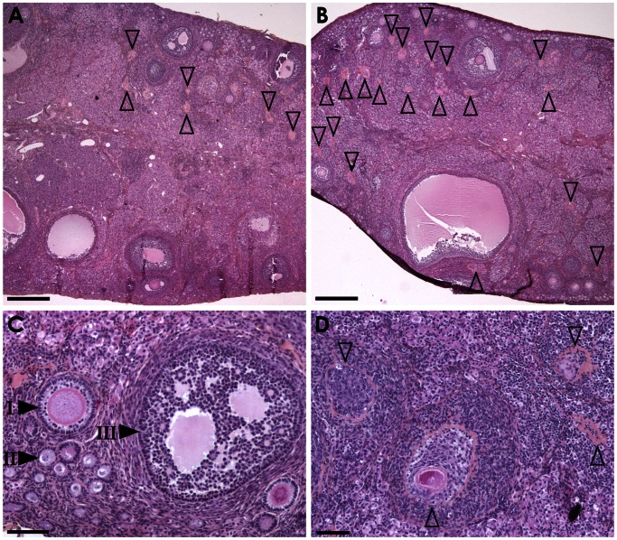
The aim of this analysis was to evaluate follicular populations in the ovary. Using intermediate ×10 magnification, a significantly higher number of atretic follicles was observed in the HH group compared to controls (51.40±4.83 vs. 39.10±5.02, respectively, P = 0.05) (Table 1, Figure 4). Using the x5 magnification, a significantly reduced number of antral follicles was observed in the HH group compared to controls (7.38±1.00 vs. 17.88±2.28, respectively, P<0.001) (Table 2, Figure 4).
Table 1. Mean number follicles (intermediate magnification).
| Type of follicle | HH diet | Control diet | P |
| Primordial | 289.00±29.64 | 340.20±37.05 | 0.14 |
| Type I | 30.60±3.51 | 29.70±3.91 | 0.43 |
| Small type II | 16.70±2.48 | 14.70±3.24 | 0.31 |
| Large type II | 12.00±2.42 | 13.50±2.18 | 0.32 |
| Antral | 9.50±1.85 | 12.20±1.49 | 0.13 |
| Atretic | 51.40±4.83 | 39.10±5.02 | 0.05 |
| Hemorrhagic | 3.60±0.65 | 6.20±0.96 | 0.02 |
| Corpus luteum | 0.00±0.00 | 0.12±0.13 | 0.16 |
| Corpus albicans | 0.60±0.26 | 0.25±0.16 | 0.12 |
Follicles are classified in 9 categories. The counting was realized using one ovary per rabbit and per group (intermediate magnification).
Figure 4. Ovarian histology.
Hematoxylin-eosin-saffron staining ovary sections of rabbits fed a control diet (left panel) or HH diet (right panel) at low (A,B) or high (C,D) magnification. Compared to the control sample (A), numerous atretic follicle remnants (open arrowheads) are scattered in the ovary parenchyma of the high fat diet-fed animal (B). With higher magnification, primary (I), secondary (II) and tertiary follicles (III) are observed in the control samples (C) whereas numerous fields in high fat diet-fed animal samples are devoid of maturing follicles and are only composed of atretic follicle remnants at different stage of involution (D). Scale bars = 500 µm (A, B) and 100 µm (C, D).
Table 2. Mean number follicles according to size (low magnification).
| Type of follicle | HH diet | Control diet | P |
| Type II | 15.50±2.27 | 12.13±2.01 | 0.14 |
| Antral | 7.38±1.00 | 17.88±2.28 | <0.001 |
| Atretic | 44.38±8.27 | 31.13±4.89 | 0.09 |
| Hemorrhagic | 3.13±0.61 | 2.13±0.69 | 0.14 |
| Corpus luteum | 0.00±0.00 | 0.25±0.16 | 0.07 |
Follicles are classified into 5 categories. The counting was realized using one ovary per rabbit and per group (low magnification).
Gene expression in the ovary
The expression of 9 genes involved in ovarian development was studied by RT-qPCR. These transcripts fell within functional categories which included: (i) steroidogenesis (HSD3B2), (ii) germ cell differentiation (VASA), (iii) apoptosis (Caspase), (iv) folliculogenesis (FOXL2, FST, GDF9, BMP15), and (v) receptors (ESR1, ESR2). Of these quantified transcripts, none showed any significant difference in expression between the 2 groups (data supplied as Data S6).
Experiment 3
Endocrine response after mating
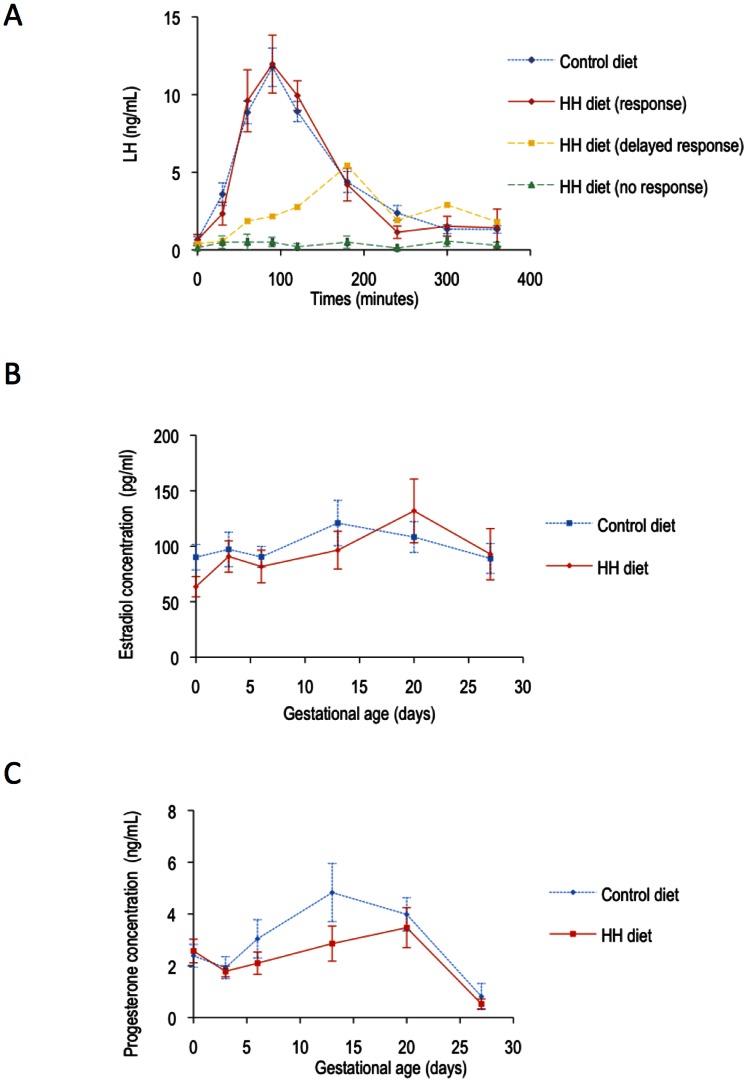
After mating at 18 weeks of age, all the control does but only 4 out of 7 HH does had a normal LH response. The three other animals had either no LH increase at all (N = 2) or a delayed response (N = 1) (Figure 5 (A)) but the difference was not significant between the two groups.
Figure 5. Hormonal response (LH response, estradiol and progesterone) after mating.
(A) Mean ±SEM serum LH concentrations (ng/mL) according to time after mating and response to mating: C animals that all responded (N = 7). HH animals that had a LH response (N = 5). – HH animal that had a delayed response (N = 1). – HH animals that did not respond (N = 2). (B) Mean ±SEM serum estradiol concentrations (pg/mL) in HH and C does at 0, 3, 6, 13, 20 and 27 days of gestation (term: 31 days). (C) Mean ±SEM serum progesterone concentration (ng/mL) in HH and C does at 0, 3, 6, 13, 20 and 27 days of gestation (term: 31 days).
During gestation, there was no significant difference between the groups for plasma estradiol and progesterone concentrations, although the hormonal peaks appeared to be delayed in HH does (Figure 5 (B and C)).
Fetal growth and offspring characteristics
All control females became pregnant whereas the two females that had no LH response did not become pregnant. The median litter size was 6 pups in the Control group and 7 pups in the HH group, with no statistical difference between groups.
On ultrasound examination, biparietal diameter, abdominal circumference and body surface area were significantly smaller in HH diet fetuses at 27 days of gestation (P = 0.012, P = 0.001; P = 0.005 respectively) (data supplied as Data S7). At birth, HH pups were significantly lighter in the HH group compared to control group (37.3±1.89 g vs. 47.77±1.63 g, respectively, P<0.0001).
Discussion
This study evaluated the effect of a diet supplemented in soybean oil and cholesterol, administered from before the age of puberty (13 weeks) on reproductive hormones, ovarian maturation and ovulation disorders, using a previously used rabbit model. In summary, although the rabbit does were not overweight, the HH diet administered from before puberty onwards affected onset of puberty, follicular growth and hormonal responses to breeding and GnRH stimulation. Although the number of antral follicles was decreased and that of atretic follicles increased in HH does, the ovarian expression of genes involved in folliculogenesis was not modified. Fertility and prolificity did not appear to be affected, although 2 does did not get pregnant as a result of a lack of LH response to mating (LH response and ovulation are induced by mating in rabbits). In contrast, fetal growth was affected with intra-uterine growth retardation observed in offspring, as previously observed [27].
The enhanced LH response at 13 weeks in the HH group indicates an early puberty onset compared to controls. An early onset of puberty [9] and of high LH pulse frequency [34], [35] has also been demonstrated in obese rats. In this model, a fat-related signal has been shown to facilitate the activation of hypothalamic GnRH release and advance the onset of puberty [6], [36]. In rhesus monkeys, administration of a high-calorie diet results in the acceleration of growth accompanied with precocious menarche [6]. In humans, the onset of puberty is influenced mainly by genetics, lifestyle, environment, nutrition and body fat [37], [38], [39]. In adults, body size parameters, such as weight or BMI, are strongly correlated with an earlier onset of puberty [37]. In the prepubertal age (5–9 years), increased subcutaneous fat and BMI are associated with increased likelihood of early (<11 years) menarche [38]. Interestingly, the studies evaluating whether nutritional habits (total, unsaturated or saturated fatty acids) could influence age of menarche are still controversial. Some studies reported that higher intake of total fat or PUFA intake in childhood were associated with earlier age at menarche [39], [40], [41], [42] while others found that a balance towards satured fat or MUFA decreased the risk of early menarche [41], [43]. In any case, early menarche constitutes a robust marker of obesity and mortality risk in adult life [44], [45]. In humans, an earlier age of menarche has been associated with an earlier age at menopause [46], [47]. In experiment 3, at 18 weeks of age, although there was no significant difference in the LH response to GnRH, 2 out of 8 HH does did not respond to mating and 1/8 had a delayed response. In experiment 1, in slightly older animals, at 22 weeks of age, the HH diet induced a significant decrease of the LH response in all females after induction of ovulation. In rats, a reduced LH surge before estrous and a reduction in plasma estradiol leading to anovulation were reported in females fed a high fat diet (45% calories from fat) [48]. In humans, a significant reduction of both amplitude and/or mean LH has been reported in obese compared to normal weight women [10], [11], [12], [14] and a recent study on 154 normal weight and 25 obese weight women showed that adiposity may delay the timing and the concentration of hormonal peaks (progesterone and LH) during the menstrual cycle [13]. In mice, neonatally undernourished is associated with delayed puberty and an impairment of the peripubertal GnRH/LH system to respond to ovariectomy [49].
Moreover, in the present study, circulating estradiol concentrations were significantly reduced in the HH group at 18 weeks, in agreement with data obtained in one obese rat model [48]. The anti apoptotic role of estradiol [50] could possibly explain the significant increase observed in the number of atretic follicles. A significant increase in plasma leptin concentrations was only observed at 22 weeks of age. Unfortunately, these females were not put down nor their body composition analysed with TOBEC at that time, so it can only be assumed that this increase in leptin is related to higher percentage of fat in these animals. It is also difficult to try and relate this increased plasma leptin to direct effects on the ovary and more work is needed to elucidate this question. During gestation both progesterone and estradiol peaks tended to be delayed in HH does, and HH fetuses were growth retarded. In women, a positive correlation has been established between plasma progesterone concentrations and weight gain during pregnancy. In obese pubertal use, intra-uterine growth retardation has also been associated with a reduced placental secretion of progesterone [51]. No association was found, however, between gestational weight gain, maternal dietary fatty acid intake and estradiol concentrations [52].
Histological analysis of the ovaries of HH does at 18 weeks of age showed a higher number of atretic follicles and remnants of atretic follicles, indicating a possible increase in apoptotic mechanisms during folliculogenesis. Unfortunately, direct numbering of atretic follicle using specific - Terminal Transferase dUTP Nick End Labeling (TUNEL) on sections of these ovaries, did not display significative results (data not shown) as this assay is focused on on-going atresia and do not reveal ended previous processes evidenced in HES staining by the presence of fibrotic foci with central hypereosinophilic remnants of the zona pellucida. Moreover no difference was found in the expression of genes involved in the ovarian development. In parallel, histological analysis also showed a significantly reduced number of antral follicles in the HH group. Previous studies showed that the administration of a polyunsaturated fatty acids (PUFA) diet to cows during the periconceptional period reduces the number of small and middle size ovarian follicles, without affecting oocyte quality or in vitro cleavage [53]. In contrast, in a sheep model, short term overnutrition increased the number of large size ovarian follicles [54]. In agreement with the present study, rat models of obesity have impaired ovarian follicular growth with increased apoptosis [21], [55], but no difference in morphology nor in the number of antral follicles [21]. In the rabbit, the ovarian cortex is thin and is characterized by a small number of primordial and small developing follicles with a higher number of mature follicles [56], [57]. In terms of reproduction, rabbit females are characterized by the fact that mating induces ovulation. Follicular growth is a continuous process with waves of maturation that guarantee mature oocytes nearly anytime. During post-natal growth, secondary, tertiary and antral follicles appear between 4 and 12 weeks of age. Basal growth until antral formation is independent of gonadotrophin secretion whereas terminal follicular development depends on FSH. Therefore, the beginning of the study (10 weeks of age) occurred during a key period when hormonal dependence was starting, between tertiary and antral follicles and it is not known whether this was an important determinant in the observed results.
Systemic alterations associated with woman obesity (hyperinsulinemia, dyslipidemia, and symptoms of chronic inflammation) extend directly into the ovarian follicular microenvironment. Overweight and obese women were shown to exhibit elevated intrafollicular insulin, triglyceride and androgens which were associated with poor reproductive outcome [58], although there was no direct relationship between serum and follicular concentrations of free fatty acids [59]. Recently, a higher concentration of inflammatory factors was also observed in the follicular fluid of infertile obese women [60]. In the present study, the increased plasma cholesterol concentrations may have induced increased oxidative stress in the ovary, leading to increased follicular atresia. Whether direct effects of the maternal diet on the oocyte and/or effects on the oviductal and uterine environment induced fetal IUGR also remains to be determined, although the very early deregulation of gene expression in the embryo with the present model suggests that the oocyte quality may be affected by the HH diet [27].
In conclusion, this paper highlights, using an animal model, the possible adverse effects of unbalanced diets on the reproductive function and possible fertility of women. Although the diet used here is rich in poly-unsaturated fatty acids whereas the diet in humans consist mainly of saturated fats, this model remains relevant for hypercholesterolemia and also gives insight in general effects of high lipid diets. Given the dramatically increasing prevalence of obesity among women of reproductive age, it is essential that women be counseled on the reproductive risks of obesity and dangerous dietary behaviors and the proven benefits of lifestyle modification. This intervention must occur in the pre-conception period.
Supporting Information
Sequences of qPCR primers.
(DOC)
Weight according to age. Mean ±SEM weight (kg) at 10, 13, 17, 23 and 27 weeks in the 2 groups (8 rabbits per groups).
(TIFF)
Serum cholesterol concentrations according to age. Mean ±SEM serum cholesterol concentration (mmol/L) in the 2 groups according to age (13, 18, 22 and 27 weeks). ***P<0.001.
(TIFF)
Serum triglycerides concentrations according to age. Mean ±SEM serum triglyceride concentration (mmol/L) in the 2 groups according to age (13, 18, 22 and 27 weeks).
(TIFF)
Body composition, organ weight and fat mass in rabbits at 18 weeks of age according to group.
(DOC)
Gene expression in the 2 groups. Relative expression of genes involved in folliculogenesis in the 2 groups.
(TIFF)
Biometric measurements made by ultrasound examination in fetuses from the 2 groups at 27 days of gestation.
(DOC)
Acknowledgments
The authors thank Stephen Besseau, Chantal Julia, and Mehdi Menai for their help for statistical analysis.
Funding Statement
No current external funding sources was obtained for this study. The work was entirely financed through internal INRA funding. The funders had no role in study design, data collection and analysis, decision to publish, or preparation of the manuscript.
References
- 1.Charles MA, Basdevant A, Eschwège E (2009) Enquête épidémiologique nationale sur le surpoids et l’obésité. Obepi 2009.
- 2. Armitage JA, Taylor PD, Poston L (2005) Experimental models of developmental programming: consequences of exposure to an energy rich diet during development. J Physiol (Lond) 565: 3–8. [DOI] [PMC free article] [PubMed] [Google Scholar]
- 3. De Lorenzo A, Del Gobbo V, Premrov M, Bigioni M, Galvano F, et al. (2007) Normal-weight obese syndrome: early inflammation? Am J Clin Nut 85: 40–45. [DOI] [PubMed] [Google Scholar]
- 4. Di Renzo L, Galvano F, Orlandi C, Bianchi A, Di Giacomo C, et al. (2010) Oxidative stress in normal-weight obese syndrome. Obesity (Silver Spring) 18: 2125–2130. [DOI] [PubMed] [Google Scholar]
- 5. Agarwal A, Aponte-Mellado A, Premkumar BJ, Shaman A, Gupta S (2012) The effects of oxidative stress on female reproduction: a review. Reprod Biol Endocrinol 10: 49. [DOI] [PMC free article] [PubMed] [Google Scholar]
- 6. Feng Li X, Lin YS, Kinsey-Jones JS, O’Byrne KT (2012) High-Fat Diet Increases LH Pulse Frequency and Kisspeptin-Neurokinin B Expression in Puberty-Advanced Female Rats. Endocrinology 153: 4422–4431. [DOI] [PubMed] [Google Scholar]
- 7. Soulis G, Kitraki E, Gerozissis K (2005) Early Neuroendocrine Alterations in Female Rats Following a Diet Moderately Enriched in Fat. Cellular and Molecular Neurobiology 25: 869–880. [DOI] [PMC free article] [PubMed] [Google Scholar]
- 8. Gonzalez-Anover P, Encinas T, Torres-Rovira L, Sanz E, Pallares P, et al. (2011) Patterns of Corpora Lutea Growth and Progesterone Secretion in Sows with Thrifty Genotype and Leptin Resistance due to Leptin Receptor Gene Polymorphisms (Iberian Pig). Reproduction in Domestic Animals 46: 1011–1016. [DOI] [PubMed] [Google Scholar]
- 9. Terasawa E, Kurian JR, Keen KL, Shiel NA, Colman RJ, et al. (2012) Body weight impact on puberty: effects of high-calorie diet on puberty onset in female rhesus monkeys. Endocrinology 153: 1696–1705. [DOI] [PMC free article] [PubMed] [Google Scholar]
- 10. Sherman BM, Korenman SG (1974) Measurement of serum LH, FSH, estradiol and progesterone in disorders of the human menstrual cycle: the inadequate luteal phase. J Clin Endocrinol Metab 39: 145–149. [DOI] [PubMed] [Google Scholar]
- 11. Grenman S, Ronnemaa T, Irjala K, Kaihola HL, Gronroos M (1986) Sex steroid, gonadotropin, cortisol, and prolactin levels in healthy, massively obese women: correlation with abdominal fat cell size and effect of weight reduction. J Clin Endocrinol Metab 63: 1257–1261. [DOI] [PubMed] [Google Scholar]
- 12. Santoro N, Lasley B, McConnell D, Allsworth J, Crawford S, et al. (2004) Body size and ethnicity are associated with menstrual cycle alterations in women in the early menopausal transition: The Study of Women’s Health across the Nation (SWAN) Daily Hormone Study. J Clin Endocrinol Metab 89: 2622–2631. [DOI] [PubMed] [Google Scholar]
- 13.Yeung EH, Zhang C, Albert PS, Mumford SL, Ye A, et al.. (2012) Adiposity and sex hormones across the menstrual cycle: the BioCycle Study. Int J Obes (Lond). [DOI] [PMC free article] [PubMed]
- 14. Jain A, Polotsky AJ, Rochester D, Berga SL, Loucks T, et al. (2007) Pulsatile luteinizing hormone amplitude and progesterone metabolite excretion are reduced in obese women. J Clin Endocrinol Metab 92: 2468–2473. [DOI] [PubMed] [Google Scholar]
- 15. Maheshwari A, Stofberg L, Bhattacharya S (2007) Effect of overweight and obesity on assisted reproductive technology–a systematic review. Hum Reprod Update 13: 433–444. [DOI] [PubMed] [Google Scholar]
- 16. Balen AH, Platteau P, Andersen AN, Devroey P, Sorensen P, et al. (2006) The influence of body weight on response to ovulation induction with gonadotrophins in 335 women with World Health Organization group II anovulatory infertility. BJOG 113: 1195–1202. [DOI] [PubMed] [Google Scholar]
- 17. Bellver J, Busso C, Pellicer A, Remohi J, Simon C (2006) Obesity and assisted reproductive technology outcomes. Reprod Biomed Online 12: 562–568. [DOI] [PubMed] [Google Scholar]
- 18. Dechaud H, Anahory T, Reyftmann L, Loup V, Hamamah S, et al. (2006) Obesity does not adversely affect results in patients who are undergoing in vitro fertilization and embryo transfer. Eur J Obstet Gynecol Reprod Biol 127: 88–93. [DOI] [PubMed] [Google Scholar]
- 19. Martinuzzi K, Ryan S, Luna M, Copperman AB (2008) Elevated body mass index (BMI) does not adversely affect in vitro fertilization outcome in young women. J Assist Reprod Genet 25: 169–175. [DOI] [PMC free article] [PubMed] [Google Scholar]
- 20. Lashen H, Ledger W, Bernal AL, Barlow D (1999) Extremes of body mass do not adversely affect the outcome of superovulation and in-vitro fertilization. Hum Reprod 14: 712–715. [DOI] [PubMed] [Google Scholar]
- 21. Sagae SC, Menezes EF, Bonfleur ML, Vanzela EC, Zacharias P, et al. (2012) Early onset of obesity induces reproductive deficits in female rats. Physiol Behav 105: 1104–1111. [DOI] [PubMed] [Google Scholar]
- 22. Kajihara T, Uchino S, Suzuki M, Itakura A, Brosens JJ, et al. (2009) Increased ovarian follicle atresia in obese Zucker rats is associated with enhanced expression of the forkhead transcription factor FOXO1. Med Mol Morphol 42: 216–221. [DOI] [PubMed] [Google Scholar]
- 23. Hamm ML, Bhat GK, Thompson WE, Mann DR (2004) Folliculogenesis is impaired and granulosa cell apoptosis is increased in leptin-deficient mice. Biol Reprod 71: 66–72. [DOI] [PubMed] [Google Scholar]
- 24. Serke H, Nowicki M, Kosacka J, Schroder T, Kloting N, et al. (2012) Leptin-deficient (ob/ob) mouse ovaries show fatty degeneration, enhanced apoptosis and decreased expression of steroidogenic acute regulatory enzyme. Int J Obes (Lond) 36: 1047–1053. [DOI] [PubMed] [Google Scholar]
- 25. Hutchinson IA, Hennessy AA, Waters SM, Dewhurst RJ, Evans AC, et al. (2012) Effect of supplementation with different fat sources on the mechanisms involved in reproductive performance in lactating dairy cattle. Theriogenology 78: 12–27. [DOI] [PubMed] [Google Scholar]
- 26. Gonzalez-Anover P, Encinas T, Torres-Rovira L, Sanz E, Pallares P, et al. (2011) Patterns of corpora lutea growth and progesterone secretion in sows with thrifty genotype and leptin resistance due to leptin receptor gene polymorphisms (Iberian pig). Reprod Domest Anim 46: 1011–1016. [DOI] [PubMed] [Google Scholar]
- 27. Picone O, Laigre P, Fortun-Lamothe L, Archilla C, Peynot N, et al. (2011) Hyperlipidic hypercholesterolemic diet in prepubertal rabbits affects gene expression in the embryo, restricts fetal growth and increases offspring susceptibility to obesity. Theriogenology 75: 287–299. [DOI] [PubMed] [Google Scholar]
- 28.Fischer B, Chavatte-Palmer P, Viebahn C, Navarete-Santos A, Duranthon V (2012) Rabbit as a reproductive model for human health. Reproduction: in press. [DOI] [PubMed]
- 29. Zheng H, Zhang C, Yang W, Wang Y, Lin Y, et al. (2009) Fat and Cholesterol Diet Induced Lipid Metabolic Disorders and Insulin Resistance in Rabbit. Exp Clin Endocrinol Diabetes 117: 400–405. [DOI] [PubMed] [Google Scholar]
- 30. Fortun-Lamothe L, Lamboley-Gauzere B, Bannelier C (2002) Prediction of body composition in rabbit females using total body electrical conductivity (TOBEC). Livestock Production Science 78: 133–142. [Google Scholar]
- 31. Bodensteiner KJ, Sawyer HR, Moeller CL, Kane CM, Pau KYF, et al. (2004) Chronic exposure to dibromoacetic acid, a water disinfection byproduct, diminishes primordial follicle populations in the rabbit. Toxicological Sciences 80: 83–91. [DOI] [PubMed] [Google Scholar]
- 32. Chavatte-Palmer P, Laigre P, Simonoff E, Chesne P, Challah-Jacques M, et al. (2008) In utero characterisation of fetal growth by ultrasound scanning in the rabbit. Theriogenology 69: 859–869. [DOI] [PubMed] [Google Scholar]
- 33. Rommers JM, Boiti C, Brecchia G, Meijerhof R, Noordhuizen J, et al. (2004) Metabolic adaptation and hormonal regulation in young rabbit does during long-term caloric restriction and subsequent compensatory growth. Animal Science 79: 255–264. [Google Scholar]
- 34. Ahima RS, Dushay J, Flier SN, Prabakaran D, Flier JS (1997) Leptin accelerates the onset of puberty in normal female mice. J Clin Invest 99: 391–395. [DOI] [PMC free article] [PubMed] [Google Scholar]
- 35. Dearth RK, Hiney JK, Dees WL (2000) Leptin acts centrally to induce the prepubertal secretion of luteinizing hormone in the female rat. Peptides 21: 387–392. [DOI] [PubMed] [Google Scholar]
- 36. Akamine EH, Marcal AC, Camporez JP, Hoshida MS, Caperuto LC, et al. (2010) Obesity induced by high-fat diet promotes insulin resistance in the ovary. Journal of Endocrinology 206: 65–74. [DOI] [PubMed] [Google Scholar]
- 37. Pierce MB, Leon DA (2005) Age at menarche and adult BMI in the Aberdeen children of the 1950s cohort study. Am J Clin Nutr 82: 733–739. [DOI] [PubMed] [Google Scholar]
- 38. Freedman DS, Khan LK, Serdula MK, Dietz WH, Srinivasan SR, et al. (2002) Relation of age at menarche to race, time period, and anthropometric dimensions: the Bogalusa Heart Study. Pediatrics 110: e43. [DOI] [PubMed] [Google Scholar]
- 39. Rogers IS, Northstone K, Dunger DB, Cooper AR, Ness AR, et al. (2010) Diet throughout childhood and age at menarche in a contemporary cohort of British girls. Public Health Nutr 13: 2052–2063. [DOI] [PubMed] [Google Scholar]
- 40. Berkey CS, Gardner JD, Frazier AL, Colditz GA (2000) Relation of childhood diet and body size to menarche and adolescent growth in girls. Am J Epidemiol 152: 446–452. [DOI] [PubMed] [Google Scholar]
- 41. Maclure M, Travis L, Willett W, MacMahon B (1991) A prospective cohort study of nutrient intake and age at menarche. Am J Clin Nutr 54: 649–656. [DOI] [PubMed] [Google Scholar]
- 42. Merzenich H, Boeing H, Wahrendorf J (1993) Dietary fat and sports activity as determinants for age at menarche. Am J Epidemiol 138: 217–224. [DOI] [PubMed] [Google Scholar]
- 43. Moisan J, Meyer F, Gingras S (1990) A nested case-control study of the correlates of early menarche. Am J Epidemiol 132: 953–961. [DOI] [PubMed] [Google Scholar]
- 44. Lakshman R, Forouhi N, Luben R, Bingham S, Khaw K, et al. (2008) Association between age at menarche and risk of diabetes in adults: results from the EPIC-Norfolk cohort study. Diabetologia 51: 781–786. [DOI] [PubMed] [Google Scholar]
- 45. Lakshman R, Forouhi NG, Sharp SJ, Luben R, Bingham SA, et al. (2009) Early age at menarche associated with cardiovascular disease and mortality. J Clin Endocrinol Metab 94: 4953–4960. [DOI] [PubMed] [Google Scholar]
- 46. Ozdemir O, Col M (2004) The age at menopause and associated factors at the health center area in Ankara, Turkey. Maturitas 49: 211–219. [DOI] [PubMed] [Google Scholar]
- 47. Cramer DW, Xu H, Harlow BL (1995) Does “incessant” ovulation increase risk for early menopause? Am J Obstet Gynecol 172: 568–573. [DOI] [PubMed] [Google Scholar]
- 48. Balasubramanian P, Jagannathan L, Mahaley RE, Subramanian M, Gilbreath ET, et al. (2012) High fat diet affects reproductive functions in female diet-induced obese and dietary resistant rats. J Neuroendocrinol 24: 748–755. [DOI] [PMC free article] [PubMed] [Google Scholar]
- 49. Caron E, Ciofi P, Prevot V, Bouret SG (2012) Alteration in neonatal nutrition causes perturbations in hypothalamic neural circuits controlling reproductive function. J Neurosci 32: 11486–11494. [DOI] [PMC free article] [PubMed] [Google Scholar]
- 50. Lund SA, Murdoch J, Van Kirk EA, Murdoch WJ (1999) Mitogenic and antioxidant mechanisms of estradiol action in preovulatory ovine follicles: relevance to luteal function. Biol Reprod 61: 388–392. [DOI] [PubMed] [Google Scholar]
- 51. Lea RG, Wooding P, Stewart I, Hannah LT, Morton S, et al. (2007) The expression of ovine placental lactogen, StAR and progesterone-associated steroidogenic enzymes in placentae of overnourished growing adolescent ewes. Reproduction 133: 785–796. [DOI] [PubMed] [Google Scholar]
- 52. Lof M, Hilakivi-Clarke L, Sandin SS, de Assis S, Yu W, et al. (2009) Dietary fat intake and gestational weight gain in relation to estradiol and progesterone plasma levels during pregnancy: a longitudinal study in Swedish women. BMC Womens Health 9: 10. [DOI] [PMC free article] [PubMed] [Google Scholar]
- 53. Zachut M, Dekel I, Lehrer H, Arieli A, Arav A, et al. (2010) Effects of dietary fats differing in n-6:n-3 ratio fed to high-yielding dairy cows on fatty acid composition of ovarian compartments, follicular status, and oocyte quality. J Dairy Sci 93: 529–545. [DOI] [PubMed] [Google Scholar]
- 54. Ying S, Wang Z, Wang C, Nie H, He D, et al. (2011) Effect of different levels of short-term feed intake on folliculogenesis and follicular fluid and plasma concentrations of lactate dehydrogenase, glucose, and hormones in Hu sheep during the luteal phase. Reproduction 142: 699–710. [DOI] [PubMed] [Google Scholar]
- 55. Jungheim ES, Schoeller EL, Marquard KL, Louden ED, Schaffer JE, et al. (2010) Diet-Induced Obesity Model: Abnormal Oocytes and Persistent Growth Abnormalities in the Offspring. Endocrinology 151: 4039–4046. [DOI] [PMC free article] [PubMed] [Google Scholar]
- 56. Kranzfelder D, Korr H, Mestwerdt W, Maurer-Schultze B (1984) Follicle growth in the ovary of the rabbit after ovulation-inducing application of human chorionic gonadotropin. Cell Tissue Res 238: 611–620. [DOI] [PubMed] [Google Scholar]
- 57. Arias-Alvarez M, GarcÌa-GarcÌa RM, Rebollar PG, Revuelta L, Mill·n P, et al. (2009) Influence of metabolic status on oocyte quality and follicular characteristics at different postpartum periods in primiparous rabbit does. Theriogenology 72: 612–623. [DOI] [PubMed] [Google Scholar]
- 58. Robker RL, Akison LK, Bennett BD, Thrupp PN, Chura LR, et al. (2009) Obese Women Exhibit Differences in Ovarian Metabolites, Hormones, and Gene Expression Compared with Moderate-Weight Women. J Clin Endocrinol Metab 94: 1533–1540. [DOI] [PubMed] [Google Scholar]
- 59. Jungheim ES, Macones GA, Odem RR, Patterson BW, Lanzendorf SE, et al. (2011) Associations between free fatty acids, cumulus oocyte complex morphology and ovarian function during in vitro fertilization. Fertil Steril 95: 1970–1974. [DOI] [PMC free article] [PubMed] [Google Scholar]
- 60. La Vignera S, Condorelli R, Bellanca S, La Rosa B, Mousavi A, et al. (2011) Obesity is associated with a higher level of pro-inflammatory cytokines in follicular fluid of women undergoing medically assisted procreation (PMA) programs. Eur Rev Med Pharmacol Sci 15: 267–273. [PubMed] [Google Scholar]
Associated Data
This section collects any data citations, data availability statements, or supplementary materials included in this article.
Supplementary Materials
Sequences of qPCR primers.
(DOC)
Weight according to age. Mean ±SEM weight (kg) at 10, 13, 17, 23 and 27 weeks in the 2 groups (8 rabbits per groups).
(TIFF)
Serum cholesterol concentrations according to age. Mean ±SEM serum cholesterol concentration (mmol/L) in the 2 groups according to age (13, 18, 22 and 27 weeks). ***P<0.001.
(TIFF)
Serum triglycerides concentrations according to age. Mean ±SEM serum triglyceride concentration (mmol/L) in the 2 groups according to age (13, 18, 22 and 27 weeks).
(TIFF)
Body composition, organ weight and fat mass in rabbits at 18 weeks of age according to group.
(DOC)
Gene expression in the 2 groups. Relative expression of genes involved in folliculogenesis in the 2 groups.
(TIFF)
Biometric measurements made by ultrasound examination in fetuses from the 2 groups at 27 days of gestation.
(DOC)


