Abstract
Attention is attracted exogenously by physically salient stimuli, but this effect can be dampened by endogenous attention settings, a phenomenon called “contingent capture”. Emotionally salient stimuli are also thought to exert a strong exogenous influence on attention, especially in anxious individuals, but whether and how top-down attention can ameliorate bottom-up capture by affective stimuli is currently unknown. Here, we paired a novel spatial cueing task with functional magnetic resonance imaging (fMRI) in order to investigate contingent capture as a function of the affective salience of bottom-up cues (face stimuli) and individual differences in trait anxiety. In the absence of top-down cues, exogenous stimuli validly cueing targets facilitated attention in low anxious participants, regardless of affective salience. However, while high anxious participants exhibited similar facilitation following neutral exogenous cues, this facilitation was completely absent following affectively negative exogenous cues. Critically, these effects were contingent on endogenous attentional settings, such that explicit top-down cues presented prior to the appearance of exogenous stimuli removed anxious individuals’ sensitivity to affectively salient stimuli. FMRI analyses revealed a network of brain regions underlying this variability in affective contingent capture across individuals, including the fusiform face area (FFA), posterior ventrolateral frontal cortex, and supplementary motor area. Importantly, activation in the posterior ventrolateral frontal cortex and the supplementary motor area fully mediated the effects observed in FFA, demonstrating a critical role for these frontal regions in mediating attentional orienting and interference resolution processes when engaged by affectively salient stimuli.
Keywords: Attention, Posner cueing paradigm, ventral attention network, contingent capture, emotion, fear
Introduction
Attention prioritizes the processing of stimuli that are relevant to our goals and well-being, thus promoting adaptive behavior. Due to their biological significance, affectively salient stimuli, such as fearful faces, may be particularly adept at attracting attention exogenously (LeDoux, 2000; Ohman et al., 2001), facilitating speeded detection among distracters during visual search (Hansen and Hansen, 1994; Ohman et al., 2001; Notebaert et al., 2011), resisting the attentional blink (Anderson and Phelps, 2001; Lim et al., 2009), and capturing attention to neutral stimuli presented at similar spatial locations (Macleod et al., 1986; Armony and Dolan, 2002; Phelps et al., 2006). Moreover, attentional prioritization of affective stimuli has been linked to individual differences in anxiety (Mathews and Mackintosh, 1998; Bar-Haim et al., 2007), as anxious individuals more rapidly detect fear-relevant stimuli and exhibit greater exogenous attentional guidance by affective cues than non-anxious participants (Fox et al., 2001; Amir et al., 2003; Salemink et al., 2007). This hypervigilance to threatening stimuli can prove maladaptive and has been linked to the etiology of anxiety disorders (Mathews and Mackintosh, 1998; Mathews and MacLeod, 2002).
However, it remains unclear whether potential affective attentional prioritization occurs automatically, regardless of context, or is contingent on endogenous attentional settings. It is well-established that top-down attention, engendered by internal goals, can moderate or even extinguish the influence of physically salient exogenous stimuli in guiding attention (Yantis and Jonides, 1990; Folk et al., 1992; Van der Stigchel et al., 2009; Theeuwes, 2010), a phenomenon known as contingent capture (Folk et al., 1992). Both endogenous attentional settings related to the spatial location of targets (Yantis and Jonides, 1990) and the features of targets (Folk et al., 1992) can mitigate the exogenous attentional influence of salient distractors, which appears to be mediated by the “ventral attention network”, including temporoparietal junction (TPJ) and ventrolateral frontal cortex (Corbetta et al., 2008). By contrast, it is not known whether endogenous attentional settings moderate the ability of affectively salient stimuli to capture attention, particularly in anxious individuals, nor by which neural mechanisms this would be achieved. To wit, could anxious individuals engage top-down attention to mitigate the bottom-up influence of affective stimuli in directing attention?
The present study sought to address this issue using a novel adaptation of Posner’s spatial cueing paradigm (Posner et al., 1980) that independently varied top-down and bottom-up drivers of spatial attention. Top-down attention was manipulated via symbolic cues that were either predictive or neutral with respect to a target stimulus location, whereas bottom-up attention was manipulated with non-predictive, sudden-onset exogenous cues (pictures of faces) that could coincide with either target or non-target locations. The use of face stimuli facilitated the tracking of perceptual cue processing, as indexed by activation of the fusiform face area (FFA) (Kanwisher et al., 1997). Critically, exogenous cues could also either be affectively neutral (a neutral face) or salient (a fearful face), thus allowing us to distinguish the effects of affective salience from those of the mere physical salience inherent in any sudden-onset stimulus (Yantis and Jonides, 1990). By assessing the behavioral effects of these manipulations as a function of individual differences in trait anxiety, we sought to delineate the neural mechanisms underlying contingent capture of attention by affective stimuli. Examining both the basic neural effects of the experimental attentional manipulations employed as well as their variation as a function of individual differences in affective contingent capture enabled a characterization of the neural regions contributing to exogenous spatial guidance of attention by affective stimuli. Mediation analyses were also conducted in order to gain insight into the mechanisms by which particular frontal regions contributed to contingent capture of attention by affective stimuli.
Methods
Participants
Twenty-five right-handed participants (13 female; mean age 24.6 years, range 19–34) provided written informed consent in accordance with the Duke University Medical Center Institutional Review Board. All participants had normal or corrected-to-normal vision and were screened by self-report for neurological or psychiatric conditions or current psychoactive medication use.
Experimental Protocol
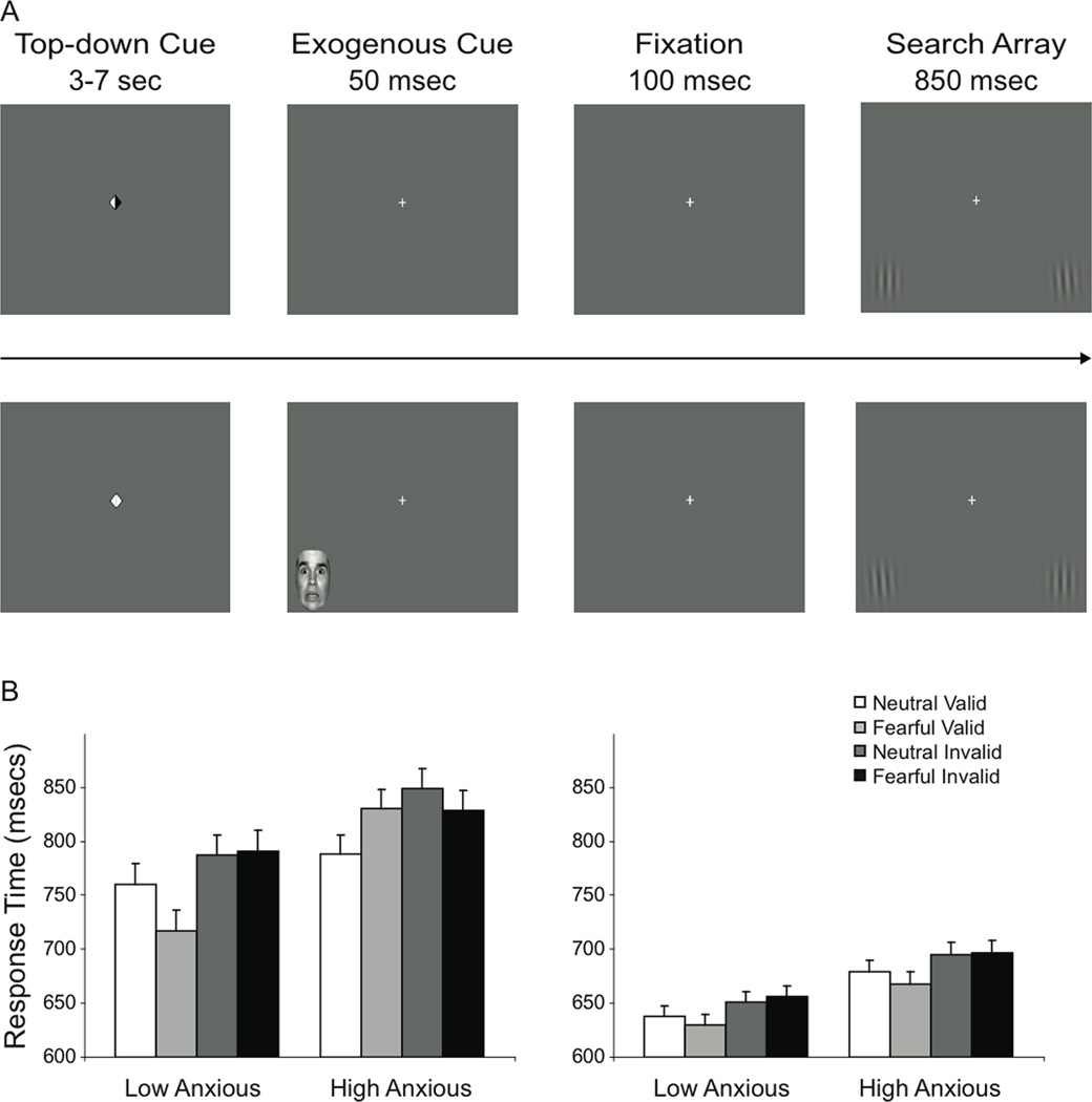
Participants completed 360 trials of the spatial cueing task across six scan runs. Each trial began with an explicit spatial cue, consisting of two centrally presented triangles, shown on a grey background (Figure 1A, “Top-down Cue”). On two-thirds of trials, one side of this cue darkened to point to the upcoming location of the target with a black arrow (informative top-down cue), and these cues were 100% predictive, always accurately indicating the location of an upcoming target stimulus. On one-third of the trials, neither side of the explicit spatial cue darkened, providing no information about the upcoming target location (uninformative top-down cue). Following a variable interval of 3–7 s during which the top-down cue remained onscreen, a 150 ms interval preceded the onset of a target search array, which was presented for 850 ms. Search arrays consisted of a white central fixation cross (1° visual angle) and two peripheral circular sinusoidal luminance gratings enveloped by a Gaussian filter (‘Gabor patches’), with a spatial frequency of 1.5 cycles/degree (Figure 1A, “Search Array”). The two gratings were presented in the lower visual field, approximately 7.4° vertical and 10.0° horizontal eccentricity from fixation and subtending approximately 4° of visual angle. On each trial, one of the two gratings, the target, was tilted either clockwise or counter-clockwise. It was the participant’s task to locate the target and indicate the direction of the tilt via a button-press, using the right index and middle fingers to respond. Note that although the informative top-down cues presented at the onset of the trial accurately signaled the target location, they did not cue a specific response, as it was still necessary for the participant to discriminate the direction of the target tilt. Thus, these cues manipulated endogenous spatial attention without priming responses.
Figure 1.
Spatial cueing task and behavior. (A) Example trials from spatial cueing task. Top panel: A trial with an informative top-down spatial cue, indicating the target will appear on the right. No exogenous cue was presented during the cue-to-target interval. The tilted Gabor patch on the right is tilted 5° counter-clockwise. Bottom panel: An uninformative top-down spatial cue. A fearful face is presented in the left potential target location during the first 50 msec of the cue-to-target interval. In this search array, the target is on the left, tilted 5° to counter-clockwise. The exogenous cue in this case was valid, as it was presented spatially coincident with the subsequent target. Not depicted here is the 3–7 second variable intertrial interval fixation period. (B) Response time data for trials, plotted as a function of exogenous cue validity and affective salience, trait anxiety, and whether top-down cueing was uninformative (left panel) or informative (right panel). Error bars represent ±1 S.E.M.
Exogenous attention was manipulated during the 150 ms interval between the offset of the top-down cue and target array onset. On two-thirds of trials, a single face stimulus was presented in one of the two potential target locations during the first 50 ms of this interval (Figure 1A, “Exogenous Cue”). Face stimuli were selected from the NimStim Set of Facial Expression1 (Tottenham et al., 2009) and consisted of five males modeling both a neutral and a fearful expression for a total of ten unique stimuli, which were counterbalanced across all conditions. We employed male faces, because previous research suggests male emotional faces to evoke stronger emotional reactions than female ones (e.g. Bradley et al., 1997). These stimuli were matched for luminance and contrast to control for basic perceptual salience and subtended approximately 3.7–4.0° of visual angle vertically and 3.4–4.0° horizontally. These face stimuli served as sudden-onset exogenous cues that were not predictive of target location. Previous research has demonstrated the efficacy of sudden-onset stimuli in exogenously guiding attention (Yantis and Jonides, 1990), and the present design enabled the independent manipulation of cue validity and affective salience while controlling for perceptual salience. Face stimuli were presented either in the location of the upcoming target (“valid” bottom-up cue) or at the alternate location (“invalid” bottom-up cue), thus potentially facilitating or distracting attention from the target location in an exogenous manner. On half of the trials, these exogenous cues were affectively neutral (a neutral face), and on the other half they were affectively salient (a fearful face). The cue stimuli were equally likely to appear in either location, and cue affect, target stimulus location, and tilt direction were fully counterbalanced across all conditions. Participants were instructed to use the top-down cues to guide their attention while ignoring these non-predictive exogenous cues. Importantly, this factorial design enabled the independent manipulation of top-down cues (informative or uninformative), exogenous cue validity (target location or alternate location), and affective salience (fearful or neutral exogenous cue). One-third of trials contained no exogenous sources of attentional guidance. These trials were included to minimize participant expectation of the exogenous cues and limit adaptation to their presence over the course of the experiment. Stimulus presentation and response recording were implemented in Presentation (Neurobehavioral Systems, Albany, CA).
To dissociate blood oxygenation level-dependent (BOLD) neural responses associated with top-down cue processing from those associated with processing of exogenous cues and targets, the duration of the top-down cue period and inter-trial interval were independently jittered, varying from 3 to 7 s in 1-s steps along a pseudoexponential distribution (Ollinger et al., 2001; Wager and Nichols, 2003), with 50% of trial periods lasting 3 s, 25% lasting 4 s, 13% lasting 5 s, 6% lasting 6 s, and 6% lasting 7 s (Egner et al., 2008). The top-down cue period was modeled separately from the exogenous cue and target search array period (see Image Analysis below).
Participants completed a separate localizer task to functionally delineate the FFA (Kanwisher et al., 1997). Participants performed a 1-back task during block-wise presentation of face and house stimuli, responding whenever two identical stimuli appeared consecutively. All face stimuli modeled neutral expressions, and none of the house images included arousing or disturbing features. Stimuli subtended approximately 10.0° of visual angle vertically and horizontally. Each block consisted of fifteen stimuli (including 1–2 repetitions), with each stimulus presented for 750 ms followed by 250 ms of fixation. The localizer task consisted of twelve blocks presented in ABAB order, each separated by 10 s of fixation.
Behavioral Data Analysis
All participants completed the Spielberger State-Trait Anxiety Inventory (STAI) prior to scanning. Participants’ state anxiety scores ranged from 20 to 37 (median = 27), and their trait anxiety scores ranged from 20 to 43 (median = 31). Participants were divided into two groups based on a median split of self-reported trait anxiety; participants with trait anxiety scores below the median had significantly lower trait anxiety scores (M = 26.2) than those with scores above the median (M = 36.2, t(23) = 6.99, p < .001). This range is consistent with published norms for nonclinical individuals in this age group (Spielberger, 1983). In addition to differing in overall trait scores, the two groups also varied in their scores on a subscale of the STAI most closely associated with anxiety as opposed to general negative affect (Bieling et al., 1998), with lower scores observed in participants below the median (M = 8.3) than those with scores above the median (M = 11.7, t(23) = 5.72, p < .001).
Response time (RT) data from correct trials was computed separately for informative and uninformative top-down cue trials for each of the exogenous cue conditions. Instead of filtering participant RTs employing a pre-specified range, median RTs were calculated to diminish the influence of extreme values. Median RTs for each condition were then submitted to a repeated-measures ANOVA with the within-subjects factors of top-down cue (uninformative or informative), exogenous cue validity (target location or alternate location), and affective salience (fearful or neutral), and the between-subjects factor of anxiety (low or high trait anxiety). Significant effects in the main ANOVA were interrogated using follow-up ANOVAs and t-tests.
Eye-movement Data
Eye-movement data were acquired during task performance to confirm that covert spatial attentional biasing during the top-down cue period was not contaminated by overt attentional shifting. All participants were explicitly instructed to maintain fixation during the top-down cue period, and eye movements were monitored during task performance by the experimenter. Eye movement data were acquired employing an MR-compatible infrared camera (MagConcepts, Palo Alto, CA) in concert with Viewpoint eye-tracking software (Arrington Research, Scottsdale, AZ). Eye-tracking data from one participant was lost due to excessive movement and from another participant due to inadequate initial calibration. To verify participants were not overtly shifting their attention from the central fixation to one of the two horizontal target positions during the top-down cue period, an area of interest (AOI) was defined subtending 2.5° of visual angle horizontally from the central fixation point. Participants were successful at maintaining fixation, with no differences observed in the number of fixations within the AOI between trials with uninformative and informative top-down spatial cues or between trials with or without exogenous cues (p’s > .8), indicating that behavioral and neural differences across these conditions were not driven by differences in overt eye-movements or gaze direction.
Image Acquisition
Images were acquired on a General Electric MR750 3.0 Tesla MRI scanner with a multi-channel (eight-coil) parallel imaging system. Whole-brain T2*-weighted images were acquired parallel to the anterior-posterior commissure axial plane using an inverse-spiral pulse sequence (Guo and Song, 2003). Each run of the attention task consisted of 362 functional volumes (repetition time 1.5 seconds; echo time 24 msec; flip angle 85°; field of view 192 × 192 mm; saturation buffer 8 volumes), recording 40 contiguous slices with 3.0 mm isotropic voxels. The FFA functional localizer employed an identical pulse sequence but consisted of 216 functional volumes. Whole-brain high-resolution T1-weighted images were acquired using a three-dimensional fast inverse-recovery-prepared SPGR sequence, producing 120 axial slices with 1.0 mm isotropic voxels.
Imaging Analysis
All imaging analyses were conducted using SPM8 (http://www.fil.ion.ucl.ac.uk/spm/software/spm8/). Each participant’s bias-corrected structural image was normalized to the Montreal Neurological Institute template brain utilizing a unified segmentation approach (Ashburner and Friston, 2005). Functional images were slice time corrected, realigned to the participant’s mean functional image, and coregistered to the structural image prior to applying the normalization transformation parameters. Functional images were then spatially smoothed utilizing a Gaussian kernel of full-width half-maximum 9 mm3 and subjected to a high-pass temporal filter of 128 s to remove low-frequency artifacts. Functional images retained their initial voxel dimensions of 3.0 mm × 3.0 mm × 3.0 mm. Functional data from one participant was excluded from analyses due to excessive movement.
Neuroimaging data were analyzed according to the assumptions of the general linear model. Timepoints corresponding to the first second of the uninformative and informative spatial cues were entered, along with timepoints modeling the one-second period from the offset of the top-down cue until the offset of the search array separately for each of the ten possible exogenous cue conditions (Uninformative or informative cue by no exogenous cue, fearful valid exogenous cue, fearful invalid exogenous cue, neutral valid exogenous cue, neutral invalid exogenous cue) and a separate regressor modeling error trials. Timepoints were convolved with the canonical hemodynamic response function, and scan session was treated as a covariate. Linear contrasts between conditions of interest were estimated for each participant individually and then entered into second-level random effects analyses. For all of the analyses of the main attention task, we applied a combined voxel-height and cluster-extent correction for multiple comparisons to guard against false-positive findings, using AlphaSim software (http://afni.nimh.nih.gov/pub/dist/doc/manual/AlphaSim.pdf). Specifically, thresholding was determined by a set of simulations that take into account the size of the search space and the estimated smoothness of the data to generate probability estimates (based on Monte-Carlo simulations) of a random field of noise producing a cluster of voxels of a given size for a set of voxels passing a given voxel-wise p-value threshold. These simulations determined that in our data set a voxel-wise threshold of p < .0025 in conjunction with a spatial extent threshold of forty voxels corresponded to a false-positive probability of p < .05 across the whole brain.
Data from the independent FFA localizer task were analyzed using regressors separately modeling the onset and duration of the face and house stimulus presentation blocks. Activation on face stimulus blocks was contrasted with activation on house stimulus blocks to functionally delineate the FFA, and participant-specific estimates of these effects were entered into a second-level analysis to identify a group-level region of interest (ROI) employing voxel-wise threshold p < .001 and an extent threshold of ten voxels due to the smaller size of the FFA.
Mediation Analysis
A mediation analysis was conducted to assess whether all neural regions activated during affective contingent capture explained similar variance in the behavioral effects of interest, or whether certain regions might subsume the effects of others. Parameter estimates were extracted from frontal ROIs as well as from the group-level FFA ROI defined in the independent localizer task. These parameter estimates were then used to compute, for each of these regions, the interaction between affective salience and type of exogenous cue. These interaction effects for each region were subsequently entered into mediation analyses (Baron and Kenny, 1986). In the primary set of analyses, each of the frontal regions was tested separately as a mediator between neural activation in the right FFA and behavioral variability. Subsequently, the right FFA was separately examined as a potential mediator between neural signal in each of the frontal regions and behavioral variability.
Results
Behavioral Data
The present study investigated how affectively salient stimuli exogenously guide attention and whether top-down attentional settings modulate this guidance. Response accuracy on the task was high (M = 93.5%) and did not differ between the low and high anxiety groups (p > .4). Response times on correct trials were analyzed using repeated-measures ANOVA with affective salience (fearful or neutral), exogenous cue validity (spatially coincident with upcoming target or not), and top-down cue (informative or uninformative) as within-subjects factors, and anxiety level (high or low) as a between-subjects factor (Figure 1B). Main effects of top-down cue (F(1,23) = 58.32, p < .001), with faster response times following informative (664 ms) compared to uninformative cues (794 ms), and of exogenous cue validity (F(1,23) = 12.03, p = .002), with faster response times to targets appearing spatially coincident with bottom-up cues (“valid” cues, 714 ms) compared to those appearing in the alternate location (“invalid” cues, 745 ms), indicated that both top-down and exogenous cues guided participants’ attention. Additionally, there was a significant interaction between affective salience, exogenous cue validity, top-down cue, and anxiety (F(1,23) = 7.87, p = .010). For trials with uninformative top-down cues (Figure 1B, left panel), there was a three-way interaction between affective salience, exogenous cue validity, and trait anxiety group (F(1,23) = 5.633, p = .026). A two-way ANOVA examining the effects of affective salience and exogenous cue validity revealed that low anxious participants responded faster following valid than invalid bottom-up cues (F(1,11) = 7.90, p = .017), but exhibited no behavioral effects of affective salience (p’s > .2), and their response times to neither valid (p > .2) nor invalid (p > .7) exogenous cues varied as a function of affective salience. Thus, only the location of the stimulus presented during the cue-to-target interval, and not its affective salience, significantly contributed to exogenous attentional cueing in low anxious participants. By contrast, high anxious participants exhibited a significant interaction between affective salience and exogenous cue (F(1,12) = 6.78, p = .023). Response times on trials with a valid bottom-up cue were significantly faster when the stimulus was neutral compared to fearful (t(12) = 2.25, p = .044); no such difference was found between neutral and fearful stimuli when the bottom-up cue was invalid (p > .4). These slowed responses following fearful stimuli demonstrate a complete attenuation of the general main effect of facilitation observed for valid exogenous cues; indeed, response times in high anxious participants to fearful stimuli did not differ based on cue validity (p > .9). However, this type of affective modulation of bottom-up attention in high anxious individuals was only found in the uninformative top-down cue condition. Response times on trials with informative spatial cues, when attention is guided endogenously, did not yield any significant effects of affective salience or trait anxiety (p’s > .2; Figure 1B, right panel), although it did produce a main effect of exogenous cue (F(1,23) = 6.55, p = .018) with faster response times to valid cues. Finally, we also ran all of the above analyses with an additional between-subject factor of participant gender; this factor did not interact with any of the effects above.
Overall, in the absence of explicit top-down cues for target location, low anxious participants show equivalent facilitation when exogenous stimuli spatially cue the location of an upcoming target, regardless of whether the stimuli are affectively salient or neutral. High anxious individuals, by contrast, show an absence of facilitation in the presence of affectively salient stimuli, as their responses are relatively delayed in this condition. However, in the presence of explicit top-down cues for target location, affectively salient and neutral stimuli similarly facilitate target detection when presented at a spatially overlapping location, even in high anxious participants, indicating that top-down attentional biasing can mitigate the intrusive effects of affectively salient stimuli for anxious participants. Thus, our behavioral data replicate the phenomenon of contingent capture (Yantis and Jonides, 1990; Folk et al., 1992) in the domain of affectively salient stimuli, and show that top-down attention settings can counteract the heightened susceptibility of high anxious individuals to attentional capture by affective stimuli.
Neuroimaging Data
General effects of top-down and bottom-up cues
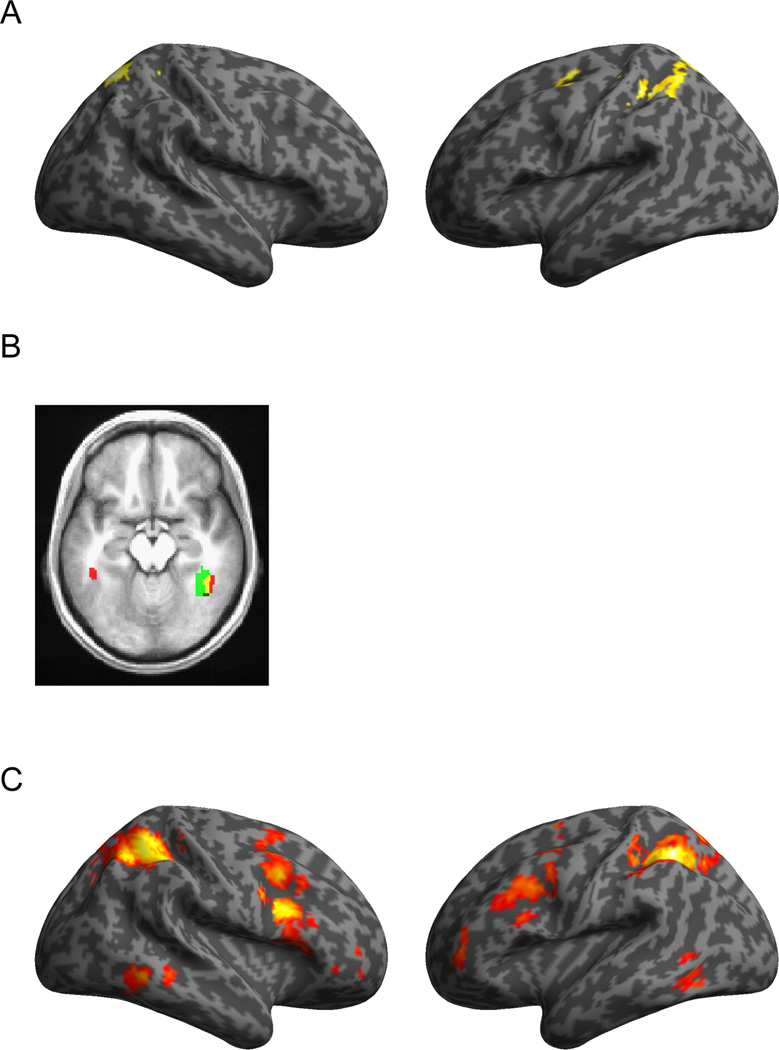
We first characterized neural regions as a function of their general responsiveness to top-down and bottom-up cues in order to assess the efficacy of our manipulation of these features in evoking activation in regions canonically associated with endogenous and exogenous attentional orienting. To assess the neural regions recruited during top-down attentional biasing, whole-brain random-effects analyses were conducted to identify the regions that exhibited greater activation in response to informative compared to uninformative top-down cues. Greater activation in response to informative spatial cues was observed in bilateral parietal regions (Figure 2A, Table 1), including the intraparietal sulcus (IPS) and a frontal region centered on the junction of the superior frontal and precentral sulci, corresponding to the left frontal eye field (FEF). These regions have been implicated in the top-down biasing of attention and are key nodes in the dorsal attention network thought to mediate endogenous orienting (Mesulam, 1999; Corbetta and Shulman, 2002). This cue-related activation is unlikely to be driven by differences in overt attentional shifting, as confirmed by results from the eye-movement data analysis (see Materials and Methods).
Figure 2.
Endogenous and exogenous influences on attention. (A) Effects of top-down cueing during the cue period (Informative > Uninformative; p < .05, corrected). (B) Effects of exogenous cue presentation in the FFA, displayed on an axial slice (z = −15) of the mean anatomical image for all subjects. Red: Group-level FFA ROI derived from independent localizer task. Green: Region in the Right Fusiform Gyrus exhibiting greater activation when exogenous cues were present than when absent (p < .05, corrected). Yellow: Overlap. (C) Effects of exogenous cue and target processing in the presence of uninformative as opposed to informative top-down cues (Uninformative > Informative; p < .05, corrected).
Table 1.
Peak activation foci for the main effects of neuroimaging analyses.
| Regions | Hemisphere | BA | MNI Coordinates (x,y,z) |
Cluster Extent (voxels) |
Z |
|---|---|---|---|---|---|
| Main effect of top-down cues (Informative > Uninformative Cue) | |||||
| Superior Parietal Lobule | R | 7 | 18, −66, 60 | 147 | 3.89 |
| Superior Frontal Gyrus | L | 8 | −24, −9, 51 | 43 | 3.84 |
| Inferior Parietal Lobule (subpeak in Superior Parietal Lobule) | L | 40 | −33, −39, 45 | 173 | 3.62 |
| Main effect of exogenous cues (Present > Absent) | |||||
| Fusiform Gyrus (subpeak in Parahippocampal Gyrus) | R | 37 | 36, −57, −6 | 170 | 4.24 |
| Middle Temporal Gyrus | R | 39 | 54, −66, 15 | 68 | 4.04 |
| Effect of top-down cues on exogenous cue (Uninformative > Informative, exogenous cue present) | |||||
| Inferior Parietal Lobule (subpeaks in Precuneus and Middle Occipital Gyrus) | L | 40 | −33, −48, 51 | 1547 | 5.68 |
| Inferior Frontal Gyrus | R | 45 | 45, 9, 33 | 686 | 5.11 |
| Superior Frontal Gyrus (subpeaks in Middle Frontal Gyrus, Inferior Frontal Junction, Inferior Frontal Gyrus, and Orbitofrontal Cortex) | L | 8 | −30, 6, 60 | 453 | 4.52 |
| Middle Temporal Gyrus | R | 37 | 54, −45, −6 | 131 | 4.49 |
| Thalamus | L | −12, −12, 3 | 100 | 4.48 | |
| Middle Frontal Gyrus | L | 10/47 | −39, 48, 6 | 96 | 3.69 |
| Inferior Frontal Gyrus | R | 10/47 | 42, 60, 0 | 78 | 3.63 |
| Inferior Temporal Gyrus | L | 37 | −51, −57, −6 | 61 | 3.56 |
| Dorsal Anterior Cingulate Cortex | R | 32/24 | 9, 18, 45 | 53 | 3.46 |
| Main effect of exogenous cue affective salience (Fearful > Neutral) | |||||
| Inferior Frontal Gyrus | L | 45 | −51, 24, 9 | 61 | 4.03 |
| Dorsomedial Prefrontal Cortex | M | 32 | 0, 51, 21 | 109 | 3.52 |
| Dorsomedial Prefrontal Cortex | M | 9/32 | 0, 42, 42 | 73 | 3.41 |
| Superior Temporal Gyrus | L | 22 | −57, 0, −12 | 45 | 3.35 |
When assessing the neural effects of the exogenous cues, results stemming from a contrast between trials where exogenous cues were present versus trials without such cues are difficult to interpret, since they could reflect the mere sensory response to additional visual stimulation or they could relate to attentional capture by these stimuli. In the context of the current experiment, this contrast nevertheless bears some interest, as it can be used to verify that the face stimuli employed as exogenous cues were in fact processed by the FFA. When exogenous cues were presented during the cue-to-target interval, greater activation was observed in visual processing regions, including right fusiform gyrus and right parahippocampal gyrus (Table 1). Activation in the right fusiform gyrus overlapped with the group-level FFA ROI defined in the independent localizer task (Figure 2B), thus confirming that the FFA was involved in processing the face stimuli employed as exogenous cues.
In order to gauge the effects of top-down attentional settings on neural processing of the exogenous cues, whole-brain random effects analyses were performed to identify regions exhibiting greater activation on trials with exogenous cues following uninformative cues compared to informative cues during the period from the onset of the exogenous cue to the offset of the search array (Figure 2C, Table 1). The identified regions included bilateral inferior parietal cortex, bilateral ventrolateral prefrontal cortex, bilateral dorsolateral prefrontal cortex, and anterior cingulate cortex. The observed enhanced neural processing of exogenous cues following uninformative compared to informative top-down cues is consistent with contingent capture, as exogenous cues were more efficacious in capturing attention in the absence of strong top-down attentional biasing. Indeed, many of the identified regions overlap closely with the putative ventral attention network thought to mediate exogenous shifting of attention (Corbetta et al., 2008). However, these activations also included components of the dorsal attention network, suggesting that endogenous attentional mechanisms were also mobilized during attentional reorienting (Corbetta et al., 2008).
Finally, on trials where exogenous cues were present, the basic effects of affective salience and cue validity were also assessed. Affectively salient exogenous cues recruited greater activation than neutral exogenous cues in the dorsomedial prefrontal cortex and left ventrolateral prefrontal cortex (Table 1), perhaps reflecting differences in attentional orienting to the stimuli. Interestingly, no differential responding was observed in the amygdala or other limbic structures. The absence of an effect of affective salience in the amygdala may be due to the short presentation time of the exogenous cues and the high attentional load of our task (Pessoa et al., 2002). Additionally, no differences were observed in neural activation as a function of bottom-up cue validity, indicating similar neural recruitment for orienting towards exogenous cues irrespective of whether or not they were subsequently followed by a target stimulus.
In sum, we documented that the processing of top-down attention cues recruited FEF and IPS foci typically associated with top-down attentional biasing (Mesulam, 1999; Corbetta et al., 2008); that the processing of bottom-up face cues elicited activity in the FFA; and that presentation of exogenous cues and target stimuli in the absence (compared to presence) of top-down cueing elicited activation in lateral frontal and parietal areas typically associated with ventral and dorsal attentional networks (Corbetta et al., 2008).
Neural regions underlying individual differences in affective contingent capture
The critical aim of the present experiment was to identify the neural regions tracking individual differences in the behavioral effects of affective exogenous attentional guidance shown in Figure 1B. Towards that end, an index of the influence of the affective exogenous cues on contingent capture was computed for each participant based on their differential response times to each condition, and this index was subsequently used as a covariate in neuroimaging analyses aimed at delineating the neural substrates of affective contingent capture. This Affective Contingent Capture Index (ACCI) represents the 3-way interaction among top-down cueing, exogenous cue validity, and affective salience ((Uninformative Cue, Fear Valid – Uninformative Cue, Neutral Valid) - (Uninformative Cue, Fear Invalid – Uninformative Cue, Neutral Invalid)) - ((Informative Cue, Fear Valid – Informative Cue, Neutral Valid) - (Informative Cue, Fear Invalid – Informative Cue, Neutral Invalid)). ACCI positively correlated with trait anxiety (Spearman’s ρ = .433, p = .030), and it was significantly larger for high anxious participants compared to low anxious participants (t(23) = 2.81, p = .010), reflecting the behavioral differences between these two groups. In other words, ACCI is a response time index that reflects the interaction between trait anxiety and the key experimental manipulation, and it should therefore provide a more sensitive tool for detecting neural correlates of individual differences in orienting to affectively salient stimuli than trait anxiety per se. However, in order to verify this assumption, we entered both ACCI and trait anxiety scores as separate covariates into the neuroimaging analyses, which allowed us to test whether ACCI would explain neural activity above and beyond that explained by trait anxiety.
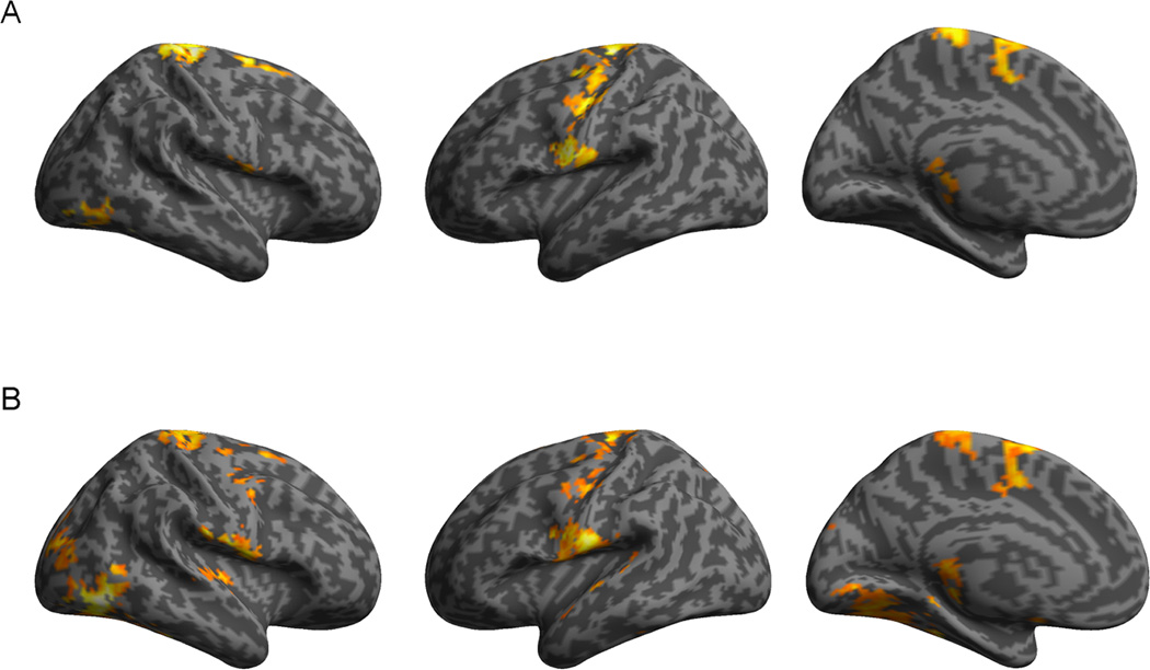
Behaviorally, the interaction between affective salience and exogenous cue validity varied across individuals on trials with uninformative cues, when endogenous attention was less directed and exogenous cues were more likely to influence attentional guidance. To isolate regions whose activation tracked this interindividual variability, the interaction between affective salience and exogenous cue validity for trials following uninformative top-down spatial cues was examined with ACCI as a between-subjects covariate. This whole-brain analysis revealed several regions whose activation positively correlated with ACCI (Figure 3A, Table 2), including bilateral dorsal frontal cortex, bilateral posterior ventrolateral frontal cortex, supplementary motor area, and extrastriate visual regions. These areas exhibited greater activation in response to the affectively salient stimuli presented spatially overlapping with upcoming targets in the absence of informative top-down spatial cues, consistent with the observed slower response times observed in high anxious participants in this condition. These neural regions, particularly posterior ventrolateral frontal cortex and supplementary motor area, have been previously implicated in attentional orienting and inhibitory control (Hampshire et al., 2010; Sharp et al., 2010; Levy and Wagner, 2011), implying that reactive control processes engaged following affectively salient stimuli may be critical to mediating individual differences in affective contingent capture. Significant activation was also observed in the right fusiform gyrus (x, y, z: 45, −45, −21), overlapping with the group-level FFA ROI, indicating that visual processing of the exogenous cues also contributed to interindividual variability in affective contingent capture. Note that neural activity in these regions was explained by ACCI above and beyond any activation attributed to trait anxiety per se, which had also been included as a covariate in the model. By contrast, we did not observe any significant activation being explained by trait anxiety above and beyond that captured by ACCI. No regions correlated negatively with ACCI.
Figure 3.
Neural correlates of affective contingent capture. Positive correlations between neural regions identified by the interaction between affective salience and exogenous cue validity [(Uninformative cue, Fearful Valid – Uninformative cue, Fearful Invalid)-(Uninformative cue, Neutral Valid – Uninformative cue, Neutral Invalid)] and the (A) Affective Contingent Capture Index (ACCI) and the (B) more focused RT covariate targeting attentional facilitation (p < .05, corrected).
Table 2.
Peak activation foci for analyses identifying neural regions underlying affective contingent capture. Analyses examined the interaction between exogenous cue validity and affective salience following uninformative top-down cues [(Uninformative Cue, Fearful Valid – Uninformative Cue, Fearful Invalid) – (Uninformative Cue, Neutral Valid – Uninformative Cue, Neutral Invalid)].
| Regions | Hemisphere | BA | MNI Coordinates (x,y,z) |
Cluster Size (voxels) |
Z |
|---|---|---|---|---|---|
| Positively correlates with Affective Contingent Capture Index | |||||
| Precentral Gyrus (subpeaks in Superior Frontal Gyrus, Middle Frontal Gyrus, Inferior Frontal Gyrus, and Supplementary Motor Area) | R | 6 | 15, −27, 75 | 1435 | 4.40 |
| Inferior Temporal Gyrus (subpeaks in Fusiform Gyrus, Lingual Gyrus, and Inferior Occipital Gyrus) | R | 37 | 51, −60, −21 | 156 | 4.28 |
| Thalamus | R | 6, −33, 6 | 89 | 3.66 | |
| Inferior Frontal Gyrus | R | 44/6 | 63, 0, 6 | 57 | 3.42 |
| Precuneus | M | 7 | 0, −63, 63 | 40 | 3.10 |
| Positively correlates with focused response time index | |||||
| Inferior Temporal Gyrus (subpeaks in Fusiform Gyrus, Lingual Gyrus, Inferior Occipital Gyrus, and Parahippocampal Gyrus) | R | 37 | 45, −66, −12 | 1470 | 4.58 |
| Cuneus (subpeaks in Superior Occipital Gyrus, Middle Occipital Gyrus, and Precuneus) | R | 19 | 12, −90, 30 | 342 | 4.36 |
| Postcentral Gyrus (subpeaks in Inferior Frontal Gyrus and Insula) | R | 43 | 63, −15, 18 | 277 | 4.32 |
| Supplementary Motor Area (subpeaks in Precentral Gyrus, and Superior Frontal Gyrus) | M | 6 | 0, 0, 75 | 1045 | 4.23 |
| Postcentral Gyrus (subpeaks in Precentral Gyrus and Inferior Frontal Gyrus) | L | 43 | −63, −15, 18 | 415 | 4.19 |
| Subgenual Anterior Cingulate Cortex | M | 25 | 3, 21, −15 | 52 | 3.82 |
| Precentral Gyrus | R | 6 | 45, −3, 45 | 76 | 3.54 |
| Precentral Gyrus | L | 6 | −39, −12, 39 | 102 | 3.52 |
Although these findings indicate that several regions, including ventrolateral frontal cortex, supplementary motor area, and FFA, play a vital role in mediating affective contingent capture, ACCI encompasses differences in response time on all experimental conditions, not merely the critical interaction of affective salience and endogenous attention. It is possible, therefore, that variability in responding in the other conditions might be bolstering our neural effects of affective contingent capture or obscuring other effects of interest. To address this possibility, we computed a more specific index of behavior focused exclusively on the critical effect: the interaction between affective salience and endogenous attentional settings, restricted to trials in which the exogenous cue was presented in the same location as the upcoming target [(Uninformative Cue, Fearful Valid – Uninformative Cue, Neutral Valid) – (Informative Cue, Fearful Valid – Informative Cue, Neutral Valid)]. This focused response time index was then entered as a between-subjects covariate with the interaction between affective salience and exogenous cue validity for trials following uninformative top-down spatial cues. This whole-brain analysis identified a network of regions exhibiting positive correlations with the response time index which overlapped with those detected employing ACCI as a covariate, namely, bilateral dorsal frontal cortex, bilateral posterior ventrolateral frontal cortex, supplementary motor area, and extrastriate visual regions (Figure 3B, Table 2). Moreover, this more focused analysis also revealed significant activation in the subgenual anterior cingulate cortex, a region that has been implicated in various forms of emotion regulation (Etkin et al., 2011). Overall, these results confirm that the findings from the analyses employing the ACCI were not driven by experimental conditions that were not germane to the behavioral findings. No regions correlated negatively with this response time covariate. Moreover, a contrast examining the interaction between affective salience and exogenous cue validity for trials following informative top-down spatial cues failed to identify any neural regions that significantly correlated with the response time covariate, indicating that, like the behavioral effects, these neural effects were specific to conditions without explicit top-down spatial biasing.
In sum, interindividual variability in affective contingent capture correlates primarily with activation variability in posterior ventrolateral frontal cortex, supplementary motor area, and the FFA. Greater activation in these regions is associated with delayed responding to targets appearing spatially coincident with fearful exogenous cues and may reflect differences in basic perceptual processing, attentional orienting or inhibitory control. Critically, these neural effects, like the behavioral effects of interest, were contingent on top-down attentional settings, present only in the absence of explicit top-down spatial cues.
Frontal regions mediate the relationship between right FFA and inter-subject behavioral variability
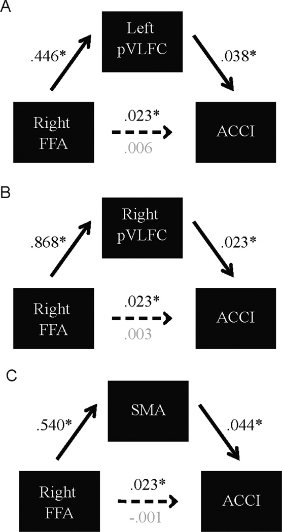
The neural regions tracking inter-subject behavioral variability in response to affectively salient exogenous cues included both regions associated with basic stimulus processing, such as the right FFA, and frontal regions typically implicated in attentional orienting or inhibitory control. As all the regions identified exhibited positive correlations with the response time indices, it remains unclear if these regions track similar properties of behavioral variability, or if some of the regions carry unique information and mediate the effects of the others. In particular, the response time data suggest that the effects of affective salience on exogenous attentional guidance are contingent on top-down attentional settings, implying that differences in attentional orienting play a critical role in mediating individual differences. Regions associated with the ventral attention network and attentional control, such as the posterior ventrolateral frontal cortex and supplementary motor area, may therefore mediate the effects of basic perceptual processing regions that also track affective contingent capture, such as the FFA. Mediation analyses were employed to determine whether or not the frontal regions that were identified in both the ACCI and the focused response time covariate analyses mediated the relationship between the right FFA and behavior, as indexed by the ACCI (see Materials & Methods). The relationship between FFA activity and behavioral variability in affective contingent capture was fully mediated by each of the three identified frontal regions: left posterior ventrolateral frontal cortex, right posterior ventrolateral frontal cortex, and supplementary motor area (Figure 4). Importantly, the converse was not true for any of these regions, as right FFA did not mediate the relationship between any of the prefrontal regions and ACCI2. Thus, activation in frontal regions subsumed the activation in the right FFA, demonstrating that signals carried in these critical nodes of the attentional orienting system play a key role in mediating the effects of affective contingent capture above and beyond the basic effects of perceptual processing in the FFA.
Figure 4.
Mediation analyses. The relationship between neural responses in right FFA and ACCI is mediated by (A) left posterior ventrolateral frontal cortex (B) right posterior ventrolateral frontal cortex and (C) Supplementary Motor Area. Numbers are unstandardized coefficients; asterisks denote p<.05.
Discussion
The present study sought to clarify the role of endogenous attention settings in modulating affective exogenous guidance of spatial attention. Employing a novel cueing paradigm, we were able to independently manipulate top-down attentional settings, exogenous cue validity, and affective salience and observe their impact on attentional orienting as a function of individual differences in anxiety. In the absence of explicit top-down spatial cues to target locations, face stimuli exogenously facilitated attention to spatially coincident targets for low anxious participants, regardless of affective expression. For high anxious participants, although neutral face stimuli facilitated attention to target locations, affectively salient face stimuli failed to generate similar facilitation, instead resulting in response times that were as slow as trials when exogenous cues served to distract attention away from target locations. Critically, these effects were contingent on top-down attentional settings; when explicit cues directed top-down attention to upcoming target locations, high anxious participants no longer exhibited differential responding to fearful faces, indicating that top-down attention can counteract the effects of exogenous affective attentional guidance. As with other forms of exogenous cueing (Folk et al., 1992), affective capture effects appear to be contingent on top-down attention settings, providing an avenue to overcome the attentional biases present in anxious individuals. Interindividual variability in this affective contingent capture tracked with neural activation in bilateral ventrolateral frontal cortex and supplementary motor area in the absence, but not the presence, of explicit top-down spatial cues, consistent with the contingent nature of behavioral affective capture effects.
High anxious participants exhibited delayed responding to fearful faces that facilitated target discrimination for low anxious participants, in contrast to some previous behavioral studies demonstrating speeded threat detection by anxious participants (Fox et al., 2001; Ohman et al., 2001). However, tasks involving target discrimination at locations spatially coincident with threat reveal deficits for high anxious, but not low anxious, participants (Salemink et al., 2007; Chajut et al., 2010). This dissociation between initial threat detection and subsequent target discrimination likely reflects differences in their relative attentional control demands. In the present task, successful performance required the visual discrimination of a subtle alteration in a non-affective stimulus (i.e., the slight tilt of one of two Gabor patches), demanding a reorientation of attentional resources from the affectively salient exogenous cue to the non-affective target stimulus. Consistent with Attentional Control Theory (Eysenck et al., 2007), highly anxious participants may have been more sensitive to the exogenous affective stimuli than low anxious participants, with the fearful stimuli potentially dominating attentional resources and generating interference with the visual discrimination task. Moreover, high anxious individuals exhibit pronounced deficits in disengaging from threatening stimuli (Fox et al., 2001; Amir et al., 2003; Koster et al., 2004; Salemink et al., 2007), further enhancing the capacity of affectively salient stimuli to disrupt attentional deployment and task performance. In contrast, on tasks involving detection of threatening stimuli (Ohman et al., 2001) or neutral stimuli presented in the same location as threatening stimuli (Mogg and Bradley, 1999), this hypersensitivity to affective stimuli and difficulty reorienting away from them would result in speeded responding. Thus, the observed differences in task performance are likely driven by enhanced demands on attentional reorienting and inhibitory processes for high anxious participants following affectively salient stimuli.
The network of neural regions that tracked individual differences in affective contingent capture is highly consistent with the enhanced capacity of affectively salient stimuli to interfere with ongoing processing for anxious participants and place enhanced demands on attentional reorienting systems. Bilateral ventrolateral frontal cortex, supplementary motor area, and the FFA all exhibited enhanced activation following presentation of affectively salient stimuli at upcoming target locations in individuals with relatively delayed responding in that condition. Importantly, signal in the ventrolateral frontal cortices and supplementary motor area fully mediated the effects of FFA, demonstrating a critical role for these frontal neural regions in mediating behavioral variability. A recent meta-analysis highlighted the role these regions play in both orienting to external stimuli and implementing inhibitory control (Levy and Wagner, 2011), consistent with enhanced attentional control demands for anxious participants when reorienting attention from affective cues towards neutral targets. Posterior ventrolateral frontal cortex has been previously implicated in attentional capture effects (de Fockert et al., 2004), exhibiting greater activation in the presence of perceptually salient distracters, and lesion evidence highlights a role for this region in resolving interference from salient distracters (Michael et al., 2006). The present study extends these findings to the domain of affective stimuli, while further demonstrating their dependence on endogenous attentional settings. Activation in the supplementary motor area is closely tied to action updating and inhibitory control processes (Aron et al., 2007; Chikazoe et al., 2009; Sharp et al., 2010), and has additionally been implicated during selective orienting and resisting interference from affectively salient distractors (Armony and Dolan, 2002; Krebs et al., 2011). Indeed, the supplementary motor area and posterior ventrolateral frontal cortex are interconnected and share projections to motor regions and the subthalamic nucleus (Aron et al., 2007; Nachev et al., 2008), rendering this network critical to implementing inhibitory control, reorienting attention, and updating action plans. Enhanced recruitment of this network likely reflects the increased interference and heightened attentional control demands generated by affectively salient stimuli for high anxious individuals. Consistent with this position, greater activation was also observed in subgenual anterior cingulate cortex, a putative emotion regulation region (Etkin et al., 2011) with strong connections to limbic structures (Beckmann et al., 2009) which could be critical in mediating affective capture effects. Crucially, increased activation was only observed in neural regions in the absence of informative top-down attentional settings, demonstrating that these regions were vital to mediating reactive attentional control during affective capture, but were not mobilized when attention was spatially directed prior to exogenous interference.
Given the critical role of the amygdala in affective processing and orienting to salient stimuli (LeDoux, 2000; Anderson and Phelps, 2001; Vuilleumier et al., 2004), it is somewhat surprising that we did not observe activation in this region or nearby voxels during our task. Despite conducting additional exploratory analyses, repeating all analyses reported here in conjunction with small-volume correction of an anatomically-defined amygdala region of interest, we identified no significant activation. Possibly the high cognitive load and short presentation time of affective stimuli contributed to the lack of differential amygdala responding, as this region has been shown to be sensitive to the availability of cognitive resources (Vuilleumier et al., 2001; Pessoa et al., 2002; Lim et al., 2008). Moreover, the timing structure of our task and the poor temporal resolution of BOLD fMRI may have diminished our ability to detect rapid, transient signals in the amygdala underlying orienting to the exogenous affective stimuli. Additionally, findings of preserved attentional capture by affectively salient stimuli in amygdala lesion patients call into question the necessity of the amygdala in mediating attentional orienting to affective stimuli (Piech et al., 2011). However, activation in the FFA was observed in response to exogenous cues, particularly in the absence of directed top-down attention. Critically, variability in the FFA was also associated with interindividual variability in attentional orienting, implicating perceptual processing of the affective exogenous cues in contributing to differences in affective capture effects. However, these differences in bottom-up processing regions were subsumed by variability in frontal regions, implying that more than variability in basic perceptual processes drove differences in affective contingent capture.
The importance of frontal regions in mediating individual differences in affective contingent capture is consistent with neurocognitive theories that highlight a critical role for executive control in anxiety disorders. Some recent theories have emphasized the importance of frontal-amygdala interactions in anxiety disorders (Bishop, 2007), highlighting the role executive function plays in modulating affective responses. The relationship between trait anxiety and affective contingent capture observed in the present study is consistent with the notion that anxious individuals are more susceptible to threatening stimuli (Macleod et al., 1986; Eysenck et al., 2007), and this hypervigilance may be central to the etiology and maintenance of anxiety disorders (Mathews and MacLeod, 2002). Our demonstration that endogenous top-down attention can overcome this sensitivity to affective stimuli points to a role for proactive engagement of top-down control as a candidate for incorporation in the development of behavioral interventions. Behavioral clinical interventions aimed at retraining anxious individuals to orient attention away from threatening stimuli have been successful in generating anxiolytic effects (Bar-Haim, 2010; Hakamata et al., 2010), and the present findings of affective contingent capture suggest that the incorporation of top-down attentional orienting into similar interventions could enhance their efficacy. Although endogenous attentional settings may need to be established prior to the appearance of affective external stimuli to diminish their capacity to exogenously direct attention, such forms of proactive control may be both highly effective in diminishing interference from external stimuli and readily implemented in ecologically valid settings (Aron, 2011).
The present study demonstrated that affective capture effects can alter exogenous attentional guidance, particularly in anxious individuals, but that these effects are contingent on endogenous attentional settings. These findings are consistent with numerous theories of anxiety emphasizing susceptibility to threat and fearful stimuli (Mathews and Mackintosh, 1998; Mathews and MacLeod, 2002; Bishop, 2007; Eysenck et al., 2007). Although the present research focused on the role of fearful stimuli in guiding attention in anxious individuals, our findings can not disambiguate whether these effects are unique to threatening stimuli or extend to other affectively salient stimuli. Additionally, similar affective contingent capture effects may potentially be present for positively valenced affective stimuli in other populations, such as individuals high in reward sensitivity. For instance, stimuli associated with large rewards can capture attention, particularly in individuals with lower self-reported inhibitory control (Anderson et al., 2011); however, the role of endogenous attentional settings in modulating these effects remains unexamined. Finally, in the present study we investigated attentional capture by single, sudden-onset stimuli. While this is a well-established means of producing attentional capture effects (Yantis & Jonides, 1990), other approaches have investigated this phenomenon in the context of simultaneously presented arrays of multiple stimuli competing for attention (e.g., Theeuwes, 1992). Whether the current findings generalize to the latter type of experimental set-up represents an interesting question for future studies.
In the present experiment, neural activation in posterior ventrolateral frontal regions and supplementary motor area mediated behavioral variability in affective contingent capture across individuals, and like the behavioral effects, demonstrated sensitivity to endogenous attentional signals. These findings indicate a critical role for these regions in mediating attentional orienting and interference resolution processes when engaged by affectively salient stimuli and provide insight into the neurocognitive mechanisms underlying anxiety.
Acknowledgments
This research was supported by NIH R01MH087610-01 (TE) and an NSF Graduate Research Fellowship (CR).
Footnotes
The authors have no competing financial interests.
Development of the MacBrain Face Stimulus Set was overseen by Nim Tottenham and supported by the John D. and Catherine T. MacArthur Foundation Research Network on Early Experience and Brain Development. Please contact Nim Tottenham at tott0006@tc.umn.edu for more information concerning the stimulus set.
Mediation analyses were also conducted examining the relationship between the right FFA and the focused response time index employed in the second between-subjects analysis. As with the analyses conducted utilizing ACCI, the relationship between responses in the right FFA and the response time measure was fully mediated by each of the three frontal regions: left posterior ventrolateral frontal cortex, right posterior ventrolateral frontal cortex, and supplementary motor area. Again, the opposite was not true; activation in the right FFA did not mediate the relationship between any of the frontal neural regions and the response time index.
References
- Amir N, Elias J, Klumpp H, Przeworski A. Attentional bias to threat in social phobia: Facilitated processing of threat or difficulty disengaging attention from threat? Behaviour Research and Therapy. 2003;41:1325–1335. doi: 10.1016/s0005-7967(03)00039-1. [DOI] [PubMed] [Google Scholar]
- Anderson AK, Phelps EA. Lesions of the human amygdala impair enhanced perception of emotionally salient events. Nature. 2001;411:305–309. doi: 10.1038/35077083. [DOI] [PubMed] [Google Scholar]
- Anderson BA, Laurent PA, Yantis S. Value-driven attentional capture. Proceedings of the National Academy of Sciences of the United States of America. 2011;108:10367–10371. doi: 10.1073/pnas.1104047108. [DOI] [PMC free article] [PubMed] [Google Scholar]
- Armony JL, Dolan RJ. Modulation of spatial attention by fear-conditioned stimuli: an event-related fMRI study. Neuropsychologia. 2002;40:817–826. doi: 10.1016/s0028-3932(01)00178-6. [DOI] [PubMed] [Google Scholar]
- Aron AR. From reactive to proactive and selective control: Developing a richer model for stopping inappropriate responses. Biological Psychiatry. 2011;69:E55–E68. doi: 10.1016/j.biopsych.2010.07.024. [DOI] [PMC free article] [PubMed] [Google Scholar]
- Aron AR, Behrens TE, Smith S, Frank MJ, Poldrack RA. Triangulating a cognitive control network using diffusion-weighted magnetic resonance imaging (MRI) and functional MRI. Journal of Neuroscience. 2007;27:3743–3752. doi: 10.1523/JNEUROSCI.0519-07.2007. [DOI] [PMC free article] [PubMed] [Google Scholar]
- Ashburner J, Friston KJ. Unified segmentation. Neuroimage. 2005;26:839–851. doi: 10.1016/j.neuroimage.2005.02.018. [DOI] [PubMed] [Google Scholar]
- Bar-Haim Y. Research Review: Attention bias modification (ABM): A novel treatment for anxiety disorders. Journal of Child Psychology and Psychiatry. 2010;51:859–870. doi: 10.1111/j.1469-7610.2010.02251.x. [DOI] [PubMed] [Google Scholar]
- Bar-Haim Y, Lamy D, Pergamin L, Bakermans-Kranenburg MJ, Van Ijzendoorn MH. Threat-related attentional bias in anxious and nonanxious individuals: A meta-analytic study. Psychological Bulletin. 2007;133:1–24. doi: 10.1037/0033-2909.133.1.1. [DOI] [PubMed] [Google Scholar]
- Baron RM, Kenny DA. The moderator mediator variable distinction in social psychological research: Conceptual, strategic, and statistical considerations. Journal of Personality and Social Psychology. 1986;51:1173–1182. doi: 10.1037//0022-3514.51.6.1173. [DOI] [PubMed] [Google Scholar]
- Beckmann M, Johansen-Berg H, Rushworth MFS. Connectivity-based parcellation of human cingulate cortex and its relation to functional specialization. Journal of Neuroscience. 2009;29:1175–1190. doi: 10.1523/JNEUROSCI.3328-08.2009. [DOI] [PMC free article] [PubMed] [Google Scholar]
- Bieling PJ, Antony MM, Swinson RP. The State--Trait Anxiety Inventory, Trait version: Structure and content re-examined. Behaviour Research and Therapy. 1998;36:777–788. doi: 10.1016/s0005-7967(98)00023-0. [DOI] [PubMed] [Google Scholar]
- Bishop SJ. Neurocognitive mechanisms of anxiety: An integrative account. Trends in Cognitive Sciences. 2007;11:307–316. doi: 10.1016/j.tics.2007.05.008. [DOI] [PubMed] [Google Scholar]
- Bradley BP, Mogg K, Millar N, Bonham-Carter C, Fergusson E, Jenkins J, Parr M. Attentional biases for emotional faces. Cognition & Emotion. 1997;11:25–42. [Google Scholar]
- Chajut E, Schupak A, Algom D. Emotional dilution of the Stroop Effect: A new tool for assessing attention under emotion. Emotion. 2010;10:944–948. doi: 10.1037/a0020172. [DOI] [PubMed] [Google Scholar]
- Chikazoe J, Jimura K, Asari T, Yamashita K, Morimoto H, Hirose S, Miyashita Y, Konishi S. Functional dissociation in right inferior frontal cortex during performance of Go/No-Go Task. Cerebral Cortex. 2009;19:146–152. doi: 10.1093/cercor/bhn065. [DOI] [PubMed] [Google Scholar]
- Corbetta M, Patel G, Shulman GL. The reorienting system of the human brain: From environment to theory of mind. Neuron. 2008;58:306–324. doi: 10.1016/j.neuron.2008.04.017. [DOI] [PMC free article] [PubMed] [Google Scholar]
- Corbetta M, Shulman GL. Control of goal-directed and stimulus-driven attention in the brain. Nature Reviews Neuroscience. 2002;3:201–215. doi: 10.1038/nrn755. [DOI] [PubMed] [Google Scholar]
- De Fockert J, Rees G, Frith C, Lavie N. Neural correlates of attentional capture in visual search. Journal of Cognitive Neuroscience. 2004;16:751–759. doi: 10.1162/089892904970762. [DOI] [PubMed] [Google Scholar]
- Egner T, Monti JMP, Trittschuh EH, Wieneke CA, Hirsch J, Mesulam MM. Neural integration of top-down spatial and feature-based information in visual search. Journal of Neuroscience. 2008;28:6141–6151. doi: 10.1523/JNEUROSCI.1262-08.2008. [DOI] [PMC free article] [PubMed] [Google Scholar]
- Etkin A, Egner T, Kalisch R. Emotional processing in anterior cingulate and medial prefrontal cortex. Trends in Cognitive Sciences. 2011;15:85–93. doi: 10.1016/j.tics.2010.11.004. [DOI] [PMC free article] [PubMed] [Google Scholar]
- Eysenck MW, Derakshan N, Santos R, Calvo MG. Anxiety and cognitive performance: Attentional control theory. Emotion. 2007;7:336–353. doi: 10.1037/1528-3542.7.2.336. [DOI] [PubMed] [Google Scholar]
- Folk CL, Remington RW, Johnston JC. Involuntary covert orienting is contingent on attentional control settings. Journal of Experimental Psychology-Human Perception and Performance. 1992;18:1030–1044. [PubMed] [Google Scholar]
- Fox E, Russo R, Bowles R, Dutton K. Do threatening stimuli draw or hold visual attention in subclinical anxiety? Journal of Experimental Psychology-General. 2001;130:681–700. [PMC free article] [PubMed] [Google Scholar]
- Guo H, Song AW. Single-shot spiral image acquisition with embedded Z-shimming for susceptibility signal recovery. Journal of Magnetic Resonance Imaging. 2003;18:389–395. doi: 10.1002/jmri.10355. [DOI] [PubMed] [Google Scholar]
- Hakamata Y, Lissek S, Bar-Haim Y, Britton JC, Fox NA, Leibenluft E, Ernst M, Pine DS. Attention Bias Modification Treatment: A meta-analysis toward the establishment of novel treatment for anxiety. Biological Psychiatry. 2010;68:982–990. doi: 10.1016/j.biopsych.2010.07.021. [DOI] [PMC free article] [PubMed] [Google Scholar]
- Hampshire A, Chamberlain SR, Monti MM, Duncan J, Owen AM. The role of the right inferior frontal gyrus: inhibition and attentional control. Neuroimage. 2010;50:1313–1319. doi: 10.1016/j.neuroimage.2009.12.109. [DOI] [PMC free article] [PubMed] [Google Scholar]
- Hansen CH, Hansen RD. Automatic emotion: Attention and facial efference. In: Niedenthal PM, Kitayama S, editors. The Heart's Eye: Emotional influences in perception and attention. Academic Press; 1994. pp. 217–243. [Google Scholar]
- Kanwisher N, Mcdermott J, Chun MM. The fusiform face area: A module in human extrastriate cortex specialized for face perception. Journal of Neuroscience. 1997;17:4302–4311. doi: 10.1523/JNEUROSCI.17-11-04302.1997. [DOI] [PMC free article] [PubMed] [Google Scholar]
- Koster EHW, Crombez G, Verschuere B, De Houwer J. Selective attention to threat in the dot probe paradigm: Differentiating vigilance and difficulty to disengage. Behaviour Research and Therapy. 2004;42:1183–1192. doi: 10.1016/j.brat.2003.08.001. [DOI] [PubMed] [Google Scholar]
- Krebs RM, Boehler CN, Egner T, Woldorff MG. The neural underpinnings of how reward associations can both guide and misguide attention. Journal of Neuroscience. 2011;31:9752–9759. doi: 10.1523/JNEUROSCI.0732-11.2011. [DOI] [PMC free article] [PubMed] [Google Scholar]
- Ledoux JE. Emotion circuits in the brain. Annual Review of Neuroscience. 2000;23:155–184. doi: 10.1146/annurev.neuro.23.1.155. [DOI] [PubMed] [Google Scholar]
- Levy BJ, Wagner AD. Cognitive control and right ventrolateral prefrontal cortex: Reflexive reorienting, motor inhibition, and action updating. Annals of the New York Academy of Sciences. 2011;1224:40–62. doi: 10.1111/j.1749-6632.2011.05958.x. [DOI] [PMC free article] [PubMed] [Google Scholar]
- Lim S-L, Padmala S, Pessoa L. Segregating the significant from the mundane on a moment-to-moment basis via direct and indirect amygdala contributions. Proceedings of the National Academy of Sciences of the United States of America. 2009;106:16841–16846. doi: 10.1073/pnas.0904551106. [DOI] [PMC free article] [PubMed] [Google Scholar]
- Lim SL, Padmala S, Pessoa L. Affective learning modulates spatial competition during low-load attentional conditions. Neuropsychologia. 2008;46:1267–1278. doi: 10.1016/j.neuropsychologia.2007.12.003. [DOI] [PMC free article] [PubMed] [Google Scholar]
- Macleod C, Mathews A, Tata P. Attentional bias in emotional disorders. Journal of Abnormal Psychology. 1986;95:15–20. doi: 10.1037//0021-843x.95.1.15. [DOI] [PubMed] [Google Scholar]
- Mathews A, Mackintosh B. A cognitive model of selective processing in anxiety. Cognitive Therapy and Research. 1998;22:539–560. [Google Scholar]
- Mathews A, Macleod C. Induced processing biases have causal effects on anxiety. Cognition & Emotion. 2002;16:331–354. [Google Scholar]
- Mesulam MM. Spatial attention and neglect: parietal, frontal and cingulate contributions to the mental representation and attentional targeting of salient extrapersonal events. Philosophical Transactions of the Royal Society of London Series B-Biological Sciences. 1999;354:1325–1346. doi: 10.1098/rstb.1999.0482. [DOI] [PMC free article] [PubMed] [Google Scholar]
- Michael GA, Garcia S, Fernandez D, Sellal F, Boucart M. The ventral premotor cortex (vPM) and resistance to interference. Behavioral Neuroscience. 2006;120:447–462. doi: 10.1037/0735-7044.120.2.447. [DOI] [PubMed] [Google Scholar]
- Mogg K, Bradley BP. Orienting of attention to threatening facial expressions presented under conditions of restricted awareness. Cognition & Emotion. 1999;13:713–740. [Google Scholar]
- Nachev P, Kennard C, Husain M. Functional role of the supplementary and pre-supplementary motor areas. Nature Reviews Neuroscience. 2008;9:856–869. doi: 10.1038/nrn2478. [DOI] [PubMed] [Google Scholar]
- Notebaert L, Crombez G, Van Damme S, De Houwer J, Theeuwes J. Signals of threat do not capture, but prioritize, attention: A conditioning approach. Emotion. 2011;11:1325–1335. doi: 10.1037/a0021286. [DOI] [PubMed] [Google Scholar]
- Ohman A, Flykt A, Esteves F. Emotion drives attention: Detecting the snake in the grass. Journal of Experimental Psychology-General. 2001;130:466–478. doi: 10.1037//0096-3445.130.3.466. [DOI] [PubMed] [Google Scholar]
- Ollinger JM, Corbetta M, Shulman GL. Separating processes within a trial in event-related functional MRI - II. Analysis. Neuroimage. 2001;13:218–229. doi: 10.1006/nimg.2000.0711. [DOI] [PubMed] [Google Scholar]
- Pessoa L, Mckenna M, Gutierrez E, Ungerleider LG. Neural processing of emotional faces requires attention. Proceedings of the National Academy of Sciences of the United States of America. 2002;99:11458–11463. doi: 10.1073/pnas.172403899. [DOI] [PMC free article] [PubMed] [Google Scholar]
- Phelps EA, Ling S, Carrasco M. Emotion facilitates perception and potentiates the perceptual benefits of attention. Psychological Science. 2006;17:292–299. doi: 10.1111/j.1467-9280.2006.01701.x. [DOI] [PMC free article] [PubMed] [Google Scholar]
- Piech RM, Mchugo M, Smith SD, Dukic MS, Van Der Meer J, Abou-Khalil B, Most SB, Zald DH. Attentional capture by emotional stimuli is preserved in patients with amygdala lesions. Neuropsychologia. 2011;49:3314–3319. doi: 10.1016/j.neuropsychologia.2011.08.004. [DOI] [PMC free article] [PubMed] [Google Scholar]
- Posner MI, Snyder CRR, Davidson BJ. Attention and the detection of signals. Journal of Experimental Psychology-General. 1980;109:160–174. [PubMed] [Google Scholar]
- Salemink E, Van Den Hout MA, Kindt M. Selective attention and threat: Quick orienting versus slow disengagement and two versions of the dot probe task. Behaviour Research and Therapy. 2007;45:607–615. doi: 10.1016/j.brat.2006.04.004. [DOI] [PubMed] [Google Scholar]
- Sharp DJ, Bonnelle V, De Boissezon X, Beckmann CF, James SG, Patel MC, Mehta MA. Distinct frontal systems for response inhibition, attentional capture, and error processing. Proceedings of the National Academy of Sciences of the United States of America. 2010;107:6106–6111. doi: 10.1073/pnas.1000175107. [DOI] [PMC free article] [PubMed] [Google Scholar]
- Spielberger CD. Manual for the State-Trait Anxiety Inventory. Palo Alto, CA: Consulting Press; 1983. [Google Scholar]
- Theeuwes J. Perceptual selectivity for color and form. Perception & Psychophysics. 1992;51:599–606. doi: 10.3758/bf03211656. [DOI] [PubMed] [Google Scholar]
- Theeuwes J. Top-down and bottom-up control of visual selection. Acta Psychologica. 2010;135:77–99. doi: 10.1016/j.actpsy.2010.02.006. [DOI] [PubMed] [Google Scholar]
- Tottenham N, Tanaka JW, Leon AC, Mccarry T, Nurse M, Hare TA, Marcus DJ, Westerlund A, Casey BJ, Nelson C. The NimStim set of facial expressions: Judgments from untrained research participants. Psychiatry Research. 2009;168:242–249. doi: 10.1016/j.psychres.2008.05.006. [DOI] [PMC free article] [PubMed] [Google Scholar]
- Van Der Stigchel S, Belopolsky AV, Peters JC, Wijnen JG, Meeter M, Theeuwes J. The limits of top-down control of visual attention. Acta Psychologica. 2009;132:201–212. doi: 10.1016/j.actpsy.2009.07.001. [DOI] [PubMed] [Google Scholar]
- Vuilleumier P, Armony JL, Driver J, Dolan RJ. Effects of attention and emotion on face processing in the human brain: An event-related fMRI study. Neuron. 2001;30:829–841. doi: 10.1016/s0896-6273(01)00328-2. [DOI] [PubMed] [Google Scholar]
- Vuilleumier P, Richardson MP, Armony JL, Driver J, Dolan RJ. Distant influences of amygdala lesion on visual cortical activation during emotional face processing. Nature Neuroscience. 2004;7:1271–1278. doi: 10.1038/nn1341. [DOI] [PubMed] [Google Scholar]
- Wager TD, Nichols TE. Optimization of experimental design in fMRI: a general framework using a genetic algorithm. Neuroimage. 2003;18:293–309. doi: 10.1016/s1053-8119(02)00046-0. [DOI] [PubMed] [Google Scholar]
- Yantis S, Jonides J. Abrupt visual onsets and selective attention - Voluntary versus automatic allocation. Journal of Experimental Psychology-Human Perception and Performance. 1990;16:121–134. doi: 10.1037//0096-1523.16.1.121. [DOI] [PubMed] [Google Scholar]


