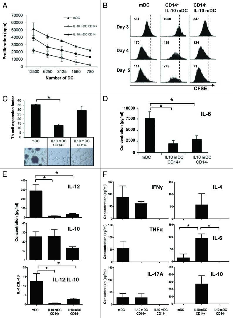
Figure 3. T-cell stimulatory characteristics of CD14+ and CD14− dendritic cells matured in the presence of interleukin-10. (A) Allostimulation of T cells by monocyte-derived dendritic cells (DCs) matured in the absence (mDCs) or in the presence (IL-10-mDCs) of interleukin (IL)-10 sorted by CD14 expression. Mean proliferation ± SEM from n = 3 mixed leukocyte reactivity (MLR) tests. (B) After 8 d of allostimulation with different mDC subsets, T cells were pulsed with CFSE and re-stimulated with anti-CD3 and anti-CD28 monoclonal antibodies, and proliferation was followed over time by CFSE dilution. Data from one representative experiment out of three are shown. (C) CD4+CD25- TH cells were stimulated with anti-CD3 monoclonal antibody (OKT3)-loaded autologous mDC populations and—after 14 d—expansion factors were measured. Data are shown as means ± SEM of n = 4 experiments, *p < 0.05. Representative microphotographs are reported (100× magnification). (D and E) Secretion of IL-6 (D) or IL-12p70 and IL-10 (E) by mDCs and IL-10-mDCs sorted by CD14 expression, upon CD40 ligation. Mean IL-12p70 and IL-10 concentrations were divided to obtain IL-12p70:IL-10 ratios for the indicated conditions. Data represent means ± SEM from n = 5 experiments, *p < 0.05. (F) After co-culturing CD4+CD25− TH cells with different OKT3-loaded autologous mDC populations for 14 d, they were re-stimulated with anti-CD3 and −CD28 monoclonal antibodies and tested for the release of interferon γ (IFNγ), tumor necrosis factor α (TNFα), IL-4, IL-6, IL-10 and IL-17 (TH1/TH2/TH17) 24 h later. Data represent means ± SD from n = 5 experiments, *p < 0.05.

An official website of the United States government
Here's how you know
Official websites use .gov
A
.gov website belongs to an official
government organization in the United States.
Secure .gov websites use HTTPS
A lock (
) or https:// means you've safely
connected to the .gov website. Share sensitive
information only on official, secure websites.
