Abstract
African American (AA) women have an elevated risk of cardiovascular disease and have been reported to have lower cardiorespiratory fitness (CRF) compared with Caucasian American (CA) women. However, little data exist that evaluate racial differences in the change in CRF following aerobic exercise training. CA (n = 264) and AA (n = 122) postmenopausal women from the Dose-Response to Exercise in Women study were randomized to 4, 8, and 12 kcal·kg body wt−1·wk−13 (KKW) of aerobic training or the control group for 6 mo. CRF was evaluated using a cycle ergometer. A greater increase in relative CRF was observed in CA compared with AA women in the 4 (CA: 1.00 vs. AA: 0.35 ml O2·kg−1·min −1, P = 0.034), 8 (CA: 1.59 vs. AA: 0.82 ml O2·kg−1·min −1, P = 0.041), and 12 (CA: 1.98 vs. AA: 0.50 ml O2·kg−1·min −1, P = 0.001) KKW groups. Similar effects were found in absolute CRF, with the exception of the 4-KKW (CA: 0.04 vs. AA: 0.02 l O2/min, P = 0.147) group. However, in categorical analyses, the percentages of women who improved in both relative (>0 ml O2·kg−1·min −1) and absolute (>0 l O2/min) CRF were not significantly different for CA and AA women in all exercise groups (all P > 0.05). AA postmenopausal women, in general, had an attenuated increase in CRF (both relative and absolute) following exercise training, but had similar response rates compared with CA women. Future studies should investigate the physiologic mechanisms responsible for this attenuated response.
Keywords: race, exercise training, African American, cardiorespiratory fitness
racial health disparities are an important public health problem (23). African American (AA) women have a higher risk of cardiovascular disease (CVD), stroke, and mortality compared with Caucasian American (CA) women (21, 23). Higher levels of cardiorespiratory fitness (CRF) are associated with reduced risk of CVD and mortality (4, 14, 20), and recent data suggest that AA women have lower CRF levels compared with their CA counterparts (11, 17, 18, 28, 31). Specifically, the Coronary Artery Risk Development in Young Adults (CARDIA) study (30) found that CRF from a maximal exercise test was higher in CA women compared with AA women. Similar findings have been reported in another epidemiological study (28), larger clinical studies (18, 31), and smaller studies in a variety of subject populations [adults (31), women (11, 17), type 2 diabetes (9)]. Thus lower CRF may contribute to CVD disparities between CA and AA women; however, racial differences in CRF have not been evaluated specifically in postmenopausal women.
CRF is inversely associated with all-cause mortality in CA and AA adults (16), and increasing CRF levels are associated with reduced risk of CVD (3). Aerobic exercise training has a well-documented effect for improving CRF, which occurs in both CA and AA individuals (8, 31). Thus increasing levels of CRF should reduce CVD risk, regardless of race. However, little data exist evaluating racial differences in the improvement of CRF as a result of aerobic exercise training between CA and AA adults.
Data from the Health, Risk Factors, Exercise Training and Genetics Family (HERITAGE) study suggest that although mean CRF [expressed in changes in maximal oxygen consumption (VO2)] increased following 20 wk of cycle ergometer training to a greater extent in CA (5.5 ml O2·kg−1·min−1) compared with AA (4.9 ml O2·kg−1·min−1) adults, no differences were observed in mean percent change in both groups (CA: 17.5%; AA: 18.7%). Since no racial differences were additionally observed in absolute CRF or in CRF corrected for lean mass, the authors concluded that race had no major effect on the change in CRF following aerobic training (31). It is important to note that in HERITAGE study's sample included both men and women and had a wide age range (17-65 yr) (5). Thus, to our knowledge, no studies have evaluated racial differences in the response to CRF following aerobic training, specifically in postmenopausal women. This has clinical importance, as postmenopausal women have a higher risk of CVD compared with premenopausal women (13).
The purpose of the present study is to determine if there are differences between CA and AA postmenopausal women in 1) baseline CRF and 2) changes in CRF following 6 mo of aerobic exercise training in the Dose-Response to Exercise in Women (DREW) trial. We hypothesized, based on the data presented above, that AA postmenopausal women would have lower CRF at baseline, but a similar increase in CRF following training compared with CA women.
METHODS
Design and Participants
The full design (25) and primary outcomes (7) for the DREW trial have been published. DREW was a randomized, controlled trial evaluating the dose response of CRF with increasingly higher doses of energy expenditure in sedentary, postmenopausal women with elevated blood pressure (BP). The protocol was reviewed and approved annually by The Cooper Institute Institutional Review Board and subsequently approved by Pennington Biomedical Research Center for continued analysis. Written, informed consent was obtained from all participants prior to screening. Women recruited for this study were overweight or obese and had elevated systolic BP. Notable exclusion criteria included the presence of significant CVD, conditions contraindicated for exercise training, elevated LDL, and significant weight loss in the previous year (25).
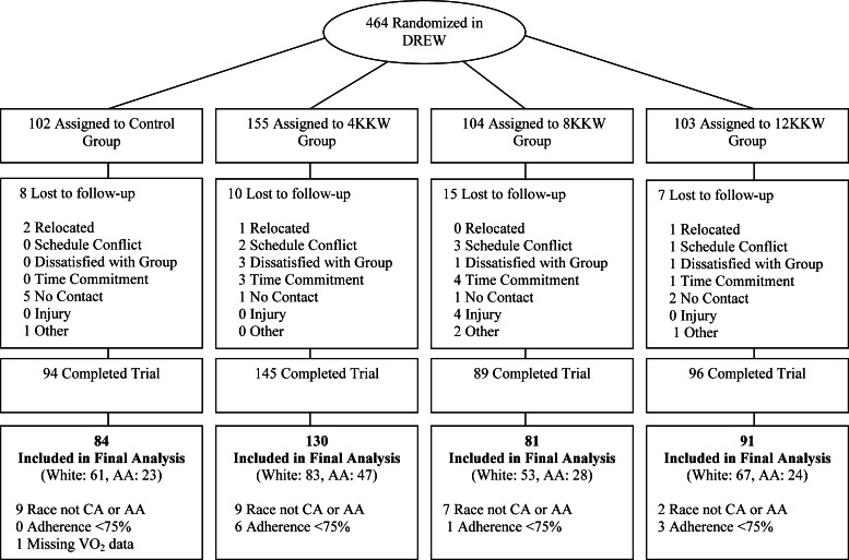
A consort diagram for the present study is shown in Fig. 1. We excluded women who were not CA or AA (n = 27), had low (≤75%) exercise-training adherence (n = 10), did not complete the trial (n = 40), or had missing exercise testing data (n = 1). Thus 264 CA and 122 AA postmenopausal women were in the analytical sample.
Fig. 1.
Consort diagram. DREW, Dose-Response to Exercise in Women trial; KKW, kcal·kg body wt−1·wk−1; AA, African American; CA, Caucasian American; VO2, maximal oxygen consumption.
Maximal Exercise Testing
Testing was performed using an Excalibur Sport electronically braked cycle ergometer (Lode, Groningen, the Netherlands). Participants cycled at 30 W for 2 min and then 50 W for 4 min, followed by increases of 20 W every 2 min until they could no longer maintain a pedal cadence of 50 revolutions/min. Respiratory gases were measured using a TrueMax 2400 metabolic measurement cart (ParvoMedics, Sandy, UT). CRF measures were calculated in relative (ml O2·kg−1·min−1) and absolute VO2 peak (l O2/min). Two fitness tests were performed on different days at baseline, and two tests were performed at follow-up. The average value for the two tests at each time point was used in the analyses, unless only one of the two tests was completed at baseline or follow-up, in which case, we used the single value. At baseline, 96% of participants completed both exercise tests. At follow-up, 95% of participants completed both exercise tests. The coefficient of variation for repeated measures of CRF (l/min) at baseline and follow-up were 5.2% and 4.7%, respectively.
Baseline Physical Activity
Baseline physical activity level was measured using an Eagle AE1620 (Accusplit, Livermore, CA) pedometer over the course of 1 wk prior to randomization.
Resting BP
Resting BP was evaluated with an automated BP unit (Colin Medical Instruments, San Antonio, TX) with the participant in the supine position.
Anthropometric Measures
Weight was measured using an electronic scale (Siemens Medical Solutions, Malvern, PA). Waist circumference was obtained following the guidelines of the Airlie Conference (22). Body mass index was calculated by dividing weight (kg) by height (m) squared.
Participant Randomization
Following baseline testing, participants were randomized to the 4-, 8-, or 12-kcal·kg body wt−1·wk−1 (KKW) or the nonexercise control group (25). The final analysis included 84 participants from the control group, 130 from the 4-KKW group, 81 from the 8-KKW group, and 91 from the 12-KKW group. The larger sample size in the 4-KKW group reflects a design feature of DREW that provided sufficient statistical power for detecting smaller anticipated changes compared with the 8- and 12-KKW groups following exercise training (25).
Exercise Training
We determined that the exercise-training energy expenditure for women in the DREW age range, associated with meeting the consensus public health guidelines, was ∼8 KKW (25). Based on this, we also determined the energy expenditure at 50% below (4 KKW) and 50% above (12 KKW) consensus public health guidelines. All groups expended 4 KKW during the 1st wk. Participants assigned to the 4-KKW treatment arm continued to expend 4 KKW for 6 mo. All of the other groups increased their energy expenditure by 1 KKW/wk until they reached the exercise dose required for their group (i.e., 8 KKW, 12 KKW). Aerobic training was performed on semirecumbent cycle ergometers and treadmills, and all sessions were supervised directly by study staff. Women in the exercise groups participated in three or four sessions/wk for 6 mo at a heart rate associated with 50% of peak VO2. Exercise training adherence was excellent in CA (99.2%) and AA (99.0%) participants. Participants in the nonexercise control group were asked to not alter their habitual physical activity level during the study.
Dietary Changes
Food frequency questionnaires were collected at baseline and follow-up in DREW to monitor dietary changes. No significant dietary changes were found following exercise training, which is reported in the DREW main outcomes paper (25).
Blinding
Separate intervention and assessment teams were maintained, and assessment staff members were blinded to the randomization of study participants.
Statistical Procedure
Statistical analyses were performed using SAS version 9.3 (SAS Institute, Cary, NC). Descriptive data were tabulated as means (SD) or frequencies (percent) as appropriate.
Baseline characteristics.
An ANOVA was performed to evaluate the significance of the racial difference in each baseline characteristic. Baseline differences between CA and AA women were found for age and maximum mean respiratory exchange ratio (RER) value from baseline exercise testing. Therefore, differences in baseline CRF between CA and AA were adjusted further for those variables.
Racial differences in the change in weight, RER, and maximum heart rate following training.
An analysis of covariance (ANCOVA) was used to evaluate the change in weight, RER, and maximum heart rate following exercise training with adjustment for baseline value. Adjusted means were reported as least squares means and 95% confidence intervals.
Evaluation for racial differences in the change in CRF.
Changes in CRF (assessed by VO2 peak) following exercise training between CA and AA postmenopausal women were compared in relative (ml O2·kg−1·min −1) and absolute terms (l/min): 1) across dose of exercise; 2) in exercisers only adjusted for exercise dose (total caloric energy expenditure during training sessions); and 3) in exercisers only adjusted for exercise dose in those that achieved a RER value >1.05 at baseline and follow-up. All linear statistical models were analyzed to compare baseline fitness and 6-mo changes in fitness with adjustment for covariates (age, baseline value, and maximum mean RER from baseline exercise test). Adjusted means were reported as least squares means and 95% confidence intervals. Statistical significance was defined as P ≤ 0.05.
Categorical analysis for change in CRF.
Women with change in relative CRF > 0 ml O2·kg−1·min −1 following exercise training were classified as relative CRF responders, and those with change in relative CRF ≤ 0 ml O2·kg−1·min−1 were classified as relative CRF nonresponders. Similarly, those with change in absolute CRF > 0 l O2/min following exercise training were classified as absolute CRF responders, and those with change in absolute CRF ≤ 0 l O2/min following exercise training were classified as absolute CRF nonresponders. A general linear model was used to compare CA with AA women with respect to the percent of responders in each study group in the entire study sample (n = 386).
Dose response to fitness.
Multiple linear regression analysis was used to test the significance of the trend of increasing CRF with higher doses of exercise (4 KKW, 8 KKW, and 12 KKW) for AA and CA participants. These analyses were performed only in the exercise groups.
Variance for change in CRF.
In exercisers only, linear regression models were used to estimate the percent of total variance for change in peak VO2 (relative and absolute) that may be attributed to race. Variables entered into the model included: exercise-training dose, age, baseline fitness, and race. Furthermore, within each training dose (4, 8, or 12 KKW) age, baseline fitness, and race were entered into regression models and analyzed analogously.
RESULTS
Baseline characteristics are summarized by race and exercise-training dose in Table 1. AA women were generally younger, had higher body weight, fasting glucose, and diastolic BP but lower waist circumference and hemoglobin values compared with CA women. The mean difference between races was not significant for systolic BP or baseline steps/day (all P > 0.05). No significant differences were observed in baseline characteristics across exercise groups or within either race group. Approximately 23% of women (both CA and AA) were taking antihypertensive medication, and 50% of CA and 32% of AA were taking hormone-replacement therapy.
Table 1.
Baseline characteristics stratified by race
| Caucasian | All (n = 264) | Control (n = 61) | 4 KKW (n = 83) | 8 KKW (n = 53) | 12 KKW (n = 67) |
|---|---|---|---|---|---|
| Age (yr) | 58.3 (6.3)* | 57.8 (6.1) | 59.5 (6.3) | 58.1 (6.3) | 57.5 (6.5) |
| Weight (kg) | 83.4 (11.6)* | 85.7 (12.9) | 82.5 (10.) | 85.1 (12.9) | 81.2 (10.5) |
| Body mass index (kg/m2) | 31.4 (3.9) | 32.2 (4.3) | 31.1 (3.4) | 32.0 (4.2) | 30.7 (3.7) |
| Waist circumference (cm) | 101.8 (11.7)* | 104.5 (12.5) | 100.1 (10.3) | 103.6 (10.3) | 100.2 (13.1) |
| Systolic blood pressure (mmHg) | 140.0 (12.9) | 141.9 (11.8) | 139.7 (11.9) | 140.2 (14.8) | 138.5 (13.5) |
| Diastolic blood pressure (mmHg) | 80.0 (8.6)* | 80.2 (8.1) | 79.5 (9.6) | 80.3 (8.1) | 80.1 (8.2) |
| Glucose (mg/dl) | 95.1 (9.4)* | 94.6 (13.1) | 95.0 (7.9) | 95.1 (7.5) | 95.7 (8.3) |
| Hemoglobin (g/dl) | 13.2 (0.9)* | 13.2 (0.8) | 13.2 (0.8) | 13.3 (1.0) | 13.1 (0.8) |
| Baseline physical activity (steps/day) | 4,930.4 (1759.5) | 5,101 (1685.9) | 4,824.3 (1,886.0) | 4,706.7 (1,634.5) | 5,082.1 (1,769.4) |
| Exercise testing data | |||||
| RER (VCO2/VO2) | 1.15 (0.07)* | 1.13 (0.06) | 1.15 (0.07) | 1.14 (0.07) | 1.16 (0.07) |
| Maximum heart rate (beats/min) | 151.8 (16.4) | 151.7 (15.2) | 150.2 (17.8) | 152.4 (16.3) | 152.9 (16.2) |
| Percentage of age-predicted maximum heart rate (%)† | 90.7 (9.2) | 90.6 (9.0) | 90.3 (9.8) | 91.0 (9.0) | 91.1 (8.9) |
| Exercise test max RER value >1.05 (%, n) | 92.1% (243)* | 93.4 (57) | 90.4 (75) | 92.5 (49) | 92.6 (62) |
| African American | All (n = 122) | Control (n = 23) | 4 KKW (n = 28) | 8 KKW (n = 28) | 12 KKW (n = 24) |
|---|---|---|---|---|---|
| Age (yr) | 54.8 (5.9) | 55.6 (5.2) | 55.4 (6.1) | 54.3 (6.8) | 53.4 (5.0) |
| Weight (kg) | 85.9 (11.9) | 87.1 (10.7) | 84.2 (13.0) | 85.8 (11.1) | 88.6 (11.5) |
| Body mass index (kg/m2) | 32.2 (3.6) | 32.4 (3.0) | 31.7 (4.0) | 32.8 (3.7) | 32.3 (3.0) |
| Waist circumference (cm) | 98.5 (11.2) | 98.3 (10.6) | 98.7 (12.2) | 99.6 (12.6) | 97.0 (8.3) |
| Systolic blood pressure (mmHg) | 138.8 (12.7) | 140.0 (11.3) | 139.0 (14.8) | 137.6 (11.1) | 138.9 (11.8) |
| Diastolic blood pressure (mmHg) | 83.2 (8.2) | 82.1 (8.1) | 83.4 (7.8) | 82.9 (7.9) | 84.1 (9.7) |
| Glucose (mg/dl) | 92.8 (10.8) | 95.8 (13.9) | 91.5 (9.3) | 92.3 (11.8) | 93.3 (9.3) |
| Hemoglobin (g/dl) | 12.6 (0.9) | 12.8 (0.6) | 12.6 (1.0) | 12.5 (0.08) | 12.6 (1.0) |
| Baseline physical activity (steps/day) | 5,087.4 (1,921.6) | 5,205.6 (1,412.0) | 4,873.8 (1,822.5) | 5,281.6 (2,177.6) | 5,139.2 (2,260.9) |
| Exercise testing data | |||||
| RER (VCO2/VO2) | 1.11 (0.07) | 1.10 (0.06) | 1.10 (0.08) | 1.12 (0.07) | 1.12 (0.07) |
| Maximum heart rate (beats/min) | 152.7 (15.8) | 152.0 (16.8) | 152.3 (15.5) | 152.4 (14.4) | 152.9 (17.5) |
| Percentage of age-predicted maximum heart rate (%)† | 89.9 (9.1) | 89.8 (8.8) | 90.1 (9.1) | 89.7 (9.0) | 89.7 (10.4) |
| Exercise test max RER value >1.05 (%, n) | 82.8 (101) | 78.3 (75) | 80.9 (38) | 85.7 (24) | 87.5 (21) |
Values are presented as mean (SD). One-way ANOVA was performed to test for significant differences among groups. KKW: kcal·kg body wt−1·wk−1; VCO2, elimination rate of carbon dioxide; VO2, maximal oxygen consumption; RER, respiratory exchange ratio.
Significantly different from African Americans;
percent of maximum heart rate calculated based on the equation from Tanaka et al. (33).
Baseline Fitness
Baseline maximal exercise tests revealed that compared with AA women, CA women, on average, had significantly higher relative CRF and maximum RER, but the racial differences in absolute CRF and maximum heart rate were not statistically significant. After adjustment for both age and maximum RER, AA women had significantly lower relative and absolute CRF compared with CA women (Table 2).
Table 2.
Evaluation for differences in baseline CRF between Caucasian and African American participants
| Caucasian | African American | P value | |
|---|---|---|---|
| Relative CRF (ml·kg−1·min−1) | |||
| Unadjusted model | 15.7 (CI: 15.5–16.1) | 15.1 (CI: 14.4–15.4) | 0.015 |
| Model adjusted for age | 15.9 (CI: 15.9–16.2) | 14.7 (CI: 14.3–15.2) | <0.001 |
| Model adjusted for age and maximum RER value | 15.8 (CI:15.5–16.1) | 15.0 (CI:14.6–15.5) | 0.011 |
| Absolute cardiorespiratory fitness (l/min) | |||
| Unadjusted model | 1.31 (CI: 1.28–1.34) | 1.28 (CI: 1.23–1.34) | 0.24 |
| Model adjusted for age | 1.33 (CI: 1.30–1.36) | 1.24 (CI: 1.20–1.28) | <0.001 |
| Model adjusted for age and maximum RER value | 1.33 (CI: 1.30–1.35) | 1.25 (CI:1.21–1.29) | 0.003 |
CRF, cardiorespiratory fitness; CI, confidence interval.
Weight, RER, and Maximum Heart Rate Outcomes
No significant race differences were observed in the change of weight, maximum RER, or maximum heart rate following exercise training (Table 3).
Table 3.
Change in weight, RER, and maximum heart rate following exercise training in Caucasian and African American women
| Caucasian | Control (n = 61) | 4 KKW (n = 83) | 8 KKW (n = 53) | 12 KKW (n = 67) |
|---|---|---|---|---|
| Δ Weight (kg) | −1.5 (CI: −2.4, −0.6) | −1.8 (CI: −2.6, −1.1) | −1.8 (CI: −2.7, −0.8) | −1.6 (CI: −2.5, −0.8) |
| Δ RER (VCO2/VO2) | −0.005 (CI: −0.013, 0.003) | −0.006 (CI: −0.013, 0.001) | −0.004 (CI: −0.013, 0.004) | −0.003 (CI: −0.011, 0.004) |
| Δ Maximum heart rate (beats/min) | −3.3 (CI:−5.5, −1.1) | −1.3 (CI: −3.2, 0.7) | −0.6 (CI: −3.0, 1.9) | −0.3 (CI: −2.4, 1.8) |
| African American | Control (n = 23) | 4 KKW (n = 28) | 8 KKW (n = 28) | 12 KKW (n = 24) |
|---|---|---|---|---|
| Δ Weight (kg) | −0.5 (CI: −2.0, 0.9) | −0.7 (CI: −1.7, 0.3) | −2.0 (CI: −3.3, −0.7) | −1.3 (CI: −2.7, 0.1) |
| Δ RER (VCO2/VO2) | −0.011 (CI: −0.024, 0.003) | −0.000 (CI: −0.010, 0.009) | −0.016 (CI: −0.028, −0.004) | −0.013 (CI: −0.026, 0.000) |
| Δ Maximum heart rate (beats/min) | −3.5 (CI: −7.2, 0.1) | −0.2 (CI: −2.8, 2.4) | 0.1 (CI: −3.3, 3.4) | −2.2 (CI: −6.0, 1.5) |
Δ, change. Results are reported as adjusted least squares means with 95% CIs.
Cardiorespiratory Fitness
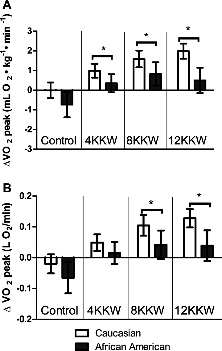
Mean changes in relative and absolute CRF following exercise training are shown by race in Fig. 2. CA women, on average, experienced significantly greater changes than AA women for relative VO2 peak in the 4-KKW (CA: 1.00 vs. AA: 0.35 ml O2·kg−1·min−1, P = 0.034), 8-KKW (CA: 1.59 vs. AA: 0.82 ml O2·kg−1·min−1, P = 0.041), and 12-KKW (CA: 1.98 vs. AA: 0.50 ml O2·kg−1·min−1, P = 0.001) groups. The race difference in mean change for relative VO2 peak approached statistical significance (P = 0.065) in the control group, with a larger decline occurring in AA women. The race differences did not vary significantly (P = 0.390) across exercise-training doses, indicating an absence of significant race by exercise-training dose interaction, whereas CA women demonstrated a more favorable main-effect change in relative VO2 peak after exercise training (P < 0.001).
Fig. 2.
Changes in cardiorespiratory fitness (CRF) in CA and AA postmenopausal women. Shown are the change in relative CRF (A) and absolute (B) CRF across doses of exercise training. Results are shown as adjusted least squares means with 95% confidence intervals. Analyses of covariance (ANCOVAs) are adjusted for baseline CRF, age, and baseline mean respiratory exchange ratio (RER) from baseline exercise testing. *Significant difference between CA and AA participants. ΔVO2, change in VO2.
CA women, on average, had a greater absolute VO2 peak in the 8-KKW (CA: 0.10 vs. AA: 0.04 l O2/min, P = 0.031) and the 12-KKW (CA: 0.13 vs. AA: 0.04 l O2/min, P = 0.003) groups but not in the 4-KKW (CA: 0.04 AA: 0.02, l O2/min, P = 0.147) group. The race difference for the change in absolute VO2 peak in the control group was not significant (P = 0.155). A significant main effect was observed for race (P = 0.001), with no significant race by exercise-training dose interaction (P = 0.486).
CRF (Adjusted for Exercise Dose)
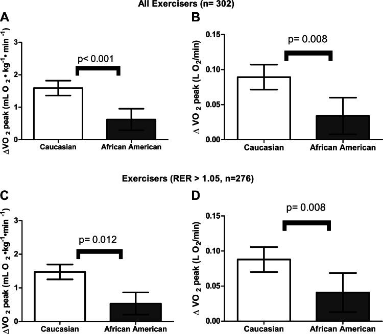
As shown in Fig. 3, CA women experienced greater improvements compared with AA women in relative and absolute CRF following exercise training (P < 0.05). Similar results were found when the analyses were restricted to women who had obtained a maximum RER value >1.05 at baseline and follow-up from exercise testing (n = 276).
Fig. 3.
Change in relative and absolute CRF in CA and AA postmenopausal exercisers (n = 302; A and B) and exercisers who obtained a RER value >1.05 at both baseline and follow-up (n = 276; C and D), adjusted for total energy expenditure during exercise training. Results are shown as adjusted least squares means with 95% confidence intervals. ANCOVAs are adjusted for baseline CRF, age, baseline mean RER from exercise testing, and total energy expenditure during 6 mo of exercise training.
Categorical Analysis for CRF
No significant race differences were observed in the percentage of participants who increased relative CRF within the control (45.9 vs. 43.5%, P = 0.822), 4-KKW (69.9 vs. 57.4%, P = 0.121), 8-KKW (86.8 vs. 82.1%, P = 0.651), and 12-KKW (85.1 vs. 75.0%, P = 0.336) groups. Similarly, race differences in the percentage of women who increased absolute CRF did not differ significantly within the control (37.7% vs. 30.4%, P = 0.519), 4-KKW (60.2 vs. 51.1%, P = 0.276), 8-KKW (75.4 vs. 78.6%, P = 0.773), and 12-KKW (82.1 vs. 70.8%, P = 0.305) groups.
Dose Response to CRF
Significant trends were observed for increasing CRF with higher exercise dose in relative fitness (P-trend < 0.001) and absolute (P-trend < 0.001) for CA women. In AA participants, no significant trend was observed for higher exercise dose and increasing CRF in either relative (P-trend = 0.62) or absolute (P-trend = 0.46) fitness. We excluded controls in this analysis, because the decrease in fitness in the AA control participants could lead to a spurious interpretation.
Percent Variance in the Change in CRF Explained by Race
The results of linear regression models (step-wise elimination) to determine the percent variance of CRF, explained by participant race, found that the coefficient of partial determination (partial r2) for race for the change in relative and absolute CRF in all exercisers was 3.8% and 3.6%, respectively. For change in relative CRF, the partial r2 for race was 1.6%, 7.2%, and 8.5% in the 4-KKW, 8-KKW, and 12-KKW groups, respectively. For change in absolute CRF, the partial r2 for race was 4.3% and 5.1% in the 8-KKW and 12-KKW groups, respectively, but was not significant in the 4-KKW group.
DISCUSSION
The primary findings of the present paper were: 1) at baseline, AA postmenopausal women had lower levels of CRF compared with CA women; 2) despite improvements in CRF in both CA and AA women, the overall magnitude of the response was generally greater in CA women; this relationship was evident after adjustment for potential confounders, such as age, baseline CRF level, and maximal RER from exercise testing; and 3) the percentage of responders to aerobic exercise training was similar in CA and AA participants, suggesting that exercise training is beneficial regardless of race. Our findings have clinical implications, as higher CRF reduces CVD and diabetes risk (2, 29), both of which are more prevalent in AA women (10, 21).
Our finding of lower baseline levels of CRF in AA compared with CA postmenopausal women is supported by large epidemiology studies (28, 30). The CARDIA study (n = 4,968) found that CRF [expressed as maximal estimated metabolic equivalents (METS) from exercise testing] was lower in young (18–30 yr) AA (9.4 METS) compared with CA (11.1 METS) women (30). Similarly, the Action for Health in Diabetes (Look AHEAD) study (n = 5,145) found that CRF was lower in AA (6.7 METS) compared with CA (7.3 METS) overweight and obese adults with type 2 diabetes (28). In our study, postmenopausal AA women had lower CRF, despite the fact that they were, on average, nearly 4 yr younger and had similar pedometer-determined physical activity levels at baseline compared with CA participants. Therefore, it is plausible that lower CRF in AA postmenopausal women may contribute to the racial disparities in risk of developing CVD and type 2 diabetes.
Following the 6 mo of aerobic training, postmenopausal AA women generally demonstrated an attenuated improvement in CRF compared with CA women, despite similar adherence to training regimens and adjustment for potential confounding factors. When CRF was expressed in relative terms, this racial difference was found in all exercise groups following the intervention. Similarly, when CRF was expressed in absolute terms, AA women demonstrated a lower change in CRF in the 8-KKW and 12-KKW groups compared with CA participants. When all exercise groups were combined, and statistical models were adjusted for the dose of exercise training, AA women demonstrated an attenuated improvement in CRF (both relative and absolute) compared with CA women following exercise training. According to Mora et al. (24), who evaluated the effect of CRF on mortality and CVD outcomes, a 1-MET increase in CRF was associated with a 17% reduction in 20-yr cardiovascular mortality risk in women. When CRF values are converted to METS, the racial difference in the change in fitness observed following exercise training at the various doses in the present study corresponds to a range of an approximate 3–7.2% reduction in cardiovascular mortality. Although the racial differences in the change in CRF observed in the present study are not large in magnitude, they may have some clinical impact, especially at higher exercise doses. In addition, the dose response of greater change in CRF with higher exercise dose was observed in CA women but not in AA women.
The HERITAGE Family Study found an attenuated improvement in relative CRF in AA (n = 198) compared with CA (n = 435), but racial differences in absolute CRF following 20 wk of aerobic training were not statistically significant. Nevertheless, the investigators concluded that race explained a small fraction of the variability in response of absolute CRF (∼1%) (6). In the present study, along with finding significant differences in absolute CRF between CA and AA participants, race explained ∼3.6% of the total variability in absolute CRF response among exercisers, with a greater influence within the higher exercise-training doses (8 KKW: 4.3%; 12 KKW: 5.1%). Differences in findings in absolute CRF between HERITAGE and DREW could be due to differences in study design (i.e., aerobic training intensity, study length, and exercise session duration). The age range and gender composition of both studies may also play a role, as HERITAGE included both men and women over a wide age range (17–65 yr), whereas DREW included postmenopausal women only. Additionally, 20% (n = 118) of The HERITAGE Family Study was composed of older adults (age ≥50 yr), of which only 13% (n = 15) were AA (31). Our data expand the findings of The HERITAGE Family Study by evaluating racial differences in CRF, specifically in postmenopausal women, which may be clinically significant, given the markedly increased CVD risk following menopause (13).
A potential mechanism that may explain the racial differences in CRF could be the higher proportion of type 1 muscle fibers observed in CA compared with AA individuals (1a, 32). Since type 1 muscle fibers are more oxidative than type 2 (32), this may contribute to the observed racial differences in baseline CRF and changes in CRF following exercise training. It could be speculated that another potential mechanism could be the lower hemoglobin levels in AAs compared with CA individuals, which have been shown in adolescents (27) and premenopausal women (12) and in the present study (Table 1). However, although lower hemoglobin levels could reduce oxygen transport to active musculature during aerobic exercise, the small differences observed in our study are unlikely to be clinically significant. Additionally, when baseline hemoglobin was included in our statistical models, it was not a significant covariate and did not alter our findings (data not shown).
Strengths of the present study are that DREW was a large, dose-response, randomized, controlled trial, and the training intervention was controlled for caloric energy expenditure. Additionally, CRF was measured twice at both baseline and follow-up with good reproducibility. Racial differences were evaluated at three doses of exercise training with similar exercise-training adherence between CA and AA women. Notable limitations are that the DREW study was not originally designed to specifically evaluate racial differences in CRF, there was a larger sample of CA compared with AA participants, and CRF values are not corrected for lean mass [since fat mass contributes little to oxygen uptake during exercise (19)]. Furthermore, it is possible that other results could be found with different exercise training intensities/modalities or in other subject populations.
In conclusion, the results of the present study suggest that AA postmenopausal women have lower CRF at baseline and an attenuated response to 6 mo of exercise training compared with CA women. However, it should be emphasized that although we found a racial difference in the magnitude of the change in CRF, the percentage of responders (increased CRF categorically) to exercise training was similar in both CA and AA women. Clinicians should strongly encourage adherence to exercise training in both CA and AA postmenopausal women due to the inverse relationship of CRF with CVD risk (15) and the increased risk of CVD following menopause (13). Future studies should evaluate whether racial differences exist in the response to CRF between CA and AA individuals in a prospective exercise-training study specifically designed to evaluate ethnic differences and investigate the physiologic mechanisms responsible for the attenuated response in AA individuals.
GRANTS
Support for this work was provided by the National Heart, Lung, and Blood Institute (Grant HL-66262) and unrestricted research support from The Coca-Cola Company. The salary and training of D. L. Swift are supported through a National Institute of Diabetes and Digestive and Kidney Diseases (T32 Postdoctoral Fellowship; Obesity: from genes to man; DK-64584-10).
DISCLOSURES
The authors have no conflict of interest to declare on the subject matter of the present paper.
AUTHOR CONTRIBUTIONS
Author contributions: D.L.S., N.M.J., C.J.L., C.P.E., T.S.C., and R.L.N. conception and design of research; C.P.E., S.N.B., and T.S.C. performed experiments; D.L.S., W.D.J., and R.L.N. analyzed data; D.L.S., N.M.J., C.J.L., C.P.E., W.D.J., S.N.B., T.S.C., and R.L.N. interpreted results of experiments; D.L.S. prepared figures; D.L.S., N.M.J., S.N.B., T.S.C., and R.L.N. drafted manuscript; D.L.S., N.M.J., C.J.L., C.P.E., W.D.J., S.N.B., T.S.C., and R.L.N. edited and revised manuscript; D.L.S., N.M.J., C.J.L., C.P.E., W.D.J., S.N.B., T.S.C., and R.L.N. approved final version of manuscript.
ACKNOWLEDGMENTS
This work was performed at The Cooper Institute, and the staff is especially commended for their efforts. We thank The Cooper Institute Scientific Advisory Board and the DREW participants.
Present address of C. P. Earnest: Dept. for Health, University of Bath, Bath, BA2 7AY, UK.
REFERENCES
- 1.Physical activity and cardiovascular health: NIH Consensus Development Panel on Physical Activity and Cardiovascular Health. JAMA 276: 241–246, 1996 [PubMed] [Google Scholar]
- 1a. Ama PF, Simoneau JA, Boulay MR, Serresse O, Theriault G, Bouchard C. Skeletal muscle characteristics in sedentary black and caucasian males. J Appl Physiol 61: 1758–1761, 1986 [DOI] [PubMed] [Google Scholar]
- 2. Blair SN, Kampert JB, Kohl HW, III, Barlow CE, Macera CA, Paffenbarger RS, Jr, Gibbons LW. Influences of cardiorespiratory fitness and other precursors on cardiovascular disease and all-cause mortality in men and women. JAMA 276: 205–210, 1996 [PubMed] [Google Scholar]
- 3. Blair SN, Kohl HW, III, Barlow CE, Paffenbarger RS, Jr, Gibbons LW, Macera CA. Changes in physical fitness and all-cause mortality: a prospective study of healthy and unhealthy men. JAMA 273: 1093–1098, 1995 [PubMed] [Google Scholar]
- 4. Blair SN, Kohl HW, III, Paffenbarger RS, Jr, Clark DG, Cooper KH, Gibbons LW. Physical fitness and all-cause mortality: a prospective study of healthy men and women. JAMA 262: 2395–2401, 1989 [DOI] [PubMed] [Google Scholar]
- 5. Bouchard C, Leon AS, Rao DC, Skinner JS, Wllmore JH, Gagnon J. Aims, design, and measurement protocol. Med Sci Sports Exerc 27: 721–729, 1995 [PubMed] [Google Scholar]
- 6. Bouchard C, Rankinen T. Individual differences in response to regular physical activity. Med Sci Sports Exerc 33: S446–S451, 2001 [DOI] [PubMed] [Google Scholar]
- 7. Church TS, Earnest CP, Skinner JS, Blair SN. Effects of different doses of physical activity on cardiorespiratory fitness among sedentary, overweight or obese postmenopausal women with elevated blood pressure. JAMA 297: 2081–2091, 2007 [DOI] [PubMed] [Google Scholar]
- 8. Duey WJ, O'Brien WL, Crutchfield AB, Brown LA, Williford HN, Sharff-Olson M. Effects of exercise training on aerobic fitness in African-American females. Ethn Dis 8: 306–311, 1998 [PubMed] [Google Scholar]
- 9. Estacio RO, Wolfel EE, Regensteiner JG, Jeffers B, Havranek EP, Savage S, Schrier RW. Effect of risk factors on exercise capacity in NIDDM. Diabetes 45: 79–85, 1996 [DOI] [PubMed] [Google Scholar]
- 10. Harris MI, Flegal KM, Cowie CC, Eberhardt MS, Goldstein DE, Little RR, Wiedmeyer HM, Byrd-Holt DD. Prevalence of diabetes, impaired fasting glucose, and impaired glucose tolerance in U.S. adults. The Third National Health and Nutrition Examination Survey, 1988–1994. Diabetes Care 21: 518–524, 1998 [DOI] [PubMed] [Google Scholar]
- 11. Hunter GR, Weinsier RL, Darnell BE, Zuckerman PA, Goran MI. Racial differences in energy expenditure and aerobic fitness in premenopausal women. Am J Clin Nutr 71: 500–506, 2000 [DOI] [PubMed] [Google Scholar]
- 12. Hunter GR, Weinsier RL, Mccarthy JP, Enette Larson-Meyer D, Newcomer BB. Hemoglobin, muscle oxidative capacity, and VO2 max in African-American and caucasian women. Med Sci Sports Exerc 33: 1739–1743, 2001 [DOI] [PubMed] [Google Scholar]
- 13. Kannel WB, Hjortland MC, McNamara PM, Gordon T. Menopause and risk of cardiovascular disease. Ann Intern Med 85: 447–452, 1976 [DOI] [PubMed] [Google Scholar]
- 14. Katzmarzyk PT, Church TS, Blair SN. Cardiorespiratory fitness attenuates the effects of the metabolic syndrome on all-cause and cardiovascular disease mortality in men. Arch Intern Med 164: 1092–1097, 2004 [DOI] [PubMed] [Google Scholar]
- 15. Kodama S, Saito K, Tanaka S, Maki M, Yachi Y, Asumi M, Sugawara A, Totsuka K, Shimano H, Ohashi Y, Yamada N, Sone H. Cardiorespiratory fitness as a quantitative predictor of all-cause mortality and cardiovascular events in healthy men and women. JAMA 301: 2024–2035, 2009 [DOI] [PubMed] [Google Scholar]
- 16. Kokkinos P, Myers J, Kokkinos JP, Pittaras A, Narayan P, Manolis A, Karasik P, Greenberg M, Papademetriou V, Singh S. Exercise capacity and mortality in black and white men. Circulation 117: 614–622, 2008 [DOI] [PubMed] [Google Scholar]
- 17. LaMonte MJ, Durstine JL, Yanowitz FG, Lim T, DuBose KD, Davis P, Ainsworth BE. Cardiorespiratory fitness and C-reactive protein among a tri-ethnic sample of women. Circulation 106: 403–406, 2002 [DOI] [PubMed] [Google Scholar]
- 18. Lavie CJ, Kuruvanka T, Milani RV, Prasad A, Ventura HO. Exercise capacity in adult African-Americans referred for exercise stress testing. Chest 126: 1962–1968, 2004 [DOI] [PubMed] [Google Scholar]
- 19. Lavie CJ, Milani RV. Metabolic equivalent (MET) inflation—not the MET we used to know. J Cardiopulm Rehabil Prev 27: 149–150, 2007 [DOI] [PubMed] [Google Scholar]
- 20. Lee CD, Blair SN, Jackson AS. Cardiorespiratory fitness, body composition, and all-cause and cardiovascular disease mortality in men. Am J Clin Nutr 69: 373–380, 1999 [DOI] [PubMed] [Google Scholar]
- 21. Lloyd-Jones D, Adams RJ, Brown TM, Carnethon M, Dai S, De Simone G, Ferguson TB, Ford E, Furie K, Gillespie C, Go A, Greenlund K, Haase N, Hailpern S, Ho PM, Howard V, Kissela B, Kittner S, Lackland D, Lisabeth L, Marelli A, McDermott MM, Meigs J, Mozaffarian D, Mussolino M, Nichol G, Roger VL, Rosamond W, Sacco R, Sorlie P, Stafford R, Thom T, Wasserthiel-Smoller S, Wong ND, Wylie-Rosett J, American Heart Association Statistics Committee and Stroke Statistics Subcommittee Heart disease and stroke statistics—2010 update. Circulation 121: e46–e215, 2010 [DOI] [PubMed] [Google Scholar]
- 22. Lohman TG, Roche AF, Martorell R. Antropometric Standardization Reference Manual. Champaign, IL: Human Kinetics Books, 1988 [Google Scholar]
- 23. Mensah GA, Mokdad AH, Ford ES, Greenlund KJ, Croft JB. State of disparities in cardiovascular health in the United States. Circulation 111: 1233–1241, 2005 [DOI] [PubMed] [Google Scholar]
- 24. Mora S, Redberg RF, Cui Y, Whiteman MK, Flaws JA, Sharrett AR, Blumenthal RS. Ability of exercise testing to predict cardiovascular and all-cause death in asymptomatic women: a 20-year follow-up of the lipid research clinics prevalence study. JAMA 290: 1600–1607, 2003 [DOI] [PubMed] [Google Scholar]
- 25. Morss GM, Jordan AN, Skinner JS, Dunn AL, Church TS, Earnest CP, Kampert JB, Jurca R, Blair SN. Dose-response to exercise in women aged 45–75 yr (DREW): design and rationale. Med Sci Sports Exerc 36: 336–344, 2004 [DOI] [PubMed] [Google Scholar]
- 27. Pivarnik JM, Bray MS, Hergenroeder AC, Hill RB, Wong WW. Ethnicity affects aerobic fitness in U.S. adolescent girls. Med Sci Sports Exerc 27: 1635–1638, 1995 [PubMed] [Google Scholar]
- 28. Ribisl PM, Lang W, Jaramillo SA, Jakicic JM, Stewart KJ, Bahnson J, Bright R, Curtis JF, Crow RS, Soberman JE. Exercise capacity and cardiovascular/metabolic characteristics of overweight and obese individuals with type 2 diabetes. Diabetes Care 30: 2679–2684, 2007 [DOI] [PubMed] [Google Scholar]
- 29. Sawada SS, Lee IM, Muto T, Matuszaki K, Blair SN. Cardiorespiratory fitness and the incidence of type 2 diabetes: prospective study of Japanese men. Diabetes Care 2003, 26: 2918–2922 [DOI] [PubMed] [Google Scholar]
- 30. Sidney S, Haskell WL, Crow R, Sternfeld B, Oberman A, Armstrong MA, Cutter GR, Jacobs DR, Savage PJ, Horn LV. Symptom-limited graded treadmill exercise testing in young adults in the CARDIA study. Med Sci Sports Exerc 24: 176–183, 1992 [PubMed] [Google Scholar]
- 31. Skinner JS, Jaskólski A, Jaskólska A, Krasnoff J, Gagnon J, Leon AS, Rao DC, Wilmore JH, Bouchard C. Age, sex, race, initial fitness, and response to training: The HERITAGE Family Study. J Appl Physiol 90: 1770–1776, 2001 [DOI] [PubMed] [Google Scholar]
- 32. Suminski RR, Mattern CO, Devor ST. Influence of racial origin and skeletal muscle properties on disease prevalence and physical performance. Sports Med 32: 667–673, 2002 [DOI] [PubMed] [Google Scholar]
- 33. Tanaka H, Monahan KD, Seals DR. Age-predicted maximal heart rate revisited. J Am Coll Cardiol 37: 153–156, 2001 [DOI] [PubMed] [Google Scholar]


