Sir,
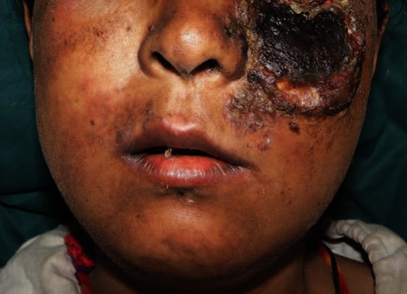
We report a case of a 28-year-old female who developed facial necrotizing fasciitis due to trivial trauma on face, which started with left-sided facial swelling, pain and fever. Later, overlying skin on that side of the face became black, showing areas of necrosis with yellowish slough and pus discharge [Figure 1]. There was neither any history of diabetes, hypertension, tuberculosis or autoimmune disorder nor any history of treatment with corticosteroids. On laboratory investigation, apart from hemoglobin of 9.6 mg% and total leukocyte count of 17000/cm with 80% polymorphs, other parameters were normal. Her enzyme-linked immunosorbent assay (ELISA) for human immunodeficiency virus (HIV) was negative. Chest X-ray, ultrasonogram of abdomen and computerized tomography (CT) scan of paranasal sinuses were normal. She underwent surgical debridement of necrotic tissue and irrigation with normal saline solution and hydrogen peroxide until bleeding tissue was encountered, after having been diagnosed with necrotizing fasciitis. Culture of debrided tissue was suggestive of Pseudomonas species sensitive to ciprofloxacin and was treated with daily dressing and injectable ciprofloxacin for 2 weeks. Considering the clinical picture and rapid development of fasciitis, antinuclear antibody (ANA) and antidouble-stranded DNA (anti-dsDNA) were done, which were strongly positive. We made a diagnosis of systemic lupus erythematosus (SLE) with necrotizing fasciitis. She was started with corticosteroids 1 mg per kg body weight. On follow-up, she was doing well with a maintenance dose of 10 mg per day.
Figure 1.

Left side of the face showing black necrotic tissue with pus discharge and slough at the margin
Necrotizing fasciitis is a rare, soft tissue infection, primarily involving the skin and superficial fascia, resulting in extensive undermining of the surrounding tissues. If untreated, it has high morbidity and mortality, and thus a high index of suspicion for the diagnosis is required.[1] The overlying skin is often erythematous, tense and tender, with three zones of demarcation: A wide peripheral zone of erythema outside a zone of tender purple skin surrounding a central black necrotic area.[2] A fetid odor signaling dead tissue is often present. Necrotizing fasciitis often affects patients with disrupted skin due to trauma, surgery, penetrating wounds or burns, but can also occur in previously healthy individuals with normal skin integrity.[3]
In our case, the lesion was rapidly progressive probably due to SLE, which was precipitated by trauma. Patients with SLE have increased risk of infection due to impaired delayed-type hypersensitivity responses (reflecting abnormal cell-mediated immunity), immunoglobulin deficiencies and various defects in other important immune effector cells, including neutrophils, macrophages and natural killer cells. Other factors such as abnormalities in the expression of complement receptors, complement factor deficiency and presence of the R131 variant of FCγRIIA on Fc receptor are also believed to contribute to the risk of infection.[4] Group A streptococcus is the most common bacteria found in cases of monomicrobial necrotizing fasciitis.[5] In our case, only Pseudomonas was cultured. The clinical presentation of patients with necrotizing fasciitis may be deceptively benign and, at onset, it may not be possible to clearly distinguish it from minor soft tissue infections. Initially, infection spreads widely along the subdermal fascial planes, with destruction occurring in the fascial and subcutaneous tissues, while little or no abnormality is evident in the skin. It is this relatively normal appearance of the skin that often causes delay in diagnosis. In our case also, there was a delay of 2 weeks between trauma and presentation. Later, occlusion of nutrient vessels results in ischemia, blistering and, eventually, gangrene. During the initial period, exploration of the wound may be necessary even before the diagnosis is clear, particularly in a toxic-appearing patient.[6] Given that an impaired immune system is a primary risk factor for developing necrotizing fasciitis and that patients with SLE are commonly immunosuppressed, it is perhaps surprising that this type of fasciitis has been reported only rarely with SLE. In this case report, it was trauma that precipitated necrotizing fasciitis.
References
- 1.Catena F, La Donna M, Ansaloni L, Agrusti S, Taffurelli M. Necrotizing fasciitis: A dramatic surgical emergency. Eur J Emerg Med. 2004;11:44–8. doi: 10.1097/00063110-200402000-00009. [DOI] [PubMed] [Google Scholar]
- 2.Mruthyunjaya B. Necrotizing faciitis: Report of case. J Oral Surg. 1981;39:60–2. [PubMed] [Google Scholar]
- 3.Jain S, Nagpure PS, Singh R, Garg D. Minor trauma triggering cervicofacial necrotizing fasciitis from odontogenic abscess. J Emerg Trauma Shock. 2008;1:114–8. doi: 10.4103/0974-2700.43197. [DOI] [PMC free article] [PubMed] [Google Scholar]
- 4.Yee AM, Ng SC, Sobel RE, Salmon JE. Fc gammaRIIA polymorphism as a risk factor for invasive pneumococcal infections in systemic lupus erythematosus. Arthritis Rheum. 1997;40:1180–2. doi: 10.1002/art.1780400626. [DOI] [PubMed] [Google Scholar]
- 5.Wong CH, Chang HC, Pasupathy S, Khin LW, Tan JL, Low CO. Necrotizing fasciitis: Clinical presentation, microbiology, and determinants of mortality. J Bone Joint Surg Am. 2003;85-A:1454–60. [PubMed] [Google Scholar]
- 6.Lille ST, Sato TT, Engrav LH, Foy H, Jurkovich GJ. Necrotizing soft tissue infections: Obstacles in diagnosis. J Am Coll Surg. 1996;182:7–11. [PubMed] [Google Scholar]


