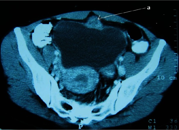
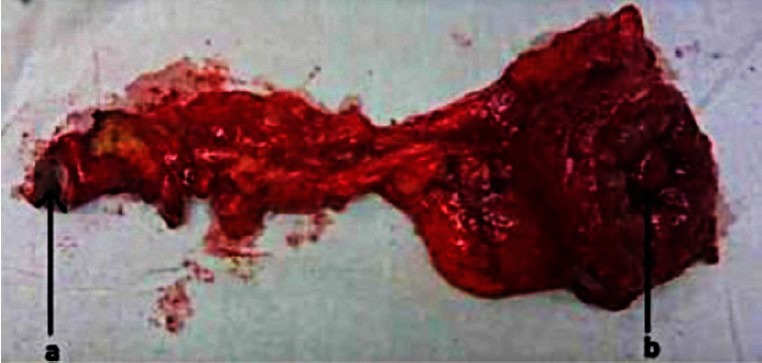
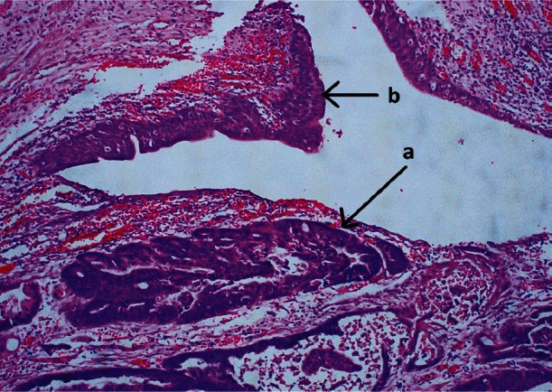
A 45 year old female patient presented with haematuria to Urology outpatient department in Colombo South Teaching Hospital, Dehiwela at Sri Lanka. The CT scan KUB showed a mass in the dome of the bladder suggestive of a urachal carcinoma due to its exophytic nature with minimal involvement of the bladder mucosa (Fig. 1). Partial cystectomy and resection of the urachus including the umbilicus (Fig. 2) showed an adenocarcinoma of enteric type (Fig. 3). The patient underwent adjuvant chemotherapy and is free of recurrences eight months after surgery.
Fig. 1.

CT KUB showing the urachal carcinoma (a).
Fig. 2.

The excised specimen with the umbilicus (a) and the tumour (b).
Fig. 3.

Urachal adenocarcinoma, intestinal type infiltrating the bladder wall (a) and the normal urothelium of the bladder (b).
Urachal carcinoma is a primary carcinoma derived from the urachal remnants. For a tumour to be classified as a urachal carcinoma, there must be a clear demarcation between the tumour and the adjacent bladder epithelium. A urachal tumour invading through the urothelium and extending into the bladder as in this patient, may be confused with a primary vesical carcinoma. In such patients, CT appearance of mainly an exophytic lesion is valuable in prompting the diagnosis of a urachal carcinoma facilitating appropriate surgery which includes removing the dome of the bladder, urachal ligament and the umbilicus. Leaving the umbilicus behind provides inadequate control and is associated with a higher risk of relapse.


