Abstract
The muscles of the leg are partitioned into three compartments (anterior, lateral, and posterior) by two intermuscular septa that have separate innervations. Anterior compartment is innervated by the deep peroneal nerve and lateral compartment is innervated by the superficial peroneal nerve. Common peroneal nerve divides into superficial and deep peroneal nerve at the neck of fibula. An unusual finding in the dividing pattern of the common peroneal nerve in the male cadaver on the left side was observed. This finding is of academic interest and clinical significance to the orthopedician operating on the proximal fibula for nerve decompression, high tibial osteotomy, and nerve transfer operations.
Keywords: Common peroneal nerve, deep peroneal nerve, osteotomy, variations
INTRODUCTION
Standard text books of anatomy and past research reports have described that the division of common peroneal nerve occurs at the neck of fibula.[1] Regional block of the superficial peroneal nerve, a branch of common peroneal nerve, allows for rapid anesthetization of the dorsum of the foot for various pathological conditions.[2] Usually surgeons are unaware of the higher division common peroneal nerve in the popliteal fossa. Such anomalies may confound clinical diagnosis or endanger the nerve during surgery or procedures such as surgical decompression of common peroneal nerve at the fibular head, percutaneous placement of wires in proximal tibia, high tibial osteotomy, and biopsy of proximal fibula.[3] The described anomaly is rare and it may be important to clinical practice.
CASE REPORT
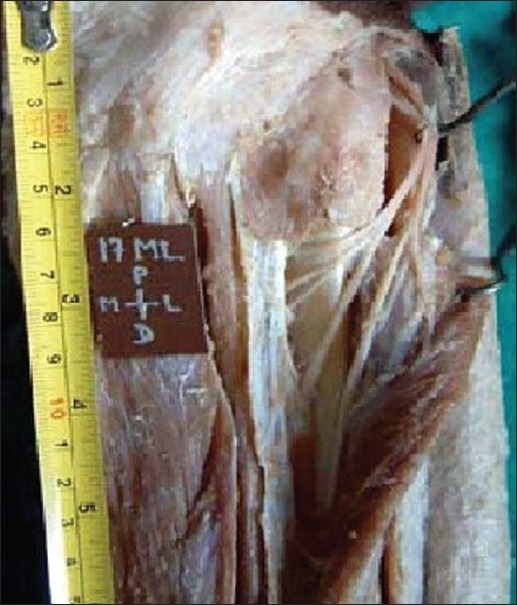
During routine cadaveric dissection of the lower limb in the Department of Anatomy, of a medical college, we observed an anomalous dividing pattern of the common peroneal nerve in the left leg of adult male cadaver. The age of the cadaver was unknown. It was observed that the common peroneal nerve was dividing in the popliteal fossa before reaching the fibular head. It divided into the superficial peroneal nerve and the deep peroneal nerve at the level of the middle of popliteal fossa, but the branches supplied crural muscles normally according to the text book pattern [Figure 1].
Figure 1.

Photograph of dissection of left leg of male cadaver showing higher division of common peroneal nerve.
DISCUSSION
Surgical procedures are commonly performed at the proximal end of fibula. Decompression of the common peroneal nerve at the fibular head is usually performed to release the fascia of the peroneus longus muscle. Understanding the anatomical distribution of the common peroneal nerve is helpful in performing a successful blockade of this nerve. Orthopedicians must be aware of higher division of common peroneal nerve while doing decompression surgery in proximal peroneal division sciatic entrapment neuropathy.[4] The findings of Saleh et al[5] suggest that the tibial nerve and common peroneal nerve leave the common synovial sheath at variable distances from the popliteal crease. Emergency practitioners and other clinicians working in acute care settings frequently encounter patients who have trauma to or pathology of the dorsum of the foot and require anesthesia for treatment and repair. Regional block of the superficial peroneal nerve allows for rapid anesthetization of the dorsum of the foot, which allows for management of lacerations, fractures, nail bed injuries, or other pathology involving the dorsum of the foot.[2]
Percutaneous placement of wires in the proximal fibula is gaining increased usage with the application of the techniques of Ilizarov, Monticelli, and Spinelli.[3] Biopsy of proximal fibula and division of fibula during high tibial osteotomy are also commonly performed procedures in which the topographical anatomy of common peroneal nerve is important.[6] Not many variations regarding the common peroneal nerve and the deep common peroneal nerve are documented in the standard anatomical textbooks. In the case, we report the common peroneal nerve dividing into superficial peroneal nerve and deep peroneal nerve at the level of the middle of popliteal fossa; and its knowledge is clinically relevant to surgeons operating on the proximal fibula in routine clinical practice.
ACKNOWLEDGMENT
Authors are grateful to the staff of Department of Anatomy GMC, Amritsar, for helping us in experimental study.
Footnotes
Source of Support: Nil.
Conflict of Interest: None declared.
REFERENCES
- 1.Sinnathamby CS. Last's Anatomy: Regional and Applied. Vol. 138. London: ELBS, Churchill Livingstone; 2001. pp. 140–1. [Google Scholar]
- 2.Tassone H, Silver MA, Raghavendra M. Nerve Block, Superficial Peroneal. Medscape Reference. [last cited on 2011 Apr 12]. Updated 2009 May: Available from: http://emedicine.medscape.com/article/83218-overview .
- 3.Stitgen SH, Cairns ER, Ebraheim NA, Neimann JM, Jackson WT. Anatomic consideration of pin placement in the proximal tibia and its relationship to the peroneal nerve. Clin Orthop Relat Res. 1992;278:134–7. [PubMed] [Google Scholar]
- 4.Congress of Neurological surgeons Neuro Wiki. Entrapment, peroneal nerve. Neuro Wiki. 2008. Apr, [Last cited on 2011 Apr 12]. Available from: http://wiki.cns.org/wiki/index.php/Entrapment,_Peroneal_Nerve .
- 5.Saleh HA, El-fark MM, Abdel GA. Anatomical variation of sciatic nerve division in the popliteal fossa and its implication in popliteal nerve blockade. Folia Morphol. 2009;68:256–9. [PubMed] [Google Scholar]
- 6.Soejima O, Ogata K, Ishinishi T, Fukahori Y, Miyauchi R. Anatomic considerations of the peroneal nerve for division of the fibula during high tibial osteotomy. Orthop Rev. 1994;23:244–7. [PubMed] [Google Scholar]


