

Abstract
Longstanding scaphoid nonunion, scaphoid malunion, and chronic scapholunate dissociation result in malalignment of the carpal bones, progressive carpal collapse, instability, and osteoarthritis of the wrist. The most commonly used procedures to treat scaphoid nonunion advanced collapse (SNAC) and scapholunate advanced collapse (SLAC) wrists are the four-corner fusion (4CF) and the proximal row carpectomy (PRC). Here we describe a different treatment option: radial styloidectomy and scaphocapitolunate (SCL) arthrodesis. This treatment option is chosen in an effort to maintain the joint contact surface and load transmission across the radiocarpal joint. Twenty patients were treated by the senior author (DLF) with this method with a mean follow-up of 4.6 years. Pain decreased in all patients, and 13 patients were pain-free postoperatively. The average Disabilities of the Arm, Shoulder, and Hand (DASH) scores decreased from 44 preoperatively to 23 postoperatively. One patient's course was complicated by nonunion, which was successfully treated with revision of the SCL arthrodesis. On follow-up radiographs, no patient had progressive osteoarthritis. This method preserves the normal ulnar-sided joints of the carpus, which are sacrificed during 4CF, and maintains a more physiologic joint surface for radiocarpal load sharing.
Keywords: SNAC, SLAC, scaphocapitolunate, wrist arthrodesis
Treatment of advanced osteoarthritis after longstanding scaphoid nonunion advanced collapse (SNAC) or chronically destabilized wrists from scapholunate advanced collapse (SLAC) is most commonly treated with four-corner fusion (4CF) or proximal row carpectomy (PRC).1,2,3,4 In each, the scaphoid is excised, reducing the cartilage contact area at the radiocarpal joint. This leads to an increased load across the lunate fossa, as the scaphoid fossa is no longer articulating with the carpus.5 Therefore, osteoarthritis is a risk, particularly in PRC.6 The rates of osteoarthritis in 4CF were recently reported at 1% by a meta-analysis,3 but only one of the eight studies included in the analysis had a greater than 5-year follow up. Additionally, two long-term studies of 4CF7,8 only follow clinical data and do not review radiologic data or report on the risk of developing progressive osteoarthritis. Therefore the rate of osteoarthritis, particularly in the long term, may be underestimated.
The scaphocapitolunate (SCL) arthrodesis is an alternate treatment option in which the majority of the scaphoid is maintained to maximize physiological loading across the radiocarpal joint, while the unaffected intact ulnar joints of the carpus remain mobile. This partial carpal fusion is performed with a radial styloidectomy as well as a partial resection of the distal scaphoid to address radial-sided osteoarthritic changes and painful radioscaphoid impingement. Although the clinical significance of progressive osteoarthritis is unknown, conceptually this procedure may be advantageous in that it maintains a more physiologic joint surface area. It is therefore an appealing treatment option, particularly in the young patient.
Etiology
SLAC is caused by a dissociation of the scaphoid and lunate, resulting in a predictable pattern of degenerative changes. This dissociation may be posttraumatic (scapholunate ligament injury), a consequence of inflammatory arthritis (particularly pseudogout), or iatrogenic if a portion of the ligament is excised (particularly after dorsal ganglion excision).
The subsequent rotatory subluxation of the scaphoid into a “flexed” position, over time, results in a degenerative arthrosis, as initially described by Watson.9 In SLAC Stage I, the distal scaphoid and radial styloid have the arthritic changes. In Stage II, the changes include the proximal radioscaphoid joint, and in Stage III, the midcarpal joint (capitolunate and scaphocapitate) is involved. The capitate may then migrate proximally between the lunate and scaphoid as the condition progresses. The lunate tilts dorsally after the uncoupling from the scaphoid, resulting in a dorsal intercalated segment instability (DISI) deformity. However, because the lunate remains congruous with the lunate facet even when dorsally tilted, the radiolunate joint is spared from arthritic changes (Fig. 1).
Fig. 1.
Posteroanterior and lateral views of a patient presented with a Stage II SLAC wrist after a failed ligament reconstruction. The DISI deformity is evident on the lateral view.
It is important to note that with Stage II and III SLAC wrists, although the proximal radioscaphoid joint is involved, in our experience the arthritis is often limited to the dorsal aspect of the scaphoid fossa and the portion of the flexed scaphoid with which it comes in contact. Thus, the normal articular surface of the scaphoid is spared, as is a large portion of the articulating aspect of the scaphoid fossa of the radius. It is because these articular surfaces remain intact despite other degenerative changes that this procedure is successful.
In SNAC wrists, the degenerative pattern is similar to SLAC wrists, involving the distal radioscaphoid joint at the radial styloid (Stage I), the proximal radioscaphoid joint (Stage II), and the midcarpal joint (Stage III) as described by Vender.10 However, the proximal aspect of the scaphoid is not involved with the degenerative process, as it is tethered to the lunate by the intact scapholunate ligament. Indeed, Moritomo et al confirmed preservation of the proximal radioscaphoid joint surface in SNAC wrists using three-dimensional computed tomography (CT) scans.11 Regardless of the location of the scaphoid fracture, the radiocarpal joint at the proximal scaphoid and lunate did not change significantly. The distal fragment of the scaphoid had degenerative changes with either the radial styloid or the dorsal lip of the radius depending on the fracture pattern. This supports the ability to preserve the scaphoid when treating SNAC wrists.
Diagnosis
SNAC and SLAC wrists are diagnosed clinically and radiographically. The patient may report a history of previous trauma, although often the patient does not recall a specific antecedent event. Some present after a failed previous intervention, such as scapholunate ligament repair or scaphoid fracture repair, although in our experience it is more common that the patient did not receive primary treatment for the wrist pathology. Occasionally, the patient presents with a secondary problem such as carpal tunnel syndrome, which can occur from a decreased volume of the carpal canal as a result of the degenerative wrist condition.
Examination
On examination, the patient usually has enlargement of the wrist secondary to synovitis and osteophyte formation. This is often accompanied by a decrease in the range of motion of the wrist, particularly extension. Tenderness may be elicited by applying pressure directly over the site of pathology. The resisted finger extension test is sensitive but not specific for proximal scaphoid pathology.12 In this test, pressure is applied to the proximal scaphoid to assess whether this results in pain. To perform the test, the patient's wrist is held in partial flexion while the patient extends the index and middle fingers against resistance. The Watson test, or scaphoid shift test, may also be useful but has limited application with advanced wrist degeneration.13
Relevant Imaging
Radiographic imaging is essential for diagnosis. We prefer four X-ray projections to evaluate the wrist: posteroanterior, lateral, supinated oblique at 45°, and pronated oblique at 45°. The images are evaluated for the patterns of arthrosis and degeneration as previously described.
Nonsurgical Treatment Options
Nonsurgical modalities include nonsteroidal anti-inflammatory drugs (NSAIDs), temporary immobilization, and intraarticular steroid injections. If these options fail, surgical treatment is discussed with the patient. Of note, when SNAC and SLAC wrists are diagnosed incidentally on X-ray in a patient without symptoms, no treatment is pursued.
Surgical Treatment and Technique
Patients are considered surgical candidates when they have significant pain and functional impairment in activities of daily living and during work after failing conservative modalities. Patients also need to have radiographic evidence of SLAC or SNAC degenerative changes in Stage II–III with a well-preserved proximal and central area of the scaphoid fossa in the radiocarpal joint. Exclusion criteria include significant cartilage loss of the scaphoid fossa with complete loss of the joint space, in which case a 4CF is performed.
The wrists are approached through a longitudinal 6–8 cm dorsal incision centered over the Lister tubercle. The wrist capsule is exposed in the interval between the third and fourth dorsal compartments, mobilizing the extensor pollicis longus radially. Following transection of the posterior interosseous nerve, the dorsal capsule is incised longitudinally in line with the skin incision at the level of the scapholunate interval extending distally to the carpometacarpal joint. Radial and ulnar capsular flaps are developed by incising the capsule transversely in an inverted t fashion, close to its radial attachment.
The cartilage of the scaphoid and radius is then inspected. If 90% of the cartilage of the scaphoid fossa is well preserved, we choose to keep the scaphoid in SLAC and SNAC wrists and proceed with the SCL arthrodesis. Otherwise, a 4CF or PRC is considered. Although arthritic changes involve the radioscaphoid joint, the changes are often localized to the dorsal aspect of the radius and the palmar aspect of the flexed scaphoid as previously discussed. In SLAC and SNAC degenerative patterns, the normal joint surface of the scaphoid is not in contact with the scaphoid fossa. Thus, by reducing the scaphoid into the sound part of the scaphoid fossa, we expect to have two surfaces of relatively normal cartilage contacting one another.
After inspection of the cartilage, the same incision is used to address the osteophytes in three areas: over the radial styloid, the dorsal distal radius, and the scaphoid. Osteophytes of the radial styloid are exposed and excised to prevent further impingement. The debridement of the radial styloid is limited to the area of osteophyte formation and bare bone at the level of the joint surface radially. Dorsal osteophytes of the distal radius, if present, are also removed. The scaphoid osteophytes are located at the volar and radial surface of the distal scaphoid abutting the radial styloid and are excised. The amount of resection depends on the magnitude of the osteophyte formation and the degenerative changes at the radial styloid and the most radial cartilage lining the scaphoid fossa. In cases of scaphoid nonunion, the distal portion of the scaphoid is removed if it is small; otherwise it is preserved and included in the arthrodesis. If the proximal pole of the scaphoid is small and avascular, we recommend resecting the proximal scaphoid and maintaining the distal pole by fusing it to the capitate, although we do not have personal experience with this situation.
Next, the carpal alignment is restored, correcting lunate and scaphoid rotation and the dorsally subluxed capitate. To achieve this, a 2 mm Kirschner wire (K-wire) is placed in the lunate and used as a “joystick” to correct this alignment (Fig. 2). Additional K-wires are then used to maintain the reduction temporarily, using a 1.6 mm K-wire first through the most ulnar and dorsal corner of the radius, then through the lunate into the capitate to maintain the alignment of the central carpal column. Next, the scaphoid is reduced out of its flexed deformity and pinned transversely to the capitate with two K-wires (Fig. 3). The articular cartilage and subchondral bone of the adjacent surfaces of the scaphoid, lunate, and capitate are then removed. The articular spaces are filled with cancellous bone graft. The arthrodesis is then stabilized with either Herbert screws (Zimmer, Warsaw, IN) or a Hub Cap plate (Acumed, Hillsboro, OR), a locking circular plate. Specifically, in SLAC cases, three Herbert screws are placed in a triangular configuration to stabilize the arthrodesis site: one spanning the scaphoid and capitate, one spanning the capitate and lunate, and one spanning the scaphoid and lunate. Alternatively, a circular plate is used for stabilization, and this practice is now our preference (Figs. 4, 5). In the SNAC cases where the distal scaphoid is preserved, the three Herbert screws are again used in a slightly different configuration: one spanning the capitate and lunate, one spanning the capitate and distal scaphoid, and a third spanning the proximal and distal scaphoid. In cases of SNAC where the distal scaphoid is removed, only two Herbert screws are used: one spanning the capitate and lunate and the other spanning the capitate and proximal scaphoid.
Fig. 2.

Intraoperative view of the same patient as in Fig. 1. Here, a 2 mm K-wire is placed in the lunate (left), and rotated as a “joystick” to reduce the lunate (right).
Fig. 3.

Intraoperative and fluoroscopic views of the temporary K-wire stabilization of the same patient as in Figs. 1, 2. The alignment of the central carpal column is maintained by driving a 1.6 mm K-wire through the most ulnar and dorsal corner of the radius through the lunate and into the capitate. Then the scaphoid is reduced out of its flexed deformity and pinned transversely across with two K-wires to the capitate.
Fig. 4.

Intraoperative view and immediate postoperative posteroanterior and lateral radiographs of the same patient as in Figs. 1–3. Distal radius bone graft was used for the SCL arthrodesis. The plate is placed after reducing and stabilizing the carpus (left). Postoperative radiographs demonstrate correction of the DISI deformity (right).
Fig. 5.

Schematic representation of the fixation options for SNAC and SLAC wrists undergoing SCL arthrodesis.
Postoperative Rehabilitation
Postoperatively, the patients are immobilized in a dorsal splint for 2 weeks, and after suture removal a short arm cast is applied for 5 weeks. After radiographic evidence of bony healing, range of motion and strengthening exercises are started under physiotherapy supervision.
Personal Outcomes and Literature Review
Degenerative osteoarthritis of the wrist secondary to SNAC or SLAC is most commonly treated with 4CF or PRC. These procedures excise the scaphoid to address arthritic degeneration and stabilize the wrist with either fusion of the midcarpal joint or excision of the remaining proximal row. SCL fusion with radial styloidectomy as described here involves a more limited excision of only the distal portion of the scaphoid and radial styloid, thus preserving the proximal scaphoid joint surface.
Twenty patients underwent SCL arthrodesis and radial styloidectomy by the senior author (DLF) from 1994 through 2010. The patients' ages ranged from 27 to 75 years old (average age, 62 years). Seven patients were treated for SNAC wrists, 12 patients for SLAC wrists, and one for degenerative joint disease following a transscapho-transcapitate perilunar dislocation. Sixteen patients had Herbert screw fixation (Figs. 6, 7), and four had circular plate fixation (Figs. 8, 9). All patients had autologous bone graft used for the arthrodesis. The mean follow-up was 4.6 years (range 2–9.6 years). Nineteen of twenty arthrodeses healed on an average of 9.6 weeks. One patient underwent a further operation 8 months after the initial operation with salvage of the SCL arthrodesis using a circular plate with an adequate result. Postoperatively, the mean active flexion-extension arc was 70° and the radioulnar deviation arc was 23°. Pain decreased in all patients, 13 of whom were pain-free postoperatively.14 The average postoperative Disabilities of the Arm, Shoulder, and Hand (DASH) score was 24 from 44 preoperatively (Table 1). Radiographically, neither radiolunate nor radioscaphoid arthritis was noted on follow-up, including the cases followed for more than 6 years.
Fig. 6.

Preoperative posteroanterior and lateral radiographs of a patient with Stage II SNAC wrist. This patient did not receive any previous treatment for his scaphoid fracure.
Fig. 7.

Posteroanterior and lateral radiographs 6.5 years after SCL arthrodesis and styloidectomy on the same patient as in Figure 6. Three Herbert screws were used for fixation. In addition to the capitolunate and scaphocapitate screws, the third screw spans the site of previous nonunion. The patient's radiocarpal joint space is maintained, and no progression of osteoarthritis is noted.
Fig. 8.

Posteroanterior and lateral radiographs of a patient presenting 25 years after a Matti-Russe procedure with a Stage III SLAC wrist.
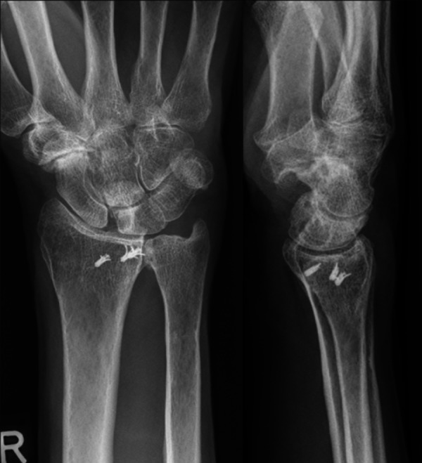
Fig. 9.

Radiographs 4.5 years after SCL arthrodesis and radial styloidectomy in the same patient as in Figure 8. A circular plate was used for fixation. The patient's radiocarpal joint space is maintained, and no progression of osteoarthritis is noted.
Table 1. Clinical and radiographic results of SCL arthrodesis.
| Preoperative | Postoperative | |
|---|---|---|
| Flexion (°) | 39 | 33 |
| Extension (°) | 42 | 37 |
| Flexion-extension arc (°) | 81 | 70 |
| Radial deviation (°) | 13 | 8 |
| Ulnar deviation (°) | 20 | 15 |
| Radioulnar deviation arc (°) | 33 | 23 |
| Capitolunate angle (°) | 22 | 2 |
| Grip strength (% opposite wrist) | 40 | 65 |
| Pain score (Fernandez) | 3.2 | 0.45 |
| DASH score | 44 | 24 |
Few studies have reported SCL fusion reported as a treatment for SLAC and SNAC wrists (Table 2).15,16,17 Overall, flexion-extension range of motion and grip strength were similar to the results obtained by the senior author. Pain was decreased in all series; however, it is important to note that differences exist between the operative techniques of these authors. Viegas excises the entire distal scaphoid, and in contrast to our technique, none of the authors describe systematic radial styloid excision, which may further decrease postoperative pain.
Table 2. Comparison of SCL arthrodesis results.
| No. of Cases | Healed | F/u Mo | Pain | ROM F/E | Strength (% contralateral) | |
|---|---|---|---|---|---|---|
| Rotman et al 1993 | 21 | 81% | 28 | Reduced-80% | 65° | 70% |
| Simmen and Bloch 1993 | 21 | 100% | 19 | Reduced-100% Pain free-81% |
57° | — |
| Viegas 1994 | 6 | 100% | 18 | Reduced-100% | 50% contralateral side | 50% |
Abbreviations: F/u, follow-up; Mo, months; ROM, range of motion; F/E, flexion/extension.
This procedure sacrifices some range of motion for a reduction in pain. Indeed, patients will have a better-functioning wrist if pain is controlled rather than restoring better wrist motion. Arthritic changes between the scaphoid and capitate, when present, are addressed with the arthrodesis. However, most of the arthritic changes in SNAC and SLAC wrists are at the distal radioscaphoid joint with impingement particularly on radial deviation. Because portions of the distal scaphoid and the radial styloid are resected, this impingement is eliminated. A limited excision of the radial styloid has been advocated to avoid detachment of the radiocarpal ligaments and destabilization of the wrist.18 Postoperative destabilization has not been observed in these patients despite a radial styloidectomy.
Excision of the entire scaphoid as performed in the PRC or 4CF may unnecessarily sacrifice the proximal scaphoid joint surface, particularly in SNAC wrists. Scaphoid excision in the 4CF or PRC reduces the cartilage contact area. This may lead to focal cartilage overload and, with time, the development of osteoarthritis of the wrist. A study of cadaveric wrists confirmed an increase in the mean total load in the lunate fossa after scaphoid excision and 4CF with no difference in the load across the triangular fibrocartilage complex (TFCC) area.5 Theoretically, degenerative osteoarthritis could occur as a consequence of the 4CF or PRC due to the smaller articular contact area. In contrast, an SCL fusion model demonstrated an increase in scaphoid contact areas by 42% accompanied by a slight decrease in the lunate contact area (by 12%).19 By preserving the proximal scaphoid, a more physiologic radiocarpal joint is maintained in SCL arthrodesis. Additionally, with respect to PRC, not only is the load increased across the lunate fossa as the only radiocarpal articulation, but the capitate articulation with the lunate fossa is also incongruous. X-ray analysis estimated the capitate to be 60% and 64% of the lunate fossa on anteroposterior (AP) and lateral views, respectively.20 Magnetic resonance imaging (MRI) evaluation of the capitate found the radius of curvature to be 37% of the lunate fossa in the coronal plane and 57% in the sagittal plane.21 Biomechanical evaluation of cadaveric wrists after PRC found the load to increase from 23.2 N/cm2 in a normal wrist at the radiolunate joint to 136.4 N/cm2 at the radiocapitate joint with an associated decrease in contact area from 2.08 cm2 to 0.30 cm2.19 Although the capitate is ∼60% of the diameter of the lunate fossa, the contact area was found to be only 14% of the lunate contact area. This may be a result of the four subfacets of the capitate, thus the capitate articular surface is not as smooth as that of the lunate.22
Although the combination of SCL arthrodesis and radial styloidectomy is an appealing treatment option in that it could theoretically prevent progressive degenerative changes of the radiocarpal joint by maintaining a more physiologic joint surface, this is difficult to ascertain, as no studies exist comparing SCL, 4CF, and PRC. Also, few studies exist with long-term follow up after PRC or 4CF.6,23,24,25
The early and midterm results of SCL have shown acceptable relief of pain and residual motion. Additionally, the senior author did not have to revise or convert any patient into a wrist fusion or a total wrist arthroplasty, suggesting that longer survival rate of this procedure may be possible. This procedure may be of particular use in the young patient.
References
- 1.Krakauer J D, Bishop A T, Cooney W P. Surgical treatment of scapholunate advanced collapse. J Hand Surg Am. 1994;19(5):751–759. doi: 10.1016/0363-5023(94)90178-3. [DOI] [PubMed] [Google Scholar]
- 2.Dacho A K, Baumeister S, Germann G, Sauerbier M. Comparison of proximal row carpectomy and midcarpal arthrodesis for the treatment of scaphoid nonunion advanced collapse (SNAC-wrist) and scapholunate advanced collapse (SLAC-wrist) in stage II. J Plast Reconstr Aesthet Surg. 2008;61(10):1210–1218. doi: 10.1016/j.bjps.2007.08.007. [DOI] [PubMed] [Google Scholar]
- 3.Mulford J S, Ceulemans L J, Nam D, Axelrod T S. Proximal row carpectomy vs four corner fusion for scapholunate (SLAC) or scaphoid nonunion advanced collapse (SNAC) wrists: a systematic review of outcomes. J Hand Surg Eur Vol. 2009;34(2):256–263. doi: 10.1177/1753193408100954. [DOI] [PubMed] [Google Scholar]
- 4.Bisneto E N, Freitas M C, Paula E J, Mattar R Jr, Zumiotti A V. Comparison between proximal row carpectomy and four-corner fusion for treating osteoarthrosis following carpal trauma: a prospective randomized study. Clinics (Sao Paulo) 2011;66(1):51–55. doi: 10.1590/S1807-59322011000100010. [DOI] [PMC free article] [PubMed] [Google Scholar]
- 5.Skie M, Grothaus M, Ciocanel D, Goel V. Scaphoid excision with four-corner fusion: a biomechanical study. Hand (NY) 2007;2(4):194–198. doi: 10.1007/s11552-007-9048-0. [DOI] [PMC free article] [PubMed] [Google Scholar]
- 6.Jebson P J, Hayes E P, Engber W D. Proximal row carpectomy: a minimum 10-year follow-up study. J Hand Surg Am. 2003;28(4):561–569. doi: 10.1016/s0363-5023(03)00248-x. [DOI] [PubMed] [Google Scholar]
- 7.Dacho A, Grundel J, Holle G, Germann G, Sauerbier M. Long-term results of midcarpal arthrodesis in the treatment of scaphoid nonunion advanced collapse (SNAC-wrist) and scapholunate advanced collapse (SLAC-wrist) Ann Plast Surg. 2006;56(2):139–144. doi: 10.1097/01.sap.0000194245.94684.54. [DOI] [PubMed] [Google Scholar]
- 8.Bain G I, Watts A C. The outcome of scaphoid excision and four-corner arthrodesis for advanced carpal collapse at a minimum of ten years. J Hand Surg Am. 2010;35(5):719–725. doi: 10.1016/j.jhsa.2010.01.025. [DOI] [PubMed] [Google Scholar]
- 9.Watson H K, Ballet F L. The SLAC wrist: scapholunate advanced collapse pattern of degenerative arthritis. J Hand Surg Am. 1984;9(3):358–365. doi: 10.1016/s0363-5023(84)80223-3. [DOI] [PubMed] [Google Scholar]
- 10.Vender M I, Watson H K, Wiener B D, Black D M. Degenerative change in symptomatic scaphoid nonunion. J Hand Surg Am. 1987;12(4):514–519. doi: 10.1016/s0363-5023(87)80198-3. [DOI] [PubMed] [Google Scholar]
- 11.Moritomo H, Viegas S F, Elder K W. et al. Scaphoid nonunions: a 3-dimensional analysis of patterns of deformity. J Hand Surg Am. 2000;25(3):520–528. doi: 10.1053/jhsu.2000.7381. [DOI] [PubMed] [Google Scholar]
- 12.Watson H K, Weinzweig J. Physical examination of the wrist. Hand Clin. 1997;13(1):17–34. [PubMed] [Google Scholar]
- 13.Watson H K, Ashmead D IV, Makhlouf M V. Examination of the scaphoid. J Hand Surg Am. 1988;13(5):657–660. doi: 10.1016/s0363-5023(88)80118-7. [DOI] [PubMed] [Google Scholar]
- 14.Fernandez D L. Radial osteotomy and Bowers arthroplasty for malunited fractures of the distal end of the radius. J Bone Joint Surg Am. 1988;70(10):1538–1551. [PubMed] [Google Scholar]
- 15.Rotman M B, Manske P R, Pruitt D L, Szerzinski J. Scaphocapitolunate arthrodesis. J Hand Surg Am. 1993;18(1):26–33. doi: 10.1016/0363-5023(93)90240-4. [DOI] [PubMed] [Google Scholar]
- 16.Simmen B R, Bloch H R. Partial arthrodesis of the carpal bones in advanced carpal collapse in chronic scapho-lunar instability and following scaphoid pseudoarthrosis [in German] Orthopade. 1993;22(1):79–85. [PubMed] [Google Scholar]
- 17.Viegas S F. Limited arthrodesis for scaphoid nonunion. J Hand Surg Am. 1994;19(1):127–133. doi: 10.1016/0363-5023(94)90236-4. [DOI] [PubMed] [Google Scholar]
- 18.Nakamura T, Cooney W P III, Lui W H. et al. Radial styloidectomy: a biomechanical study on stability of the wrist joint. J Hand Surg Am. 2001;26(1):85–93. doi: 10.1053/jhsu.2001.20963. [DOI] [PubMed] [Google Scholar]
- 19.Viegas S F, Patterson R M, Peterson P D. et al. Evaluation of the biomechanical efficacy of limited intercarpal fusions for the treatment of scapho-lunate dissociation. J Hand Surg Am. 1990;15(1):120–128. doi: 10.1016/s0363-5023(09)91118-2. [DOI] [PubMed] [Google Scholar]
- 20.Imbriglia J E, Broudy A S, Hagberg W C, McKernan D. Proximal row carpectomy: clinical evaluation. J Hand Surg Am. 1990;15(3):426–430. doi: 10.1016/0363-5023(90)90054-u. [DOI] [PubMed] [Google Scholar]
- 21.Hawkins-Rivers S, Budoff J E, Ismaily S K, Noble P C, Haddad J. MRI study of the capitate, lunate, and lunate fossa with relevance to proximal row carpectomy. J Hand Surg Am. 2008;33(6):841–849. doi: 10.1016/j.jhsa.2008.02.021. [DOI] [PubMed] [Google Scholar]
- 22.Zhu Y L, Xu Y Q, Ding J, Li J, Chen B, Ouyang Y F. Biomechanics of the wrist after proximal row carpectomy in cadavers. J Hand Surg Eur Vol. 2010;35(1):43–45. doi: 10.1177/1753193409344527. [DOI] [PubMed] [Google Scholar]
- 23.DiDonna M L, Kiefhaber T R, Stern P J. Proximal row carpectomy: study with a minimum of ten years of follow-up. J Bone Joint Surg Am. 2004;86-A(11):2359–2365. [PubMed] [Google Scholar]
- 24.Kitzinger H B, Löw S, Karle B, Lanz U, Krimmer H. The posttraumatic carpal collapse—long-term results after midcarpal fusion [in German] Handchir Mikrochir Plast Chir. 2003;35(5):282–287. doi: 10.1055/s-2003-43116. [DOI] [PubMed] [Google Scholar]
- 25.Ferreres A, Garcia-Elias M, Plaza R. Long-term results of lunocapitate arthrodesis with scaphoid excision for SLAC and SNAC wrists. J Hand Surg Eur Vol. 2009;34(5):603–608. doi: 10.1177/1753193409105683. [DOI] [PubMed] [Google Scholar]