

Abstract
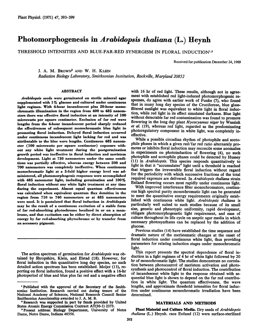
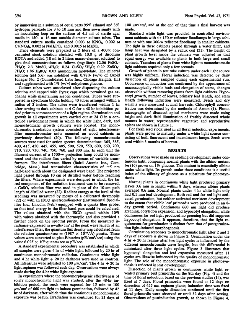
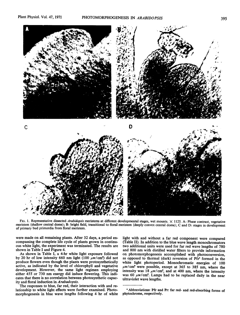
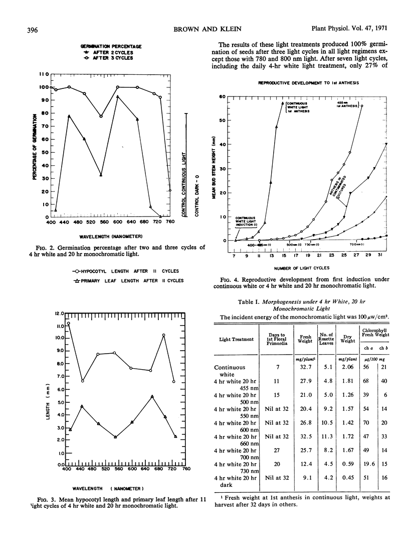
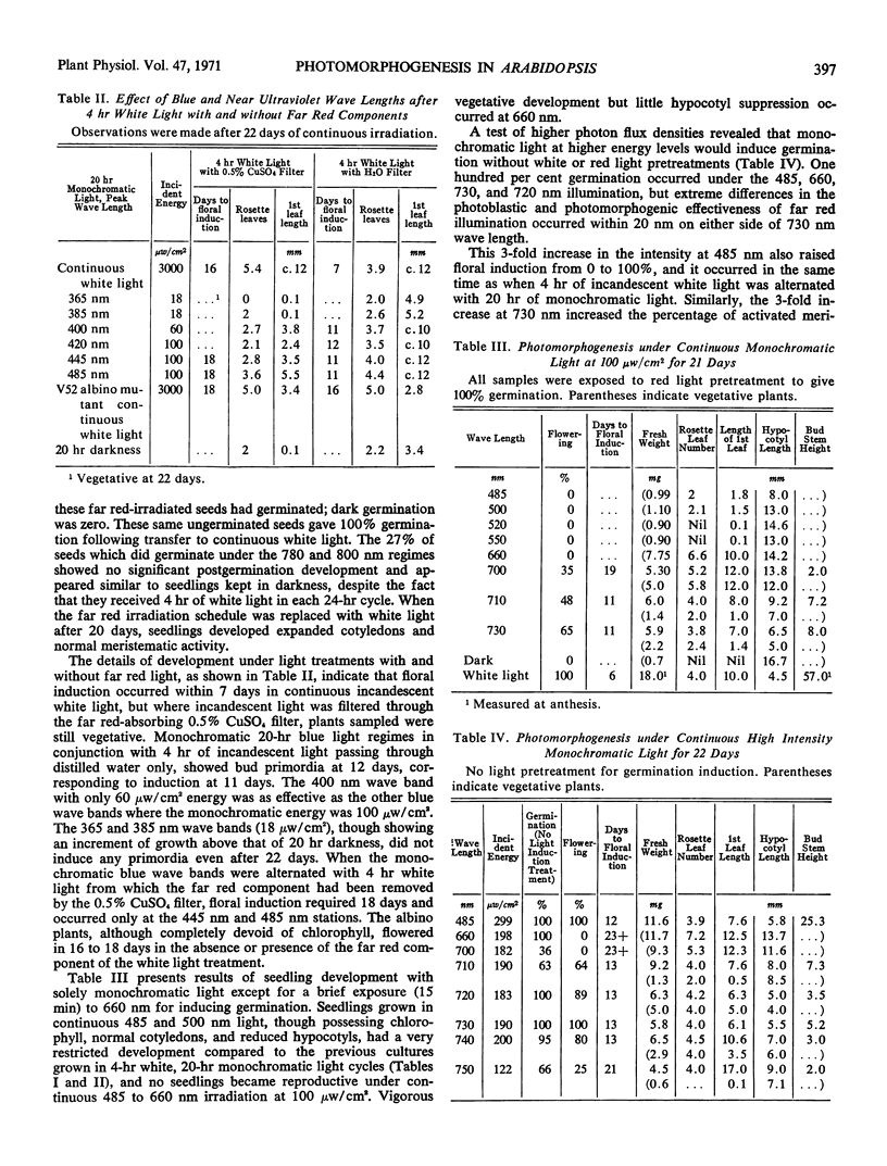
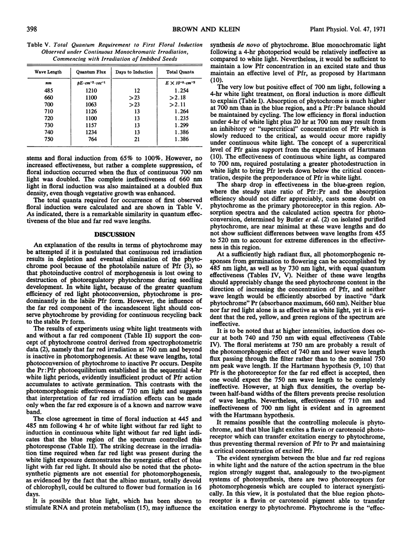
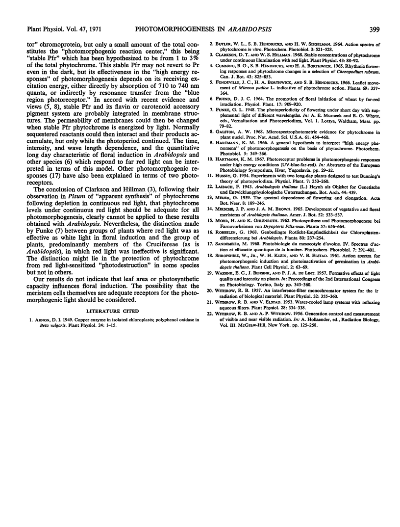
Arabidopsis seeds were germinated on sterile mineral agar supplemented with 1% glucose and cultured under continuous light regimes. With 4-hour incandescent plus 20-hour monochromatic illumination in the region from 400 to 485 nanometers there was effective floral induction at an intensity of 100 microwatts per square centimeter. Exclusion of far red wave lengths from the 4-hour incandescent period sharply reduced the effectiveness of subsequent monochromatic blue light in promoting floral induction. Delayed floral induction occurred under continuous incandescent light lacking far red and was attributable to the blue wave lengths. Continuous 485 nanometer (100 microwatts per square centimeter) exposure without any white light treatment during the postgermination growth period was ineffective in floral induction and meristem development. Light at 730 nanometers under the same conditions was partially effective, whereas energy between 500 and 700 nanometers was completely ineffective. When continuous monochromatic light at a 3-fold higher energy level was administered, all photomorphogenic responses were accomplished with 485 nanometer light, including germination and 100% floral induction without any white light treatment at any time during the experiment. Almost equal quantum effectiveness was calculated when equivalent quantum flux densities in the region from 710 to 740 nanometers or at 485 nanometers were used. It is postulated that floral induction in Arabidopsis may be the result of a continuous excitation of a stable form of far red-absorbing phytochrome localized in or on a membrane, and that excitation can be either by direct absorption of energy by far red-absorbing phytochrome or by transfer from an accessory pigment.
Full text
PDF
Images in this article
Selected References
These references are in PubMed. This may not be the complete list of references from this article.
- Arnon D. I. COPPER ENZYMES IN ISOLATED CHLOROPLASTS. POLYPHENOLOXIDASE IN BETA VULGARIS. Plant Physiol. 1949 Jan;24(1):1–15. doi: 10.1104/pp.24.1.1. [DOI] [PMC free article] [PubMed] [Google Scholar]
- Clarkson D. T., Hillman W. S. Stable concentrations of phytochrome in pisum under continuous illumination with red light. Plant Physiol. 1968 Jan;43(1):88–92. doi: 10.1104/pp.43.1.88. [DOI] [PMC free article] [PubMed] [Google Scholar]
- Galston A. W. Microspectrophotometric evidence for phytochrome in plant nuclei. Proc Natl Acad Sci U S A. 1968 Oct;61(2):454–460. doi: 10.1073/pnas.61.2.454. [DOI] [PMC free article] [PubMed] [Google Scholar]
- Withrow R. B. An Interference-Filter Monochromator System for the Irradiation of Biological Material. Plant Physiol. 1957 Jul;32(4):355–360. doi: 10.1104/pp.32.4.355. [DOI] [PMC free article] [PubMed] [Google Scholar]
- Withrow R. B., Elstad V. Water-cooled Lamp Systems with Refluxing Aqueous Filters. Plant Physiol. 1953 Apr;28(2):334–338. doi: 10.1104/pp.28.2.334. [DOI] [PMC free article] [PubMed] [Google Scholar]