Abstract
North America and the neotropics harbor nearly all species of plethodontid salamanders. In contrast, this family of caudate amphibians is represented in Europe and Asia by two genera, Speleomantes and Karsenia, which are confined to small geographic ranges. Compared to neotropical and North American plethodontids, mortality attributed to chytridiomycosis caused by Batrachochytrium dendrobatidis (Bd) has not been reported for European plethodontids, despite the established presence of Bd in their geographic distribution. We determined the extent to which Bd is present in populations of all eight species of European Speleomantes and show that Bd was undetectable in 921 skin swabs. We then compared the susceptibility of one of these species, Speleomantes strinatii, to experimental infection with a highly virulent isolate of Bd (BdGPL), and compared this to the susceptible species Alytes muletensis. Whereas the inoculated A. muletensis developed increasing Bd-loads over a 4-week period, none of five exposed S. strinatii were colonized by Bd beyond 2 weeks post inoculation. Finally, we determined the extent to which skin secretions of Speleomantes species are capable of killing Bd. Skin secretions of seven Speleomantes species showed pronounced killing activity against Bd over 24 hours. In conclusion, the absence of Bd in Speleomantes combined with resistance to experimental chytridiomycosis and highly efficient skin defenses indicate that the genus Speleomantes is a taxon unlikely to decline due to Bd.
Introduction
With more than 430 species, the family Plethodontidae comprises the majority of extant urodelan species and has experienced a marked evolutionary radiation in North, Central and northern South America [1]. In the rest of the world, this family is confined to the Maritime Alps, the central Apennine mountains in continental Italy and Sardinia in Europe (the genus Speleomantes, containing 8 species), and to South Korea (1 species, Karsenia koreana). The European plethodontids are closely related to the North American genus Hydromantes [1] and occupy an area well known to be infected by the amphibian pathogenic fungus Batrachochytrium dendrobatidis (Bd), one of the known drivers underlying global amphibian declines [2]–[6]. In Italy and France, the two countries where the genus Speleomantes occurs, the aggressive lineage of the pathogen, the Bd global panzootic lineage (BdGPL), also occurs [7]. Both infection and mortality due to Bd has been reported for both countries, including localities where Speleomantes sp. are endemic [3], [4], [6], [8]. Although habitat alteration has taken its toll on Speleomantes populations, enigmatic declines that would match chytridiomycosis driven declines witnessed elsewhere have not been reported. Indeed, these salamanders are among the most abundant vertebrates in suitable habitats [9]. The skin is of vital importance to plethodontid salamanders, which rely exclusively on cutaneous respiration. Chytridiomycosis dramatically disturbs the skin function [10], [11] and thus compromises respiration. Therefore, chytrid infections in Speleomantes should result in rapid killing of the plethodontid host, as has been hypothesized to be the case for several declining neotropical plethodontids and demonstrated in some, but not all [12], North American species [13]–[16]. Although different infection protocols have been used in these studies, they clearly demonstrate that some New World plethodontid species are easily colonized by the fungus.
Hitherto no suspected chytridiomycosis associated declines in Speleomantes have been observed, even in a region where chytridiomycosis occurs. This leads us to hypothesize first that prevalence of lethal cutaneous infections, such as infections by Bd, are low in European plethodontid salamanders. For this purpose, we determined to what extent Bd is present in populations of all eight species of Speleomantes.
Susceptibility to clinical chytridiomycosis, however, varies greatly among plethodontid species. In amphibian hosts, differences in host susceptibility have been attributed to the presence of fungicidal skin microbiota [17]–[19], antimicrobial peptides (reviewed in Rollins-Smith [20]), host genetics [21]–[23] and/or environmental factors (e.g. [8]), which may affect invasion of amphibian skin by the pathogen [24]. This leads us to hypothesize that resistance of European plethodontid salamanders is due to skin defenses that efficiently cope with Bd infection. Subsequently, we then examined susceptibility of Speleomantes strinatii to experimental infection with a global panzootic lineage strain of Bd (BdGPL isolate IA2011, [7]). Finally, we determined to what extent skin secretions of Speleomantes species are capable of killing Bd.
Materials and Methods
All animal experiments were conducted according to biosecurity and ethical guidelines and approved by the ethical committee of the Faculty of Veterinary Medicine, Ghent University (EC2011-073). All species involved in this study are protected as defined in Annex IV of the EU Habitats Directive (Council Directive 92/43/EEC on the Conservation of natural habitats and of wild fauna and flora). The entire experiment was submitted and approved by the Italian Ministry of Environment that issued permits to SS (issue numbers: DPN-2010-0010807 and PNM-2012-0007331). In Italy, state permits are valid over the entire country, since wildlife is a public property (national law 157/92). In addition however, when salamanders were sampled inside Protected Areas, local permits were also obtained from the “Parco Regionale Frasassi and Gola della Rossa” (permit number 3774/2012) the “Parco Regionale delle Alpi Apuane” (permit number DD.5/2012) and “Parco Regionale Alpi Marittime” (permit DD.165a/2011 issued to DO and FO). Permit PNM-2012-0007331 was also valid to capture animals (outside Protected Areas) to be used in experimental infections at Ghent University (EC2011-073). Since the study did not involve work on living animals in Italian laboratories, authorisation from the Italian Ministry of Health was not required.
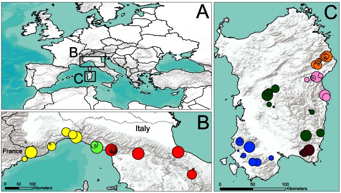
Between December 2004 and September 2012, we sampled 921 specimens including examples of all 8 recognized species of Speleomantes (Speleomantes ambrosii, S. flavus, S. genei, S. imperialis, S. italicus, S. sarrabusensis, S. strinatii, S. supramontis) at 65 localities in mainland Italy and southern France (351 samples) and Sardinia (570 samples) ( Fig. 1 , Table S1). Samples were collected by rubbing the abdomen, feet and the ventral side of the tail at least 10 times as has been described by Van Rooij et al. [25] using a rayon tipped swab (160 C, Copan Italia S.p.A., Brescia, Italy). The sex of the adults was recorded for the three continental species (S. italicus, S. ambrosii and S. strinatii) by checking for the presence of the typical male mental gland [9] and the ratio females: males: juveniles was approximately 1∶1∶1. DNA from the swabs was extracted in 100 µl PrepMan Ultra (Applied Biosystems, Foster City, CA, USA), according to Hyatt et al. [26]. DNA samples were diluted 1∶10 and quantitative PCR (qPCR) assays were performed in duplicate on a CFX96 Real Time System (BioRad Laboratories, Hercules, CA, USA). Amplification conditions, primer and probe concentrations were according to Boyle et al. [27]. Within each assay, 1 positive control sample containing Bd DNA from a naturally infected and deceased Costa Rican Eleutherodactylus sp. as template and 3 negative control samples with HPLC water as template were included. Samples were considered positive for Bd when a clear log-linear amplification was observed, when the number of genomic equivalents (GE) of Bd, defined as the measure of infection, was higher than the detection limit of 0.1 GE and when amplification that met both of the previous criteria was observed in both replicates. In case of conflict between both replicates of the same sample, the sample was run again in duplicate. To control and estimate inhibition, a subset of samples negative for the presence of Bd (n = 84) was retested under the same conditions as described above, but with an exogenous internal positive control (VIC™ probe, Life technologies, Austin, TX, USA) included as described by Hyatt et al. [26]. The Bayesian 95% credible interval for prevalence was estimated as described by Lötters et al. [28].
Figure 1. Sampling locations for Bd in Europe.
The boxed areas in the larger map of Europe (A) show the geographic locations. Expanded maps show the collection sites in southeastern France, mainland Italy (B) and Sardinia (C). For map (B), the colours represent the species Speleomantes strinatii (yellow), S. ambrosii (bright green) and S. italicus (red). For map (C), the colours represent the species S. genei (blue), S. sarrabusensis (purple), S. imperialis (dark green), S. supramontis (pink) and S. flavus (orange). Localities are indicated by symbols proportional to sample size.
We assayed susceptibility to chytridiomycosis in five male subadult S. strinatii by experimentally exposing them to a controlled dose of Bd. Ten captive bred juvenile Alytes muletensis were used as susceptible, positive control animals [29]. The animals were housed individually in plastic boxes (20×10×10 cm) lined with moist tissue, provided with PVC-tubes as shelter and kept at 18°C. Crickets were provided as food items ad libitum. All animals were sampled for the presence of Bd before inoculation using the method described above. A virulent isolate of the global panzootic lineage of Bd (BdGPL IA2011) [7], isolated in 2011 in the Spanish Pyrenees and capable of causing severe chytridiomycosis in urodelans was used in this study. All animals were exposed to a single dose of 1 ml of distilled water containing 105 zoospores/ml. Skin swabs were collected weekly for 4 weeks and processed as described above to determine the infection load for Bd each animal exhibited over the course of the experiment. After termination of the experiment, all animals were treated with voriconazole [30]. The S. strinatii specimens are still kept following strict biosecurity guidelines at the clinic for Exotic Animals and Avian Diseases (Faculty of Veterinary Medicine, Ghent University) for further follow-up.
To determine the extent to which skin secretions of Speleomantes are capable of killing Bd zoospores, skin secretions were collected non-invasively from wild individuals of 7 of the 8 Speleomantes species (Table S2) and processed within 1 h (S. strinatii), 48 h (S. ambrosii, S. italicus, S. flavus, S. supramontis, S. genei) or 72 h (S. sarrabusensis). For this purpose, a microbiological inoculating loop was gently rubbed over the dorsal tail until white skin secretions accumulated on the loop. Prior to sampling sterile loops were cut off, stored individually in sterile vials and the total weight of each vial was determined. Collected skin secretions were weighed to the nearest 0.1 mg by subtracting the weight of the vial and the inoculation loop from the total weight. Varying amounts of skin secretions were collected per Speleomantes specimen, ranging from 3.1 to 32.7 mg. Collected skin secretions were not further diluted prior exposure to Bd zoospores. Loops with secretions were incubated in a zoospore suspension. To keep the ratio between the amount of skin secretions and Bd zoospores added constant, 10 µl of zoospore suspension containing 106 zoospores/ml distilled water was added per mg skin secretion. Samples were incubated for 24 h at 20°C. At 0 and 24 h of incubation, the number of viable zoospores was assessed using qPCR on the zoospores that were pretreated with ethidium monoazide (EMA, Sigma-Aldrich Inc., Bornem, Belgium) as described by and validated in Blooi et al. [31]. Viable/death differentiation is obtained by covalent binding of EMA to DNA in dead Bd by photoactivation. EMA penetrates only dead Bd with compromised membranes and DNA covalently bound to EMA cannot be PCR amplified [32]. In brief, at 0 and 24 h of incubation a 5 µl aliquot was taken from each sample, transferred into a 24-well plate and 195 µl TGhL broth (tryptone, gelatin hydrolysate, lactose) was added to each well for its protective effect on viable Bd zoospores during EMA treatment. Negative controls for skin secretion activity consisted of zoospore suspensions not exposed to skin secretions in order to quantify the ‘natural’ loss of viability in Bd zoospores, while positive controls were heat-killed zoospores. Five µl of a 1 mg/ml stock solution of EMA in dimethyl formamide was added to 200 µl zoospore suspension in TGhL broth to obtain a final concentration of 25 µg/ml EMA, incubated for 10 minutes, protected from light and exposed to a 500 W halogen light at 20 cm distance for 5 minutes. Then, samples were washed by centrifugation (5000 rpm, 5 min, 20°C), the supernatant was discarded and the pellet was suspended in HPLC water. In parallel, the total amount of Bd zoospores in all samples, including controls, was enumerated using exactly the same procedure as described above, only 5 µl HPLC water was added to each sample instead of EMA. DNA extraction and qPCR were then done as described above. Killing activity was expressed as log(10) reduction of viable spores in a given sample compared to the negative controls.
Results
None of the 921 skin swabs collected from any Speleomantes sp. tested positive for Bd. The Bayesian 95% credible interval for the observed prevalence of 0% is (0.0000, 0.0040). However, in 12 out of 84 samples tested (14%) PCR-inhibition occurred that could not be abolished by diluting the samples 1/100. Extrapolated to the total number of individual salamanders tested, this reduces the reliable number of Bd-negative salamanders to 789 with a corresponding 95% credible interval of (0.0000, 0.0047).
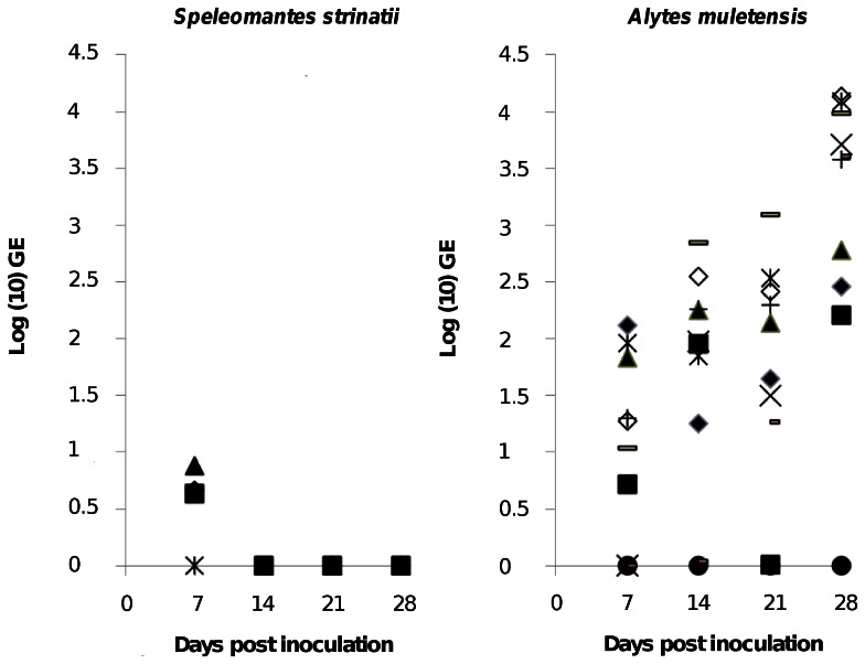
None of the experimental animals tested positive for Bd prior to the exposure. Over the four week infection period, 9/10 Alytes muletensis developed marked infection with increasing Bd loads. Of the 5 inoculated Speleomantes strinatii, 3 animals exhibited weak infections (small GE value) at 7 days post infection (dpi) with an average of 5.5±1.8 GE per swab. At 14 dpi, one salamander was borderline positive (0.2 GE per swab) but at 21 and 28 dpi all salamanders tested negative for Bd. In contrast, A. muletensis exhibited median GE counts of 91 at 21 dpi and 3920 at 28 dpi ( Fig. 2 ). No clinical signs were noticed in the infected salamanders. For animal welfare reasons, A. muletensis were treated at 4 weeks pi using voriconazole to clear infection, as described by Martel et al. [30].
Figure 2. Experimental infection of Speleomantes strinatii with Bd.
Infection loads are represented as log (10) genomic equivalents (GE) of Bd in skin swabs from Speleomantes strinatii (left panel) and compared with Alytes muletensis (right panel) serving as positive control animals, up to four weeks post experimental inoculation with Bd. Each symbol represents an individual animal.
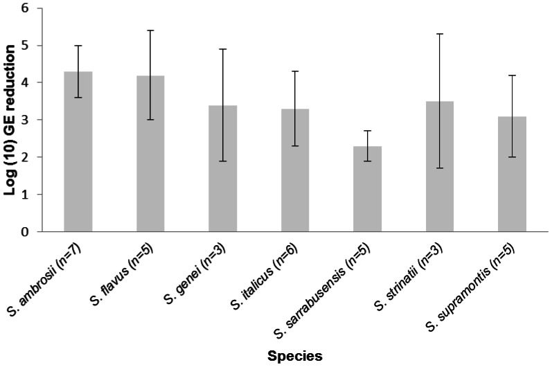
Skin secretions of all Speleomantes species were capable of efficiently killing Bd zoospores ( Fig. 3 ). Exposure to skin secretions resulted in a 200 to 20000 fold reduction of the number of viable spores within 24 h post exposure.
Figure 3. Killing activity of skin secretions of Speleomantes species against Bd.
Killing activity of Speleomantes skin secretions at physiological concentrations is expressed as log(10) viable spores of Bd added to the skin secretions –log(10) viable spores recovered 24 h later. Results are presented as mean genomic equivalents of Bd (GE) ± standard error (SEM); n = sample size.
Discussion
Bd infections appear to be highly uncommon, if not absent, in adult and juvenile Speleomantes of all species and throughout their range. This observation is strengthened by the recent publication of Chiari et al. [33], reporting absence of Bd in Sardanian S. flavus, S. genei, S. imperialis, S. sarrabusensis and S. supramontis (n = 143). The genus Speleomantes occupies an ecological niche highly suitable to Bd colonization, persistence and spread due to relatively low preferred body temperatures (<18.5°C, reviewed by Lanza et al. [9]) and higher humidity environments. Contacts that would facilitate interspecific transmission are also common because at some locations salamanders occur outside of caves and are found under retreat sites with other species that are capable of carrying infections (FP personal observations, see also [3], [4], [6]). Intraspecific transmission would also be highly likely, as courtship involves intimate contact, salamanders crowd together in summer retreats, and juveniles also exhibit highly aggregated distribution patterns at certain times of the year [34]. Thus, substantial opportunity exists for Bd for introduction into Speleomantes populations and to amplify rapidly once introduced, but neither of these seems to have occurred to any significant degree. Field studies of other amphibian species have reported low prevalence or absence of detectable infection in species that show seasonal fluctuations in prevalence 35–37. We find this an unlikely explanation for the observed absence of infection. Moreover, for the present study samples were taken from December to September and over multiple years. Seasonally mediated fluctuations are associated with strong variation in environmental metrics that influence Bd growth and reproduction [35], [36], [38] suggesting that more stable environments should result in more consistent patterns of infection (see [39]). However, recent evidence suggests that variable temperatures may be most favorable for Bd driven declines, probably due to temperature drop induced zoospore release [40]. Variation of prevalence should be minimized in the cave-dwelling Speleomantes sp., where relatively stable cave temperatures and moisture regimes should buffer against environmentally mediated changes in prevalence. Further, our experimental results indicate the infection is unlikely in at least one species and the majority of Speleomantes species are equipped with the tools to resist infection.
Exposing S. strinatii to a highly virulent Bd strain and in a manner that resulted in potentially lethal infection in a susceptible host did not result in persistent infection of the salamanders. It is possible that the low GE values detected until two weeks post inoculation in S. strinatii represent dead Bd cells or some form of Bd DNA contamination rather than active infection. Thus it is possible that S. strinatii is extremely efficient at blocking epidermal colonization by Bd even when exposed to a highly concentrated and strong dose of Bd zoospores, but our experimental design prevents us from distinguishing between this and rapid clearing of infection. Since successful epidermal colonization by Bd requires keratinocyte invasion [22], [41], [42], we hypothesized that Speleomantes skin contains highly effective fungicidal properties that prevent skin invasion. Indeed, we showed that Speleomantes skin secretions were very efficient in killing Bd zoospores as assessed using a recently developed and highly reproducible assay [31]. Factors present in the skin secretions that account for the observed Bd killing need further identification but probably include antimicrobial peptides (AMP) [43]–[45] and/or bacterially produced metabolites [17], [18], [46]. AMPs that play a defensive role against invasion by pathogenic microorganisms have been described for other Ambystomidae and plethodontid species [47]–[49] but not characterized. Hitherto, only the antifungal metabolites 2,4-diacetylphloroglucinol, indol-3-carboxaldehyde and violacein have been identified that are secreted by symbiotic bacteria residing on the skin of plethodontid species Plethodon cinereus and Hemidactylium scutatum [17], [46]. Moreover, these metabolites may work synergistically with AMPs to inhibit colonization of the skin by Bd [50]. Further characterization of such AMPs and the composition of microbial skin communities, combined with the study of their assessment in plethodontid species and/or populations can open new perspectives for further understanding factors mediating resistance towards chytridiomycosis, its control and mitigation. In addition, a study of microbial skin communities has the potential to direct probiotic conservation strategies for susceptible species in the area.
The apparent absence of Bd and chytridiomycosis driven declines in Speleomantes throughout their range, lack of colonization or sustained infection in experimentally infected animals and pronounced Bd killing capacity of Speleomantes skin secretions together suggest the genus Speleomantes to be refractory to Bd infection and thus resistant to chytridiomycosis. Resistance to chytridiomycosis would at least in part explain the localized persistence of the genus Speleomantes in the presence of highly virulent BdGPL strains in Europe. This situation differs markedly from some of the plethodontids in North America that are susceptible to infection and those in the Neotropics that underwent recent and sharp chytridiomycosis driven declines upon the arrival of Bd [15], [16], [51]–[55]. While our results are preliminary evidence that the genus Speleomantes is a low-risk taxon for decline due to chytridiomycosis, we recommend additional studies that further investigate the risk Bd may pose to this unique amphibian taxon.
Supporting Information
Overview of the sampled Speleomantes species, sampling localities, sample size and sampling dates. Seconds have been removed from coordinates to prevent illegal collection.
(DOCX)
Overview of the sampled Speleomantes species for collection of skin secretions and respective sampling localities. Seconds have been removed from coordinates to prevent illegal collection
(DOCX)
Acknowledgments
We thank in particular Paolo Casale, Giacomo Bruni, Sandro Casali, David Fiacchini, Carlo Torricelli and Olivier Gerriet for field assistance.
Funding Statement
Funding for this work was provided to PVR by research grant BOF08/24J/004 from Ghent University and to MB by a Dehousse grant from the Royal Zoological Society of Antwerp, Belgium. No additional external funding was received for this study. The funders had no role in study design, data collection and analysis, decision to publish, or preparation of the manuscript.
References
- 1. Vieites DR, Nieto Román S, Wake MH, Wake DB (2011) A multigenic perspective on phylogenetic relationships in the largest family of salamanders, the Plethodontidae. Mol Phylog Evol 59: 623–635. [DOI] [PubMed] [Google Scholar]
- 2. Garner TWJ, Walker S, Bosch J, Hyatt AD, Cunningham AA, et al. (2005) Chytrid fungus in Europe. Emerg Infect Dis 11: 1639–1641. [DOI] [PMC free article] [PubMed] [Google Scholar]
- 3. Bovero S, Sotgiu G, Angelini C, Doglio S, Gazzaniga E, et al. (2008) Detection of chytridiomycosis caused by Batrachochytrium dendrobatidis in the endangered Sardinian newt (Euproctus platycephalus) in southern Sardinia, Italy. J Wildl Dis 44: 712–715. [DOI] [PubMed] [Google Scholar]
- 4. Bielby J, Bovero S, Sotgiu G, Tessa G, Favelli M, et al. (2009) Fatal chytridiomycosis in the Tyrrhenian painted frog. Ecohealth 6: 27–32. [DOI] [PubMed] [Google Scholar]
- 5. Fisher MC, Garner TWJ, Walker SF (2009) Global Emergence of Batrachochytrium dendrobatidis and amphibian chytridiomycosis in space, time, and host. Ann Rev Microbiol 63: 291–310. [DOI] [PubMed] [Google Scholar]
- 6.Tessa G, Angelini C, Bielby J, Bovero S, Giacona C, et al. (2012) The pandemic pathogen of amphibians, Batrachochytrium dendrobatidis, in Italy. Ital J Zool. In press.
- 7. Farrer RA, Weinert LA, Bielby J, Garner TWJ, Balloux F, et al. (2011) Multiple emergences of genetically diverse amphibian infecting chytrids include a globalized hypervirulent recombinant lineage. Proc Natl Acad Sci USA 108: 18732–18736. [DOI] [PMC free article] [PubMed] [Google Scholar]
- 8. Walker SF, Bosch J, Gomez V, Garner TWJ, Cunningham AA, et al. (2010) Factors driving pathogenicity vs. prevalence of amphibian panzootic chytridiomycosis in Iberia. Ecol Lett 13: 372–382. [DOI] [PubMed] [Google Scholar]
- 9. Lanza B, Pastorelli C, Laghi P, Cimmaruta R (2005) A review of systematics, taxonomy, genetics, biogeography and natural history of the genus Speleomantes Dubois, 1984 (Amphibia, Caudata, Plethodontidae). Atti Museo Civico Storia Naturale 52: 5–135. [Google Scholar]
- 10. Voyles J, Young S, Berger L, Campbell C, Voyles WF, et al. (2009) Pathogenesis of Chytridiomycosis, a cause of catastrophic amphibian declines. Science 326: 582–585. [DOI] [PubMed] [Google Scholar]
- 11. Brutyn M, D’Herde K, Dhaenens M, Van Rooij P, Verbrugghe E, et al. (2012) Batrachochytrium dendrobatidis zoospore secretions rapidly disturb intercellular junctions in frog skin. Fungal Genet Biol 49: 830–837. [DOI] [PubMed] [Google Scholar]
- 12. Keitzer SC, Goforth R, Pessier AP, Johnson AJ (2011) Survey for the pathogenic chytrid fungus Batrachochytrium dendrobatidis in southwestern North Carolina salamander populations. J Wildl Dis 47: 455–8. [DOI] [PubMed] [Google Scholar]
- 13. Chinnadurai SK, Cooper D, Dombrowski DS, Poore MF, Levy MG (2009) Experimental infection of native North Carolina salamanders with Batrachochytrium dendrobatidis . J Wildl Dis 45: 631–636. [DOI] [PubMed] [Google Scholar]
- 14. Vazquez VM, Rothermel BB, Pessier AP (2009) Experimental infection of North American plethodontid salamanders with the fungus Batrachochytrium dendrobatidis . Dis Aquat Organ 84: 1–7. [DOI] [PubMed] [Google Scholar]
- 15.Weinstein SB (2009) An aquatic disease on a terrestrial salamander: individual and population level effects of the amphibian chytrid fungus, Batrachochytrium dendrobatidis, on Batrachoseps attenuatus (Plethodontidae). Copeia 653–660.
- 16. Cheng TL, Rovito SM, Wake DB, Vredenburg VT (2011) Coincident mass extirpation of neotropical amphibians with the emergence of the infectious fungal pathogen Batrachochytrium dendrobatidis . Proc Natl Acad Sci USA 108: 9502–9507. [DOI] [PMC free article] [PubMed] [Google Scholar]
- 17. Brucker RM, Harris RN, Schwantes CR, Gallaher TN, Flaherty DC, et al. (2008) Amphibian chemical defense: antifungal metabolites of the microsymbiont Janthinobacterium lividum on the salamander Plethodon cinereus . J Chem Ecol 34: 1422–1429. [DOI] [PubMed] [Google Scholar]
- 18. Harris RN, Lauer A, Simon MA, Banning JL, Alford RA (2009) Addition of antifungal skin bacteria to salamanders ameliorates the effects of chytridiomycosis. Dis Aquat Organ 83: 11–16. [DOI] [PubMed] [Google Scholar]
- 19. Becker MH, Harris RN (2010) Cutaneous bacteria of the Redback salamander prevent morbidity associated with a lethal disease. PLoS ONE 5: e10957. [DOI] [PMC free article] [PubMed] [Google Scholar]
- 20. Rollins-Smith LA (2009) The role of amphibian antimicrobial peptides in protection of amphibians from pathogens linked to global amphibian declines. Biochim Biophys Acta 1788: 1593–1599. [DOI] [PubMed] [Google Scholar]
- 21. Tobler U, Schmidt BR (2010) Within- and among-population variation in chytridiomycosis-induced mortality in the toad Alytes obstetricans . PLoS ONE 5(6): e10927. [DOI] [PMC free article] [PubMed] [Google Scholar]
- 22. Savage AE, Zamudio KR (2011) MHC genotypes associate with resistance to a frog-killing fungus. Proc Natl Acad Sci USA 108: 16705–16710. [DOI] [PMC free article] [PubMed] [Google Scholar]
- 23. Luquet E, Garner TW, Lena JP, Bruel C, Joly P, et al. (2012) Genetic erosion in wild populations makes resistance to a pathogen more costly. Evolution 66: 1942–1952. [DOI] [PubMed] [Google Scholar]
- 24. Van Rooij P, Martel A, D’Herde K, Brutyn M, Croubels S, et al. (2012) Germ tube mediated invasion of Batrachochytrium dendrobatidis in amphibian skin is host dependent. PLoS ONE 7: e41481. [DOI] [PMC free article] [PubMed] [Google Scholar]
- 25. Van Rooij P, Martel A, Nerz J, Voitel S, Van Immerseel F, et al. (2011) Detection of Batrachochytrium dendrobatidis in Mexican bolitoglossine salamanders using an optimal sampling protocol. Ecohealth 8: 237–243. [DOI] [PubMed] [Google Scholar]
- 26. Hyatt AD, Boyle DG, Olsen V, Boyle DB, Berger L, et al. (2007) Diagnostic assays and sampling protocols for the detection of Batrachochytrium dendrobatidis . Dis Aquat Organ 73: 175–192. [DOI] [PubMed] [Google Scholar]
- 27. Boyle DG, Boyle DB, Olsen V, Morgan JA, Hyatt AD (2004) Rapid quantitative detection of chytridiomycosis (Batrachochytrium dendrobatidis) in amphibian samples using real-time Taqman PCR assay. Dis Aquat Organ 60: 141–148. [DOI] [PubMed] [Google Scholar]
- 28. Lötters S, Kielgast J, Sztatecsny M, Wagner N, Schulte U, et al. (2012) Absence of infection with the amphibian chytrid fungus in the terrestrial Alpine salamander, Salamandra atra . Salamandra 48: 58–62. [Google Scholar]
- 29. Muijsers M, Martel A, Van Rooij P, Baert K, Vercauteren G, et al. (2012) Antibacterial therapeutics for the treatment of chytrid infection in amphibians: Columbus’s egg? BMC Vet Res 8: 175. [DOI] [PMC free article] [PubMed] [Google Scholar]
- 30. Martel A, Van Rooij P, Vercauteren G, Baert K, Van Waeyenberghe L, et al. (2010) Developing a safe antifungal treatment protocol to eliminate Batrachochytrium dendrobatidis from amphibians. Med Mycol 49: 143–149. [DOI] [PubMed] [Google Scholar]
- 31. Blooi M, Martel A, Vercammen F, Pasmans F (2013) Combining ethidium monoazide treatment with real-time PCR selectively quantifies viable Batrachochytrium dendrobatidis cells. Fungal Biol 117: 156–162. [DOI] [PubMed] [Google Scholar]
- 32. Rudi K, Moen B, Dromtorp SM, Holck AL (2005) Use of ethidium monoazide and PCR in combination for quantification of viable and dead cells in complex samples. Appl Environ Microbiol 71: 1018–1024. [DOI] [PMC free article] [PubMed] [Google Scholar]
- 33. Chiari Y, van der Meijden A, Mucedda M, Wagner N, Veith M (2013) No detection of the pathogen Batrachochytrium dendrobatidis in Sardanian cave salamanders, genus Hydromantes . Ampibia-Reptilia 34: 136–141. [Google Scholar]
- 34. Salvidio S, Pastorino MV (2002) Spatial segregation in the European plethodontid salamander Speleomantes strinatii in relation to age and sex. Amphibia-Reptilia 23: 505–510. [Google Scholar]
- 35. Kriger KM, Hero JM (2007) Large scale seasonal variation in the prevalence and severity of chytridiomycosis. J Zool 271: 352–359. [Google Scholar]
- 36. Kinney VC, Heemeyer JL, Pessier AP, Lannoo MJ (2011) Seasonal pattern of Batrachochytrium dendrobatidis infection and mortality in Lithobates areolatus: affirmation of Vredenburg’s “10,000 Zoospore Rule”. PLoS ONE 6: e16708. [DOI] [PMC free article] [PubMed] [Google Scholar]
- 37. Martel A, Fard MS, Van Rooij P, Jooris R, Boone F, et al. (2012) Road-killed common toads (Bufo bufo) in Flanders (Belgium) reveal low prevalence of ranaviruses and Batrachochytrium dendrobatidis. . J Wildl Dis 48: 835–839. [DOI] [PubMed] [Google Scholar]
- 38. Longo AV, Burrowes PA, Joglar RL (2010) Seasonality of Batrachochytrium dendrobatidis infection in direct-developing frogs suggests a mechanism for persistence. Dis Aquat Organ 92: 253–260. [DOI] [PubMed] [Google Scholar]
- 39. Chatfield MWH, Moler P, Richards-Zawacki CL (2012) The amphibian chytrid fungus, Batrachochytrium dendrobatidis, in fully aquatic salamanders from southeastern North America. PLoS ONE 7: e44821. [DOI] [PMC free article] [PubMed] [Google Scholar]
- 40. Raffel TR, Romansic JM, Halstead NT, McMahon TA, Venesky MD, et al. (2012) Disease and thermal acclimation in a more variable and unpredictable climate. Nat Clim Change. 3: 146–151. [Google Scholar]
- 41. Longcore JE, Pessier AP, Nichols DK (1999) Batrachochytrium dendrobatidis gen et sp nov, a chytrid pathogenic to amphibians. Mycologia 91: 219–227. [Google Scholar]
- 42. Berger L, Hyatt AD, Speare R, Longcore JE (2005) Life cycle stages of the amphibian chytrid Batrachochytrium dendrobatidis . Dis Aquat Organ 68: 51–63. [DOI] [PubMed] [Google Scholar]
- 43. Woodhams DC, Ardipradja K, Alford RA, Marantelli G, Reinert LK, et al. (2007) Resistance to chytridiomycosis varies among amphibian species and is correlated with skin peptide defenses. Anim Conserv 10: 409–417. [Google Scholar]
- 44. Ramsey JP, Reinert LK, Harper LK, Woodhams DC, Rollins-Smith LA (2010) Immune Defenses against Batrachochytrium dendrobatidis, a fungus linked to global amphibian declines, in the South African clawed frog, Xenopus laevis . Infect Immun 78: 3981–3992. [DOI] [PMC free article] [PubMed] [Google Scholar]
- 45. Rollins-Smith LA, Ramsey JP, Pask JD, Reinert LK, Woodhams DC (2011) Amphibian immune defenses against chytridiomycosis: impacts of changing environments. Integr Comp Biol 51: 552–562. [DOI] [PubMed] [Google Scholar]
- 46. Brucker RM, Baylor CM, Walters RL, Lauer A, Harris RN, et al. (2008) The identification of 2,4-diacetylphloroglucinol as an antifungal metabolite produced by cutaneous bacteria of the salamander Plethodon cinereus . J Chem Ecol 34: 39–43. [DOI] [PubMed] [Google Scholar]
- 47. Fredericks LP, Dankert JR (2000) Antibacterial and hemolytic activity of the skin of the terrestrial salamander, Plethodon cinereus. J Exp Zool 287: 340–345. [PubMed] [Google Scholar]
- 48. Davidson EW, Parris M, Collins JP, Longcore JE, Pessier AP, et al. (2003) Pathogenicity and transmission of chytridiomycosis in tiger salamanders (Ambystoma tigrinum). Copeia 3: 601–607. [Google Scholar]
- 49. Sheafor B, Davidson EW, Parr L, Rollins-Smith L (2008) Antimicrobial peptide defenses in the salamander Ambystoma tigrinum, against emerging amphibian pathogens. J Wildl Dis 44: 226–236. [DOI] [PubMed] [Google Scholar]
- 50. Myers JM, Ramsey JP, Blackman AL, Nichols EA, Minbiole KPC, et al. (2012) Synergistic inhibition of the lethal fungal pathogen Batrachochytrium dendrobatidis: the combined effect of symbiotic bacterial metabolites and antimicrobial peptides of the frog Rana muscosa . J Chem Ecol 8: 958–965. [DOI] [PubMed] [Google Scholar]
- 51. Parra-Olea G, Garcia-Paris M, Wake DB (1999) Status of some populations of Mexican salamanders (Amphibia: Plethodontidae). Rev Biol Trop 47: 217–223. [Google Scholar]
- 52. Gaertner JP, Forstner MRJ, O’Donnell L, Hahn D (2009) Detection of Batrachochytrium dendrobatidis in Endemic Salamander Species from Central Texas. Ecohealth 6: 20–26. [DOI] [PubMed] [Google Scholar]
- 53. Rovito SM, Parra-Olea G, Vasquez-Almazan CR, Papenfuss TJ, Wake DB (2009) Dramatic declines in neotropical salamander populations are an important part of the global amphibian crisis. Proc Natl Acad Sci USA 106: 3231–3236. [DOI] [PMC free article] [PubMed] [Google Scholar]
- 54. Hossack BR, Adams MJ, Grant EHC, Pearl CA, Bettaso JB, et al. (2010) Low Prevalence of chytrid fungus (Batrachochytrium dendrobatidis) in amphibians of US headwater streams. J Herpet 44: 253–260. [Google Scholar]
- 55. Caruso NM, Lips KR (2012) Truly enigmatic declines in terrestrial salamander populations in Great Smoky Mountains National Park. Divers Distrib 19: 38–48. [Google Scholar]
Associated Data
This section collects any data citations, data availability statements, or supplementary materials included in this article.
Supplementary Materials
Overview of the sampled Speleomantes species, sampling localities, sample size and sampling dates. Seconds have been removed from coordinates to prevent illegal collection.
(DOCX)
Overview of the sampled Speleomantes species for collection of skin secretions and respective sampling localities. Seconds have been removed from coordinates to prevent illegal collection
(DOCX)


