Abstract
Werner Syndrome (WS) is a rare autosomal recessive disorder caused by mutations in the WRN gene. WRN helicase, a member of the RecQ helicase family, is involved in various DNA metabolic pathways including DNA replication, recombination, DNA repair and telomere maintenance. In this study, we have characterized the G574R missense mutation, which was recently identified in a WS patient. Our biochemical experiments with purified mutant recombinant WRN protein showed that the G574R mutation inhibits ATP binding, and thereby leads to significant decrease in helicase activity. Exonuclease activity of the mutant protein was not significantly affected, whereas its single strand DNA annealing activity was higher than that of wild type. Deficiency in the helicase activity of the mutant may cause defects in replication and other DNA metabolic processes, which in turn could be responsible for the Werner syndrome phenotype in the patient. In contrast to the usual appearance of WS, the G574R patient has normal stature. Thus the short stature normally associated with WS may not be due to helicase deficiency.
Keywords: Werner syndrome, missense mutation, loss of helicase, ATP binding defect
1. Introduction
Werner Syndrome (WS) is a rare autosomal recessive disorder characterized by accelerated aging [1–4]. The clinical phenotype of WS has been well summarized as a “caricature of aging” [1, 2]. Typically, WS patients are normal at birth and begin to exhibit accelerated aging in the late teens with short stature (lack of growth spurt), atrophic skin, loss of subcutaneous fat, and premature graying or loss of the hair [5, 6]. Subsequently, the patients often develop common age-related disorders including bilateral ocular cataracts, type 2 diabetes mellitus, hypogonadism, osteoporosis, atherosclerosis and cancers. Cancer is the most common cause of death, and multiple cancers and sarcomas are not uncommon in WS [7–11]. Persistent leg ulcers associated with soft tissue calcification around Achilles tendons are highly characteristic to WS [3]. Cells derived from WS patients show increased DNA deletions, translocations, chromosomal breaks, and display replicative defects, including an elongated S-phase and premature senescence [12–14]. The WRN gene, encoding Werner protein (WRN), has been identified as a cause of WS. WRN belongs to the RecQ helicase family, members of which are ubiquitously conserved from bacteria to humans [15], and has been implicated in various DNA metabolic pathways, including DNA replication, recombination, DNA repair, transcription and telomere maintenance [15–17]. It should be noted that in addition to WS, other diseases caused by mutations in RecQ helicases, such as Bloom syndrome (BS) (BLM-mutations) and Rothmund-Thomson syndrome (RTS) (RECQL4-mutations), share a marked propensity for developing specific neoplasms.
To date, over 80 WRN disease mutations have been reported from around the world [5, 18–20]. These mutations were found across the entire WRN gene, and include: (a) nonsense mutations that change an amino acid codon to a stop codon and cause the termination of protein translation; (b) insertions and/or deletions (indels), which lead to reading frameshifts and subsequent termination of protein translation; (c) substitutions at splice junctions that cause the skipping of exons and a subsequent frameshift; (d) missense mutations that cause amino acid changes in the protein; (e) genomic rearrangements spanning multiple exons and introns [18]. Most of the patient mutations result in truncations of the WRN protein, eliminating the C-terminal nuclear localization signal (NLS) [21]. This renders the protein unable to enter the nucleus, making it functionally null. In addition, most of the small indels and splicing mutations identified in WS are expected to trigger rapid nonsense mediated decay of mutant mRNAs [22]. This explanation is likely sufficient for why these truncation mutations lead to the loss of enzymatic activities at the cellular level, and why WS patients exhibit similar phenotypes regardless of the location of the truncation mutations. However, this hypothesis cannot account for the WS patients who have missense mutations. These amino acid substitutions could have an effect on one of the enzymatic activities, on protein stability and/or on the sub-cellular distribution of the WRN protein. Indeed, the studies of WRN single nucleotide polymorphisms (SNPs) have demonstrated a connection with cancer susceptibility [10, 23]. Thus, it is particularly important to analyze the missense mutations found in WS patients.
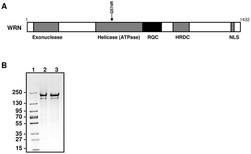
WRN protein (1,432 amino acids) contains multiple domains, including helicase (ATPase), exonuclease, RecQ C-terminal (RQC), and helicase-and-RNaseD-like-C-terminal (HRDC) domains (Fig. 1A). WRN exhibits DNA-dependent ATPase, ATP dependent 3′ → 5′ DNA helicase, single strand DNA annealing and exonuclease activities. The enzyme is able to resolve a variety of DNA substrates, including forks, flaps, displacement loops (D-loops), bubbles, Holliday junctions and G-quadruplexes (G4), all of which represent intermediates in DNA replication and repair (recently reviewed in [15]). Post-translational modifications of WRN modulate its enzymatic activity, thereby regulating its roles in multiple DNA metabolic processes [24].
Figure 1.
WS missense mutation, G574R. (A) Domain structure of WRN protein. Position of the G574R is indicated by arrow. Functional domains are indicated below the structure. The position of G574R is indicated by arrow. (B) SDS-PAGE of purified WRN variants. Lane 1, molecular marker; lane 2, WRN wild type; lane 3, WRN G574R.
Recent genetic studies have reported new missense mutations, such as a c.1720G>A, p.Gly574Arg, along with small insertions/deletions, a deep intronic mutation that creates a new exon, a splice consensus mutation, and genomic rearrangements [18]. Here, we have characterized the biochemical properties of a missense change, c.1720G>A, p.Gly574Arg, identified in a patient with a clinical diagnosis of Werner syndrome. This amino acid is highly conserved and lies just upstream of the nucleotide binding Walker A motif in the ATPase domain. We report that recombinant WRN G574R exhibits significantly decreased helicase and slightly decreased exonuclease activity, as compared to the wild type WRN. The mutant protein displays more efficient strand annealing activity. ATP binding analysis clearly demonstrates that the loss of the helicase activity is due to the lack of ATP binding. Based on our biochemical findings, we discuss possible cellular defects caused by the G574R mutation in relation to the clinical features seen in the patient.
2. Materials and methods
2.1. Plasmid construction and protein purification
6xHis-WRN-FLAG/pFastBac1-InteinCBDAla construct was used for generation of baculovirus expressing WRN wild type, as described previously [25]. Using this plasmid as a template, glycine 574 was substituted with arginine by site-directed mutagenesis methods. The mutagenic primers were designed such that the codon for Gly (GGA) is changed to that for Arg (AGA). The following primers are used for the mutagenesis: 5′-AGATATGGAAAGAGTTTGTGCTTC-3′, in which the mutation site are underlined, and 5′-AGTTGCCATGACAGCAACATTATC-3′. The nucleotide sequences were confirmed by sequencing. Mutagenic primers were purchased form Integrated DNA Technologies (Coralville, IA).
Recombinant baculoviruses were generated using Bac-to-Bac® Baculovirus Expression System (Invitrogen), as described previously [25]. Overexpression and purification of WRN wild type and mutant protein were performed as described previously [25]. Protein concentration was determined using Bradford reagent (Bio-Rad), and protein purity was analyzed on SDS-PAGE.
2.2. Helicase assay
Helicase unwinding assay was performed as described previously [26]. Reactions (20 μl) contained 0.5 nM substrate, 2.5 mM ATP and the indicated concentrations of WRN wild type and mutant protein in 40 mM Tris-HCl, pH 8.0, 50 mM NaCl, 5 mM MgCl2, 100 μg/ml BSA, 2 mM ATP, 1 mM DTT. Reactions were carried out at 37°C for 30 min, and terminated by the addition of 10 μl of SDS stop solution (2% SDS, 50 mM EDTA, 30% glycerol, 0.1% bromophenol blue, 0.1% xylene cyanol). Products were separated on 8% non-denaturing polyacrylamide gel. Radiolabeled DNA was visualized using Typhoon phosphorimager, (Typhoon 9400, GE Healthcare) and quantified using ImageQuant software (Molecular Dynamics). For normal forked duplex substrates, following oligonucleotides were used: 5′-TTTTTTTTTTTTTTTGAGTGTGGTGTACATGCACTAC-3′, and 5′-GTAGTGCATGTACACCACACTCTTTTTTTTTTTTTTT-3′. Following oligonucleotides were used for blocked substrate: 5′-T^TTTTTTTTTTTTTTGAGTGTGGTGTACATGCACTA^C-3′, and 5′-G^TAGTGCATGTACACCACACTCTTTTTTTTTTTTTT^T-3′, which contain phosphorothioate likage (^) to avoid the degradation by exonuclease. Radiolabeled substrates were prepared as described previously [27].
2.3. Exonuclease assay
Exonuclease assays were performed as described previously with modifications [28, 29]. Reactions (10 μl) were performed in buffer (40 mM Tris-HCl, pH 8.0, 4 mM MgCl2, 5 mM DTT, 2 mM ATP, 10% glycerol and 0.1 mg/ml BSA) containing 5′ overhang DNA substrate (0.5 nM) and WRN. Samples were incubated at 37°C for 15 min. Reactions were terminated by addition of equal volume of formamide stop dye (98% formamide, 10 mM EDTA, and 0.1% bromophenol blue). Products were heat-denatured for 5 min at 95 °C, loaded on 14% denaturing polyacrylamide gels, visualized using Typhoon phosphorimager (GE Healthcare), and quantified using ImageQuant software (Molecular Dynamics).
2.4. Single strand DNA annealing assay
DNA strand annealing assays were performed as described previously [6, 30]. C80 and G80 oligonucleotides were used. Reactions (20 μL) were carried out in 20 mM Tris-HCl, pH 7.5, 2 mM MgCl2, 40 μg/mL BSA and 1 mM DTT at 37°C for 15 min with 0.5 nM of each oligonucleotide, one of which was 5′-32P-end-labeled. Protein concentrations used in the reactions were indicated in figure legends. Reactions were stopped by the addition of stop buffer (50 mM EDTA, 1% SDS and 50% glycerol) and immediately loaded onto 8% non-denaturing polyacrylamide gel. Radiolabeled DNA was detected using Typhoon Imager (GE Healthcare), and percentage of annealed oligonucleotide was quantified using ImageQuant software (Molecular Dynamics).
2.5. ATPase assay
ATPase assays were performed as described previously [27]. Buffers for standard ATPase reactions contained 20 mM HEPES-NaOH, pH 8.0, 0.05 mM ATP, 40 μg/ml BSA, 1 mM DTT. ATPase reactions employed WRN and 12.5 μCi of [γ-32P]-ATP. Reactions were incubated for 1 h at 30°C with 150 ng of nucleic acid (M13 ssDNA), and stopped by the addition of 5 μl of 0.5 M EDTA. ATP hydrolysis was analyzed by polyethyleneimine thin-layer chromatography using 1 M formic acid, 0.8 M LiCl mobile phase. The results were analyzed using Typhoon phosphorimager (GE Healthcare) and ImageQuant software (Molecular Dynamics).
2.6. ATP photo-crosslinking assay
1 μg of purified protein was incubated in 10 ul of reaction containing helicase buffer (40 mM Tris-HCl, pH 8.0, 50 mM NaCl, 5 mM MgCl2, 100 μg/ml BSA, 1 mM DTT) with 25 μM ATP and 2 μCi of [γ-32P]-ATP (3000 Ci/mmol) for 10 min at 37°C. Reactions were spotted onto parafilm, placed on ice and irradiated for 5 min using a UV-Stratalinker 2400 (Stratagene). The covalent protein-ATP complex was separated from free ATP by electrophoresis in a 4–15 % SDS-polyacrylamide gel (Bio-rad). Radiolabeled species were detected using Typhoon imager (GE Healthcare) and quantified using ImageQuant software (Molecular Dynamics). The gel was subsequently stained with Coomassie brilliant blue to determine the size of the protein.
2.7. Homology modeling
A model structure of WRN helicase and RQC domain (533–1052 a.a.) was built using sequence alignment and the structure of human RECQL1 Mg-ADP bound form (PDB code: 2V1X) as a template. Briefly, sequence alignment of helicase domains of RecQ helicases was established using CLUSTALW [31] and combined with the structure-based alignment of RECQL1 (PDB code: 2V1X) and E. coli RecQ (PDB code: 1OYY) using CE [32, 33] to improve the alignment. Based on this alignment, WRN helicase model was built and evaluated using SWISS-MODEL (Swiss Institute of Bioinfomatics) [34, 35].
3. Results
3.1. Werner syndrome patient
The patient was a 40 year-old German female with no known consanguinity. She was born with normal height (50 cm) and weight (2.88 kg). A physical examination revealed bilateral ocular cataracts, tight atrophic skin, premature graying and loss of hair, a hoarse voice, flat feet, thin limbs and overall appearance of accelerated aging. She also had a thyroid enlargement due to epithelial hyperplasia. Her height was normal,167 cm (Z score 0.0), and weight was 50 kg (BMI 17.9). She had a history of meningioma and adenoma of the liver. Sequencing of WRN exons revealed a heterozygous change, c.1720G>A (p.G574R) in exon 14 and another heterozygous change, c.1982-1G>A, in intron 17. The latter is expected to cause skipping of exon 18 (r.1982_2088del107) followed by a frameshift (p.I662fs) [18].
3.2. WRN activity is affected by the mutation
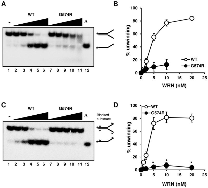
To analyze the biochemical properties of the G574R mutant derived from this WS patient, we have successfully purified the mutant protein employing a purification scheme recently developed for the wild type protein (Fig. 1B) [25]. Helicase assays were performed using a forked duplex DNA substrate, and as shown in Fig. 2A and 2B, the DNA unwinding activity of G574R was significantly lower than that of the wild type. Since the degradation products (smear) appeared on the gel with increased concentration of mutant protein (Fig. 2A, lane 10, 12), indicating that the mutant protein may contain exonuclease activity, we could not quantify the 20 nM concentration point of mutant. To analyze the helicase activity more quantitatively, we used specific substrates containing phosphorothioate linkages at both ends of the oligomer DNA, which prevent exonuclease degradation. As expected, the degradation products observed in Fig. 2A were not present on this gel (Fig. 2C). The quantitative results clearly demonstrated that the helicase activity of G574R was significantly deficient (Fig. 2C and 2D). Collectively, our results imply that the helicase defect in the mutant protein potentially could underlie the WS disease.
Figure 2.
Helicase activity of WRN mutant protein is defected. (A) G574R exhibits lower helicase activity on forked duplex. 1, 2, 5, 10, 20 nM WRN wild type (lane 2 to 6) or WRN G574R (lane 7 to 11) were mixed with 0.5 nM substrate, and reactions were carried out as described Materials and methods. (B) Quantitative analysis of (A). (C) G574R exhibits lower helicase activity on forked duplex. 1, 2, 5, 10, 20 nM WRN wild type (lane 2 to 6) or WRN G574R (lane 7 to 11) were mixed with 0.5 nM blocked substrate, and reactions were carried out as described Materials and methods. (D) Quantitative analysis of (C). Experiments were repeated at least three times, error bars represent ± SD. * represents P value (P<0.001) analyzed with the Student’s t-test.
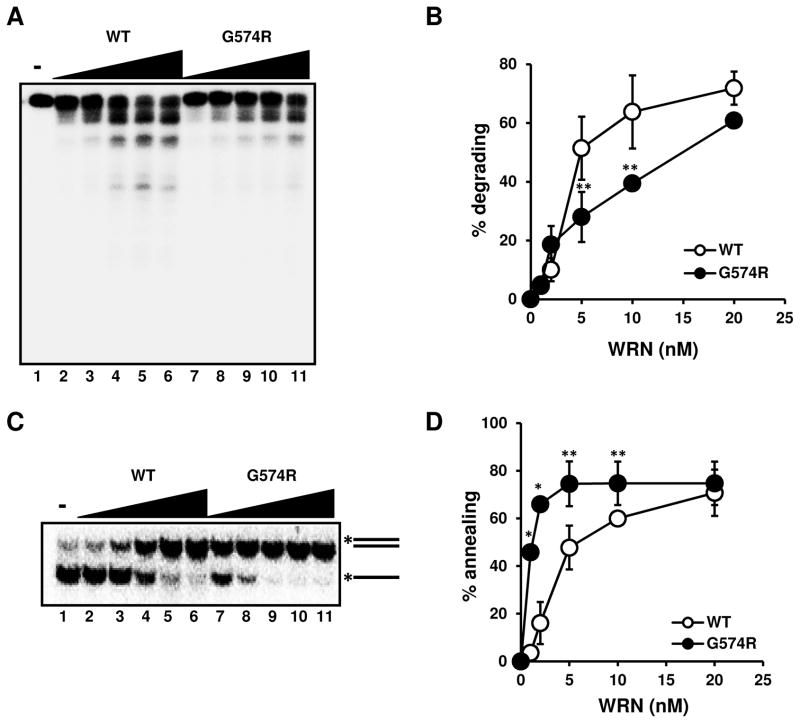
Next, we examined the exonuclease activity of the purified WRN mutant protein. The results indicated that WRN G574R exhibited significantly weaker exonuclease activity (especially when 5 and 10 nM WRN concentrations were used), but comparable to the wild type at lower and higher protein concentrations (Fig. 3A and 3B). This finding suggests that the mutation has a significant but modest effect on WRN exonuclease.
Figure 3.
Exonuclease and strand annealing activities are affected by the mutation. (A) G574R exhibits slightly lower exonuclease activity. 1, 2, 5, 10, 20 nM WRN wild type (lane 2 to 6) or WRN G574R (lane 7 to 11) were mixed with 0.5 nM substrate, and reactions were carried out as described Materials and methods. (B) Quantitative analysis of (A). (C) G574R exhibits more efficient single strand DNA annealing activity. 1, 2, 5, 10, 20 nM WRN wild type (lane 2 to 6) or WRN G574R (lane 7 to 11) were mixed with 0.5 nM substrate, and reactions were carried out as described Materials and methods. (D) Quantitative analysis of (C). All experiments were repeated at least three times, error bars represent ± SD. * and ** represent P value (P<0.001 and P<0.05) analyzed with the Student’s t-test.
We then examined the DNA strand annealing activity of the WRN mutant. Interestingly, G574R exhibited more efficient strand annealing activity than wild type WRN, and the differences were significant except at the highest concentration of WRN (20 nM) (Fig. 3C and 3D). This may be due to the lack of helicase activity, which counteracts strand annealing by binding and unwinding the intermediate products, such as partial DNA duplex.
3.3. ATP binding deficiency leads to loss of ATP hydrolysis
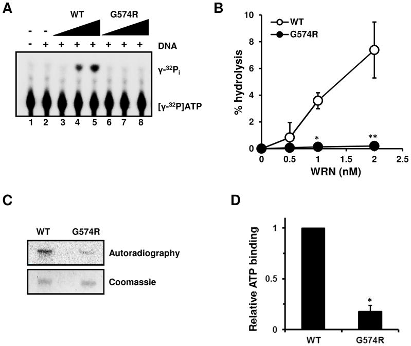
Among the RecQ helicases, ATP binding is thought to be an important regulatory factor underlying both helicase and strand annealing activities [30, 36, 37]. Therefore, we hypothesized that the substitution of Gly574 to Arg would affect ATP-Mg binding. To explore this hypothesis, we tested the ATPase and ATP binding activity of the wild type and mutant protein. The results showed that the ATPase activity of G574R was significantly lower than that of the wild type under all conditions tested (Fig. 4A and 4B), and that its ATP binding affinity was decreased to approximately 20% of the wild type (Fig. 4C and 4D). These results clearly suggest that the decrease in helicase activity of the mutant is due to the loss of ATP binding, and lack of ATP hydrolysis.
Figure 4.
The ATP binding and hydrolysis of WRN G574R. (A) G574R exhibits significantly lower ATPase activity than the wild type. 0.5, 1, 2 nM WRN wild type (lane 3 to 5) or WRN G574R (lane 6 to 8) were used for the reaction. (B) Quantitative analysis of (A). Experiments were repeated at least three times, error bars represent ± SD. * and ** represent P value (P<0.001, P<0.05) analyzed with the Student’s t-test. (C) G574R has decreased the ATP binding ability. ATP binding was measured using the ATP photo-crosslinking assay described in Materials and methods. The reaction products were loaded on SDS-PAGE, subjected to autoradiography (top) and stained with Coomassie brilliant blue (bottom). (D) Quantitative analysis of (C). Experiments were repeated at least three times, error bars represent ± SD. * represents P value (P<0.001) analyzed with the Student’s t-test.
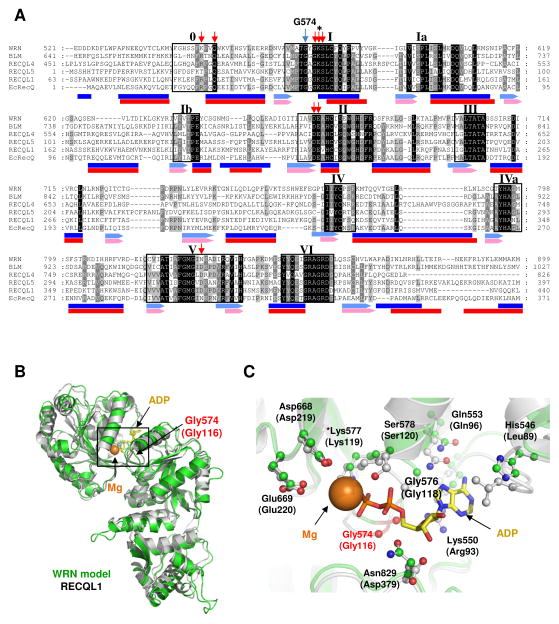
To further understand the impact of the mutation, we used a bioinformatic approach to compare the amino acid sequences of the helicase (ATPase) domains among the RecQ helicases. When the amino acid sequence of the WRN helicase domain was compared with those of other RecQ helicases, Gly574 could be identified as a conserved amino acid just upstream of the Walker A ATPase motif (GSK) of the RecQ helicase domain (Fig. 5A). Thus, the Gly at this position may be quite important for its function at the molecular level. To further identify the molecular mechanism of this mutation, we created a model structure of the WRN helicase and RQC domains using the sequence alignment of the helicase domains from several RecQ helicases (Fig. 5A). As shown in Fig. 5B, the overall structure of the WRN model fits well into the RECQL1 structure (PDB ID; 2V1X). When focusing on the local conformation around the Mg-ADP complex (Fig. 5C), most of the amino acid residues contacting Mg-ADP, except His546 (Leu89 in RECQL1) appear to be spatially well conserved. These amino acid residues are also well conserved at the primary sequence level (Fig. 5A, indicated by red arrow), suggesting a conservative binding mode for Mg-ATP in RecQ helicases.
Figure 5.
Structural insights on the G574R mutation. (A) Alignment of the amino acid sequence of the ATPase domain of RecQ helicases. EcRecQ; E. coli RecQ helicase. While blue arrow indicates the position of G574R, red arrows indicate the amino acid residues in contact with Mg-ADP in RECQL1 structure. * represents the conserved Lys in Walker A motif. Secondary structures of RECQL1 (PDB ID: 2V1X) and E. coli RecQ (PDB ID: 1OYY) are indicated below the sequences in blue and red, respectively. (B) Modeled structure of the WRN helicase domain. WRN model (light green) and RECQL1 (gray) structures are superimposed. (C) Local conformation around ATP (ADP) binding sites. Corresponding amino acid residues involved in Mg-ADP binding in RECQL1 structure are indicated with stick and ball.
4. Discussion
4.1. Defect of WRN protein function in G574R
Our results demonstrate that the G574R mutation strongly inhibits ATP binding, which leads to the abolishment of ATPase and ATPase-dependent helicase activity of WRN. Consistently, a previous biochemical study has demonstrated that the ATPase activity is essential for WRN helicase activity, and WRN protein with a K577M substitution within the ATPase domain, which eliminates ATP hydrolysis, lacks helicase activity [38]. We also found that the exonuclease activity is slightly lower in the G574R mutant protein. Although the isolated WRN exonuclease fragment can function independently as an exonuclease [39], it evidently requires another domain, such as the helicase domain, as shown in the present study, for optimal function. We showed recently that mutations in the WRN RQC domain, R993A and F1037A, abolished WRN exonuclease activity, suggesting that the RQC domain, via its DNA binding properties, is critical for WRN exonuclease activity [25]. Thus, the results of our study support the importance of other WRN functional domains for optimal exonuclease activity. Further, ATP hydrolysis may play a role in the exonuclease activity of WRN since WRN exonuclease activity on duplex DNA substrates containing recessed 3′ ends can be stimulated by ATP [40].
4.2. Enhanced DNA strand annealing activity in G574R
Interestingly, WRN G574R was found to exhibit higher strand annealing activity than wild type. For WRN and BLM, the strand annealing activity is inhibited in the presence of ATP, suggesting that ATP binding modulates their helicase and annealing activities [41]. Biochemical studies on RECQL1 and RECQL5 have also shown that ATP binding is a switch that converts the protein from annealing to helicase function [36, 37]. Although the in vivo relevance of WRN’s strand annealing activity still remains unclear, several studies have indicated the significance of single strand annealing in recombination and double strand break (DSB) repair [42–44]. WRN is significantly involved in the DSB repair pathway [28, 29]. Thus, excessive strand annealing activity of G574R may lead to the accumulation of recombination intermediates in that pathway.
4.3. Possible structural property of Gly574
To address the role of Gly574 in WRN protein, we built a model structure of WRN ATPase (helicase) with RQC domain, and superimposed it with the RECQL1 Mg-ADP bound structure (PDB ID: 2V1X) (Fig. 5B). According to this model, Gly574 is located in the glycine-rich loop, which supports ADP and Mg binding. The local conformation around the glycine-rich loop is well conserved between RECQL1 and the WRN model structure (Fig. 5C). Since the change in size of the amino acid side chain is quite drastic following the mutation from Gly to Arg, it is likely that the G574R substitution inhibits the stable binding of Mg-ATP due to steric hindrance. Our ATP binding analysis (Fig. 4B and 4C) clearly supports this hypothesis. It has been reported that an infrequent WRN polymorphism, R834C, occurring close to the ATP contacting residue, Asn829 (Fig. 5C), reduces WRN helicase and exonuclease activity due to decreased ATP hydrolysis [45]. The authors of the study used WRN-containing immune precipitates for the assays rather than purified recombinant protein. They concluded that the helicase and exonuclease activities of the WRN R834C mutant protein were significantly reduced [45]. Thus the conclusions from the studies on R834C resemble our findings with G574R. However, we additionally showed enhancement of strand annealing activity in G574R, suggesting that the accumulation of unusual recombination intermediates may be a cause of WS as proposed above. Based on the biochemical assay results and from our modeling, it appears that both G574R and R834C mutations may impact the binding pocket for Mg-ATP, and therefore G574R and R837C mutations both lead to a ATP hydrolysis defects.
4.4. Possible cellular and organismal defects in G574R
A pathogenic missense mutation, c.1720G>A, p.Gly574Arg, has been identified in a compound heterozygous patient of German origin [18]. Except for an absence of short stature, the clinical phenotype of the patient carrying G574R as a compound heterozygous mutation was similar to that of other WS patients with truncation mutations. Short stature is one of the cardinal signs of WS and has been seen in 94.7% of the molecularly confirmed WS cases [5]. Our biochemical studies clearly demonstrate that the G574R mutation results in severely decreased ATP binding, and thereby causes a significant decrease in DNA unwinding activity of the WRN mutant protein. The biochemical properties of WRN G574R found in this study were quite similar to those of K577M, which has been well characterized as a helicase dead mutant [38, 46]. A study on the K577M mutation in the mouse model reported that the tail fibroblasts from K577M-WRN mice showed hypersensitivity to 4-nitroquinoline-1-oxide (4NQO), and reduced replicative potential [47]. Another research group also demonstrated that the lagging strand synthesis during telomere replication was affected when the WRN helicase activity was abolished by the K577M mutation [48, 49]. It has been shown using a yeast system that the WRN helicase, but not its exonuclease activity, is genetically required for the restoration of top3 slow growth phenotype [50]. On the other hand, WRN helicase activity is not required for WRN recruitment to the DSBs [51]. Based on these studies, it is likely that WRN helicase activity is particularly important for the replicative processes.
It is possible that the absence of short stature in the patient carrying the G574R mutation is related to the residual activities of WRN. WRN plays an important role in replication and interacts with major proteins in this pathway, including POLδ, PCNA and FEN1 [52–54]. In this individual, G574R, full length WRN protein is still present in the nucleus whereas most WS patients lack WRN expression in the nucleus. Thus, the G574R patient might be able to sustain normal growth by retaining enough WRN associated activity and/or protein:protein interactions. Further, WRN interacts with other RecQ helicases [55, 56], and thus other RecQ helicases could play roles in the growth pathway as a backup to WRN helicase, and might promote normal growth in this patient. Kamath-Loeb et al. previously reported on the biochemical characteristics of the R834C polymorphism [45], and some of their results were similar to what we report here with G574R. Unfortunately, there is no information about the clinical phenotype of the R837C individual. The defect in helicase activity in G574R does not hinder growth in this individual, and thus the helicase defect may not be the major cause of growth arrest in WS. It may rather be caused by incomplete protein interactions of WRN. WRN is involved in many protein interactions that are not only critical for replication and growth (PCNA, RPA, POLδ, FEN1), but also critical for various DNA repair pathways, such as DSB repair. A critical protein interaction may be deficient in G574R and thus the clinical phenotype of G574R individual may partially resemble the WRN null phenotype. We hypothesize that the G574R missense mutation leads to helicase deficiency which impacts DNA replication and repair, and that these functional defect are responsible for the WS phenotype. However, our proposal above is made based on only one individual, and should therefore be considered speculative.
Additional case studies and biochemical studies are needed to fully characterize the phenotypes of WS due to the missense mutations, and to understand how they differ from WS caused by the truncation mutations.
Highlights.
WRN G574R exhibits significantly decreased helicase activity.
G574R displays decreased exonuclease and increased strand annealing activities.
The loss of the helicase activity of G574R is due to the lack of ATP binding.
The Werner syndrome patient carrying G574R has normal stature, and this is unusual.
The short stature normally associated with WS may not be due to helicase deficiency.
Acknowledgments
We would like to thank Drs. Chandrika Canugovi and Huiming Lu for critically reading this manuscript. This research was supported entirely by the Intramural Research Program of the NIH, National Institute on Aging and NIH grants, CA078088 and AG033313 (JO).
Abbreviations
- WS
Werner Syndrome
- WRN
Werner protein
- NLS
nuclear localization signal
- ATP
adenosine triphosphate
- SNP
single nucleotide polymorphism
Footnotes
Conflict of Interest statement
The authors declare no conflict of interest.
Publisher's Disclaimer: This is a PDF file of an unedited manuscript that has been accepted for publication. As a service to our customers we are providing this early version of the manuscript. The manuscript will undergo copyediting, typesetting, and review of the resulting proof before it is published in its final citable form. Please note that during the production process errors may be discovered which could affect the content, and all legal disclaimers that apply to the journal pertain.
References
- 1.Epstein CJ, Martin GM, Schultz AL, Motulsky AG. Werner’s syndrome a review of its symptomatology, natural history, pathologic features, genetics and relationship to the natural aging process. Medicine (Baltimore) 1966;45:177–221. doi: 10.1097/00005792-196605000-00001. [DOI] [PubMed] [Google Scholar]
- 2.Goto M. Hierarchical deterioration of body systems in Werner’s syndrome: implications for normal ageing. Mech Ageing Dev. 1997;98:239–254. doi: 10.1016/s0047-6374(97)00111-5. [DOI] [PubMed] [Google Scholar]
- 3.Takemoto M, Mori S, Kuzuya M, Yoshimoto S, Shimamoto A, Igarashi M, Tanaka Y, Miki T, Yokote K. Diagnostic criteria for Werner syndrome based on Japanese nationwide epidemiological survey. Geriatrics & gerontology international. 2012 doi: 10.1111/j.1447-0594.2012.00913.x. [DOI] [PubMed] [Google Scholar]
- 4.Oshima J, Martin GM, Hisama FM. In: Werner Syndrome. Pagon RA, Bird TD, Dolan CR, Stephens K, Adam MP, editors. GeneReviews; Seattle (WA): 1993. [Google Scholar]
- 5.Huang S, Lee L, Hanson NB, Lenaerts C, Hoehn H, Poot M, Rubin CD, Chen DF, Yang CC, Juch H, Dorn T, Spiegel R, Oral EA, Abid M, Battisti C, Lucci-Cordisco E, Neri G, Steed EH, Kidd A, Isley W, Showalter D, Vittone JL, Konstantinow A, Ring J, Meyer P, Wenger SL, von HA, Wollina U, Schuelke M, Huizenga CR, Leistritz DF, Martin GM, Mian IS, Oshima J. The spectrum of WRN mutations in Werner syndrome patients. Hum Mutat. 2006;27:558–567. doi: 10.1002/humu.20337. [DOI] [PMC free article] [PubMed] [Google Scholar]
- 6.Muftuoglu M, Oshima J, von KC, Cheng WH, Leistritz DF, Bohr VA. The clinical characteristics of Werner syndrome: molecular and biochemical diagnosis. Hum Genet. 2008;124:369–377. doi: 10.1007/s00439-008-0562-0. [DOI] [PMC free article] [PubMed] [Google Scholar]
- 7.Frank B, Hoffmeister M, Klopp N, Illig T, Chang-Claude J, Brenner H. Colorectal cancer and polymorphisms in DNA repair genes WRN, RMI1 and BLM. Carcinogenesis. 2010;31:442–445. doi: 10.1093/carcin/bgp293. [DOI] [PubMed] [Google Scholar]
- 8.Futami K, Ishikawa Y, Goto M, Furuichi Y, Sugimoto M. Role of Werner syndrome gene product helicase in carcinogenesis and in resistance to genotoxins by cancer cells. Cancer Sci. 2008;99:843–848. doi: 10.1111/j.1349-7006.2008.00778.x. [DOI] [PMC free article] [PubMed] [Google Scholar]
- 9.Hsu JJ, Kamath-Loeb AS, Glick E, Wallden B, Swisshelm K, Rubin BP, Loeb LA. Werner syndrome gene variants in human sarcomas. Mol Carcinog. 2010;49:166–174. doi: 10.1002/mc.20586. [DOI] [PMC free article] [PubMed] [Google Scholar]
- 10.Wirtenberger M, Frank B, Hemminki K, Klaes R, Schmutzler RK, Wappenschmidt B, Meindl A, Kiechle M, Arnold N, Weber BH, Niederacher D, Bartram CR, Burwinkel B. Interaction of Werner and Bloom syndrome genes with p53 in familial breast cancer. Carcinogenesis. 2006;27:1655–1660. doi: 10.1093/carcin/bgi374. [DOI] [PubMed] [Google Scholar]
- 11.Goto M, Miller RW, Ishikawa Y, Sugano H. Excess of rare cancers in Werner syndrome (adult progeria). Cancer epidemiology, biomarkers & prevention: a publication of the American Association for Cancer Research, cosponsored by the American Society of Preventive. Oncology. 1996;5:239–246. [PubMed] [Google Scholar]
- 12.Fukuchi K, Martin GM, Monnat RJ., Jr Mutator phenotype of Werner syndrome is characterized by extensive deletions. Proc Natl Acad Sci USA. 1989;86:5893–5897. doi: 10.1073/pnas.86.15.5893. [DOI] [PMC free article] [PubMed] [Google Scholar]
- 13.Martin GM, Sprague CA, Epstein CJ. Replicative life-span of cultivated human cells. Effects of donor’s age, tissue, and genotype. Lab Invest. 1970;23:86–92. [PubMed] [Google Scholar]
- 14.Salk D, Au K, Hoehn H, Martin GM. Cytogenetics of Werner’s syndrome cultured skin fibroblasts: variegated translocation mosaicism. Cytogenet Cell Genet. 1981;30:92–107. doi: 10.1159/000131596. [DOI] [PubMed] [Google Scholar]
- 15.Rossi ML, Ghosh AK, Bohr VA. Roles of Werner syndrome protein in protection of genome integrity. DNA Repair (Amst) 2010;9:331–344. doi: 10.1016/j.dnarep.2009.12.011. [DOI] [PMC free article] [PubMed] [Google Scholar]
- 16.Bohr VA. Rising from the RecQ-age: the role of human RecQ helicases in genome maintenance. Trends Biochem Sci. 2008;33:609–620. doi: 10.1016/j.tibs.2008.09.003. [DOI] [PMC free article] [PubMed] [Google Scholar]
- 17.Brosh RM, Jr, Bohr VA. Human premature aging, DNA repair and RecQ helicases. Nucleic Acids Res. 2007;35:7527–7544. doi: 10.1093/nar/gkm1008. [DOI] [PMC free article] [PubMed] [Google Scholar]
- 18.Friedrich K, Lee L, Leistritz DF, Nurnberg G, Saha B, Hisama FM, Eyman DK, Lessel D, Nurnberg P, Li C, Garcia F, Kets VCM, Schmidtke J, Cruz VT, Van den Akker PC, Boak J, Peter D, Compoginis G, Cefle K, Ozturk S, Lopez N, Wessel T, Poot M, Ippel PF, Groff-Kellermann B, Hoehn H, Martin GM, Kubisch C, Oshima J. WRN mutations in Werner syndrome patients: genomic rearrangements, unusual intronic mutations and ethnic-specific alterations. Hum Genet. 2010;128:103–111. doi: 10.1007/s00439-010-0832-5. [DOI] [PMC free article] [PubMed] [Google Scholar]
- 19.Goto M, Imamura O, Kuromitsu J, Matsumoto T, Yamabe Y, Tokutake Y, Suzuki N, Mason B, Drayna D, Sugawara M, Sugimoto M, Furuichi Y. Analysis of helicase gene mutations in Japanese Werner’s syndrome patients. Hum Genet. 1997;99:191–193. doi: 10.1007/s004390050336. [DOI] [PubMed] [Google Scholar]
- 20.Uhrhammer NA, Lafarge L, Dos Santos L, Domaszewska A, Lange M, Yang Y, Aractingi S, Bessis D, Bignon YJ. Werner syndrome and mutations of the WRN and LMNA genes in France. Human mutation. 2006;27:718–719. doi: 10.1002/humu.9435. [DOI] [PubMed] [Google Scholar]
- 21.Suzuki T, Shiratori M, Furuichi Y, Matsumoto T. Diverged nuclear localization of Werner helicase in human and mouse cells. Oncogene. 2001;20:2551–2558. doi: 10.1038/sj.onc.1204344. [DOI] [PubMed] [Google Scholar]
- 22.Yamabe Y, Sugimoto M, Satoh M, Suzuki N, Sugawara M, Goto M, Furuichi Y. Down-regulation of the defective transcripts of the Werner’s syndrome gene in the cells of patients. Biochemical and biophysical research communications. 1997;236:151–154. doi: 10.1006/bbrc.1997.6919. [DOI] [PubMed] [Google Scholar]
- 23.Wang Z, Xu Y, Tang J, Ma H, Qin J, Lu C, Wang X, Hu Z, Wang X, Shen H. A polymorphism in Werner syndrome gene is associated with breast cancer susceptibility in Chinese women. Breast Cancer Res Treat. 2009;118:169–175. doi: 10.1007/s10549-009-0327-z. [DOI] [PubMed] [Google Scholar]
- 24.Kusumoto R, Muftuoglu M, Bohr VA. The role of WRN in DNA repair is affected by post-translational modifications. Mech Ageing Dev. 2007;128:50–57. doi: 10.1016/j.mad.2006.11.010. [DOI] [PubMed] [Google Scholar]
- 25.Tadokoro T, Kulikowicz T, Dawut L, Croteau DL, Bohr VA. DNA binding residues in the RQC domain of Werner protein are critical for its catalytic activities. Aging (Albany NY) 2012 doi: 10.18632/aging.100463. [DOI] [PMC free article] [PubMed] [Google Scholar]
- 26.Opresko PL, Otterlei M, Graakjaer J, Bruheim P, Dawut L, Kolvraa S, May A, Seidman MM, Bohr VA. The Werner syndrome helicase and exonuclease cooperate to resolve telomeric D loops in a manner regulated by TRF1 and TRF2. Mol Cell. 2004;14:763–774. doi: 10.1016/j.molcel.2004.05.023. [DOI] [PubMed] [Google Scholar]
- 27.Brosh RM, Jr, Opresko PL, Bohr VA. Enzymatic mechanism of the WRN helicase/nuclease. Methods Enzymol. 2006;409:52–85. doi: 10.1016/S0076-6879(05)09004-X. [DOI] [PubMed] [Google Scholar]
- 28.Cooper MP, Machwe A, Orren DK, Brosh RM, Ramsden D, Bohr VA. Ku complex interacts with and stimulates the Werner protein. Genes Dev. 2000;14:907–912. [PMC free article] [PubMed] [Google Scholar]
- 29.Li B, Comai L. Functional interaction between Ku and the werner syndrome protein in DNA end processing. J Biol Chem. 2000;275:39800. doi: 10.1074/jbc.C000289200. [DOI] [PubMed] [Google Scholar]
- 30.Machwe A, Xiao L, Groden J, Matson SW, Orren DK. RecQ family members combine strand pairing and unwinding activities to catalyze strand exchange. J Biol Chem. 2005;280:23397–23407. doi: 10.1074/jbc.M414130200. [DOI] [PubMed] [Google Scholar]
- 31.Aiyar A. The use of CLUSTAL W and CLUSTAL X for multiple sequence alignment. Methods Mol Biol. 2000;132:221–241. doi: 10.1385/1-59259-192-2:221. [DOI] [PubMed] [Google Scholar]
- 32.Guda C, Lu S, Scheeff ED, Bourne PE, Shindyalov IN. CE-MC: a multiple protein structure alignment server. Nucleic Acids Res. 2004;32:W100–W103. doi: 10.1093/nar/gkh464. [DOI] [PMC free article] [PubMed] [Google Scholar]
- 33.Shindyalov IN, Bourne PE. Protein structure alignment by incremental combinatorial extension (CE) of the optimal path. Protein Eng. 1998;11:739–747. doi: 10.1093/protein/11.9.739. [DOI] [PubMed] [Google Scholar]
- 34.Guex N, Peitsch MC. SWISS-MODEL and the Swiss-PdbViewer: an environment for comparative protein modeling. Electrophoresis. 1997;18:2714–2723. doi: 10.1002/elps.1150181505. [DOI] [PubMed] [Google Scholar]
- 35.Schwede T, Kopp J, Guex N, Peitsch MC. SWISS-MODEL: An automated protein homology-modeling server. Nucleic Acids Res. 2003;31:3381–3385. doi: 10.1093/nar/gkg520. [DOI] [PMC free article] [PubMed] [Google Scholar]
- 36.Garcia PL, Liu Y, Jiricny J, West SC, Janscak P. Human RECQ5beta, a protein with DNA helicase and strand-annealing activities in a single polypeptide. EMBO J. 2004;23:2882–2891. doi: 10.1038/sj.emboj.7600301. [DOI] [PMC free article] [PubMed] [Google Scholar]
- 37.Sharma S, Sommers JA, Choudhary S, Faulkner JK, Cui S, Andreoli L, Muzzolini L, Vindigni A, Brosh RM., Jr Biochemical analysis of the DNA unwinding and strand annealing activities catalyzed by human RECQ1. J Biol Chem. 2005;280:28072–28084. doi: 10.1074/jbc.M500264200. [DOI] [PubMed] [Google Scholar]
- 38.Gray MD, Shen JC, Kamath-Loeb AS, Blank A, Sopher BL, Martin GM, Oshima J, Loeb LA. The Werner syndrome protein is a DNA helicase. Nat Genet. 1997;17:100–103. doi: 10.1038/ng0997-100. [DOI] [PubMed] [Google Scholar]
- 39.Perry JJ, Yannone SM, Holden LG, Hitomi C, Asaithamby A, Han S, Cooper PK, Chen DJ, Tainer JA. WRN exonuclease structure and molecular mechanism imply an editing role in DNA end processing. Nat Struct Mol Biol. 2006;13:414–422. doi: 10.1038/nsmb1088. [DOI] [PubMed] [Google Scholar]
- 40.Kamath-Loeb AS, Shen JC, Loeb LA, Fry M. Werner syndrome protein. II. Characterization of the integral 3′ --> 5′ DNA exonuclease. The Journal of biological chemistry. 1998;273:34145–34150. doi: 10.1074/jbc.273.51.34145. [DOI] [PubMed] [Google Scholar]
- 41.Machwe A, Lozada EM, Xiao L, Orren DK. Competition between the DNA unwinding and strand pairing activities of the Werner and Bloom syndrome proteins. BMC Mol Biol. 2006;7:1. doi: 10.1186/1471-2199-7-1. [DOI] [PMC free article] [PubMed] [Google Scholar]
- 42.Cheok CF, Wu L, Garcia PL, Janscak P, Hickson ID. The Bloom’s syndrome helicase promotes the annealing of complementary single-stranded DNA. Nucleic acids research. 2005;33:3932–3941. doi: 10.1093/nar/gki712. [DOI] [PMC free article] [PubMed] [Google Scholar]
- 43.Yan H, McCane J, Toczylowski T, Chen C. Analysis of the Xenopus Werner syndrome protein in DNA double-strand break repair. The Journal of cell biology. 2005;171:217–227. doi: 10.1083/jcb.200502077. [DOI] [PMC free article] [PubMed] [Google Scholar]
- 44.Hickson ID. RecQ helicases: caretakers of the genome. Nature reviews Cancer. 2003;3:169–178. doi: 10.1038/nrc1012. [DOI] [PubMed] [Google Scholar]
- 45.Kamath-Loeb AS, Welcsh P, Waite M, Adman ET, Loeb LA. The enzymatic activities of the Werner syndrome protein are disabled by the amino acid polymorphism R834C. J Biol Chem. 2004;279:55499–55505. doi: 10.1074/jbc.M407128200. [DOI] [PubMed] [Google Scholar]
- 46.Opresko PL, Laine JP, Brosh RM, Jr, Seidman MM, Bohr VA. Coordinate action of the helicase and 3′ to 5′ exonuclease of Werner syndrome protein. J Biol Chem. 2001;276:44677–44687. doi: 10.1074/jbc.M107548200. [DOI] [PubMed] [Google Scholar]
- 47.Wang L, Ogburn CE, Ware CB, Ladiges WC, Youssoufian H, Martin GM, Oshima J. Cellular Werner phenotypes in mice expressing a putative dominant-negative human WRN gene. Genetics. 2000;154:357–362. doi: 10.1093/genetics/154.1.357. [DOI] [PMC free article] [PubMed] [Google Scholar]
- 48.Crabbe L, Verdun RE, Haggblom CI, Karlseder J. Defective telomere lagging strand synthesis in cells lacking WRN helicase activity. Science. 2004;306:1951–1953. doi: 10.1126/science.1103619. [DOI] [PubMed] [Google Scholar]
- 49.Crabbe L, Jauch A, Naeger CM, Holtgreve-Grez H, Karlseder J. Telomere dysfunction as a cause of genomic instability in Werner syndrome. Proc Natl Acad Sci USA. 2007;104:2205–2210. doi: 10.1073/pnas.0609410104. [DOI] [PMC free article] [PubMed] [Google Scholar]
- 50.Aggarwal M, Brosh RM. WRN helicase defective in the premature aging disorder Werner syndrome genetically interacts with topoisomerase 3 and restores the top3 slow growth phenotype of sgs1 top3. Aging (Albany NY) 2009;1:219–233. doi: 10.18632/aging.100020. [DOI] [PMC free article] [PubMed] [Google Scholar]
- 51.Lan L, Nakajima S, Komatsu K, Nussenzweig A, Shimamoto A, Oshima J, Yasui A. Accumulation of Werner protein at DNA double-strand breaks in human cells. J Cell Sci. 2005;118:4153–4162. doi: 10.1242/jcs.02544. [DOI] [PubMed] [Google Scholar]
- 52.Szekely AM, Chen YH, Zhang C, Oshima J, Weissman SM. Werner protein recruits DNA polymerase delta to the nucleolus. Proceedings of the National Academy of Sciences of the United States of America. 2000;97:11365–11370. doi: 10.1073/pnas.97.21.11365. [DOI] [PMC free article] [PubMed] [Google Scholar]
- 53.Brosh RM, Jr, von Kobbe C, Sommers JA, Karmakar P, Opresko PL, Piotrowski J, Dianova I, Dianov GL, Bohr VA. Werner syndrome protein interacts with human flap endonuclease 1 and stimulates its cleavage activity. The EMBO journal. 2001;20:5791–5801. doi: 10.1093/emboj/20.20.5791. [DOI] [PMC free article] [PubMed] [Google Scholar]
- 54.Lebel M, Spillare EA, Harris CC, Leder P. The Werner syndrome gene product co-purifies with the DNA replication complex and interacts with PCNA and topoisomerase I. The Journal of biological chemistry. 1999;274:37795–37799. doi: 10.1074/jbc.274.53.37795. [DOI] [PubMed] [Google Scholar]
- 55.von Kobbe C, Karmakar P, Dawut L, Opresko P, Zeng X, Brosh RM, Jr, Hickson ID, Bohr VA. Colocalization, physical, and functional interaction between Werner and Bloom syndrome proteins. The Journal of biological chemistry. 2002;277:22035–22044. doi: 10.1074/jbc.M200914200. [DOI] [PubMed] [Google Scholar]
- 56.Popuri V, Huang J, Ramamoorthy M, Tadokoro T, Croteau DL, Bohr VA. RECQL5 plays co-operative and complementary roles with WRN syndrome helicase. Nucleic acids research. 2012 doi: 10.1093/nar/gks1134. [DOI] [PMC free article] [PubMed] [Google Scholar]


