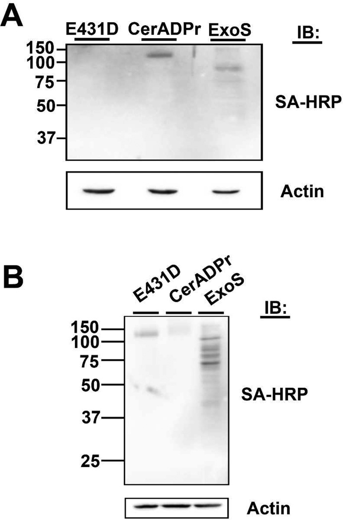
Figure 6. CerADPr ADP-ribosylates the 120KDa target within HeLa cells.
(A) HeLa cells were transfected with pEGFP-CerADPr, pEGFP-CerADPr(E431D), or pExoS for 2 hr. Cells were treated with 200 ng/ml of tetanolysin and then incubated with biotin-NAD for 2.5 hr. Cell lysates were resolved by SDS-PAGE and biotin-ADP-ribose incorporation was visualized by immunoblotting with streptavidin-HRP. Actin was used as a loading control. (B) HeLa cells were treated with 200 ng/ml of tetanolysin, then incubated with 3 µg/ml recombinant CerADPr, CerADPr(E431D), or ExoS for 2 hr. Cell lysates prepared and then incubated with biotin-NAD and recombinant CerADPr for an hour. Lysates were resolved by SDS-PAGE, transferred to PVDF membranes, where biotin-ADP-ribose incorporation was visualized with streptavidin-HRP. Actin was used as a loading control.

