

Abstract
Fractures of the scapula are relatively uncommon. Fractures specific to the scapular body comprise 35–65% of these fractures. Currently, 99% of all isolated scapular body fractures are being treated nonoperatively with an immobilizing sling or brace and some form of manual therapy with an 86% success rate. We present the conservative management of three patients with comminuted fractures involving the scapular body that were managed in chiropractic settings. Residual disabilities in these three patients as measured by a standardized outcome tool were 2%, 5% and 23% after 3 years, 2 years, and 6 years respectively.
Keywords: scapula, fractures, comminuted, chiropractic, case management
Abstract
Les fractures de la scapula sont relativement communes. Les fractures spécifiques au corps de la scapula représentent entre 35 et 65 % de ces fractures. Actuellement, 99 % des fractures isolées du corps de la scapula se traitent sans intervention chirurgicale, simplement en immobilisant la partie concernée avec une écharpe ou un appareil orthopédique et à l’aide de thérapies manuelles, avec un taux de réussite de 86 %. Nous présentons la gestion conservatrice de trois patients touchés avec des fractures comminutives touchant le corps de la scapula, qui sont traités par manipulations chiropratiques. Les incapacités résiduelles de ces trois patients mesurées par un instrument standardisé sont de 2 %, 5 % et 23 % après 3 ans, 2 ans et 6 ans respectivement.
Keywords: scapula, fractures, comminutives, chiropratique, gestion de cas
Introduction
Fractures of the scapula are relatively uncommon. Fractures specific to the scapular body comprise 35–65% of these fractures.1–4 Currently, 99% of all isolated scapular body fractures are being treated non-operatively with an immobilizing sling or brace and some form of manual therapy with an 86% success rate.5 Scapular fractures have been a subject of investigation since Desault’s treatise of 1805.6 Scapular fractures constitute only 1% of all fractures, 3% of shoulder-girdle injuries, and only 5% of all shoulder fractures.7 The infrequent occurrence of scapular fractures has been attributed to its protection anteriorly by the rib cage and thoracic cavity, a thick covering of soft tissue including eighteen muscular origins and insertions, and a wide range of mobility that allows for considerable dissipation of traumatic forces.8,9
Scapular fractures occur most frequently in the 3rd and 4th decades of life with 64–90% occurring in men.3,10,11 While isolated body fractures are most frequently caused by falls from different heights,12 the most common cause of scapular body fractures in multi-trauma patient groups are road traffic accidents.2, 13
The Disabilities of the Arm, Shoulder and Hand (DASH) outcome measure used in these case studies is a 30-item, self-report questionnaire designed to measure the physical function and symptoms in patients with any or several musculoskeletal disorders of the upper limb. The questionnaire (www.dash.iwh.on.ca) was jointly developed by the Institute for Work and Health and the American Academy of Orthopaedic Surgeons (AAOS) and was designed to help grade the disability experienced by people with upper-limb disorders and also to monitor changes in symptoms and function over time. Testing has shown that the DASH performs well in both these roles.14
The purpose of this paper is to describe both the identification and conservative management of three cases of comminuted scapular body fractures which presented at three different chiropractic centers.
Case Reports
Case 1
History
A 43 year old male manual laborer presented to a chiropractic community outreach clinic with right shoulder pain eight days after slipping on ice and striking his right shoulder on cement blocks.
Physical Examination
This patient was able to remove his shirt but expressed mild discomfort with arm movements. Considerable contusion was evident overlying the inferior angle of the scapula and posterolateral thoracic wall. Decreased shoulder ranges of motion were accompanied by pain to palpation of the acromioclavicular (AC) joint evoking clinical suspicion of an AC joint sprain or separation. Crepitus was elicited over the acromion process and at the superior aspect of the shoulder during active abduction and external rotation. Shallow breathing and decreased normal lung sounds on inspiration and expiration in the right apical as well as upper anterior and posterior regions were observed with no significant signs of respiratory distress.
Diagnostic Imaging
Chest and shoulder radiographs were obtained and revealed comminuted scapular fracture with isolation and antero-medial displacement of the GH joint, separation at the superior aspect of the acromioclavicular joint, displaced fractures of ribs 3, 4 and 6–9 on the right, pneumothorax, and passive/relaxation atelectasis (Figure 1A).
Management
The patient was transferred to the emergency department of a local hospital, where blood testing and a metabolic panel were interpreted as normal. The patient was then prescribed analgesic medication, a sling, and an orthopedic referral. The orthopedic consultant recommended against surgery, but advised the patient to continue immobilizing the shoulder in the sling for 6 weeks and then follow up. Approximately 3 weeks post injury, he presented back to the chiropractic community outreach clinic for treatment. Management consisted primarily of myofascial release of the trapezius and cervicothoracic paraspinal musculature, as well as diversified chiropractic adjustments to the cervical spine and instrument assisted (activator) adjustments as needed and tolerable in the thoracic spine. Treatments were administered approximately once per week for 16 weeks.
Outcome
Four months after the initial injury, follow up radiographs were taken and demonstrated a reduction in the displacement of the fracture fragment composed of the inferior scapular body and angle (Figure 1B). Re-examination at this time indicated some improvement in both active and passive right shoulder ranges of motion and normal shoulder orthopedic testing, but reduced muscle strength of the rotator cuff and deltoid musculature (4/5) and continued mild edema, bruising and tenderness overlying the scapula. However, with continued treatment, two months later, this patient was pain free, with near normal ranges of motion of the shoulder and strength of the rotator cuff musculature. Despite the temporary disability leave he was given by his orthopedist initially, this patient continued with light duty throughout his treatments and was able to return to work fully afterward with only slight soreness at the end of the day. On follow up, three years after the initial injury, this patient completed the Disabilities of the Shoulder, Arm and Hand (DASH) outcome questionnaire which indicated disability of less than 2%.14
Case 2
History
A 53 year old Caucasian male businessman presented to the emergency room with right posterior shoulder pain sustained when an unexpected acceleration of a boat caused him to lose his balance and fall backward – striking his right scapula directly on the gunwale of the boat. Radiographs (Figure 2A) and CT images (Figure 2B) of the chest and shoulder were performed revealing a comminuted fracture of the right mid and lower scapular body with irregular fracture planes and multiple distracted, angulated and foreshortened fracture fragments as well as additional linear fractures through the base of the acromion and coracoid processes. The acromioclavicular and glenohumeral joints were intact with no dislocation and there was no evidence of pneumothorax or pleural effusion. The patient was fitted for a brace, prescribed analgesic medication to alleviate his symptoms and referred to an orthopedist. During this visit one week later, in agreement with the ER orthopedist, he was advised against surgical intervention due to the delicate muscular envelope of the scapula and scheduled for re-checks two and six weeks later. Additionally, an MRI was scheduled demonstrating a partial thickness tear of the supraspinatus tendon, fluid in the subacromial/subdeltoid bursa compatible with shoulder impingement and moderate degenerative changes at the acromioclavicular joint encroaching upon the supraspinatus muscle tendon complex.
The patient presented for a chiropractic evaluation three weeks following the incident to begin conservative management of his injuries. At that time, the patient had already resumed working, discontinued his analgesic medication, and was beginning to feel more comfortable with his arm out of the brace.
Physical Examination
Initial examination revealed limitations with active shoulder range of motion in all directions (flexion 100°, extension 15°, abduction 20°, adduction 15°, external rotation 0°, internal rotation 40°). Passive ranges of motion were limited only in extension, abduction, and external rotation to 40°, 90° and 0° respectively. Neurologic examinations were unremarkable, with normal deep tendon reflexes, and no motor or sensory loss with the exception of an inability to test deltoid or bicep strength on the right due to pain. Peripheral upper extremity pulses were symmetrical and of normal amplitude. At this time the DASH outcome questionnaire score indicated an 81% disability.14
Management
This patient was treated eighteen times over an eleven week period. Therapy began with early progressive passive range of motion and Codman exercises and progressed to active movements under supervision as tolerated. Complementary therapies included electric muscle stimulation, therapeutic ultrasound and class IV laser treatment. After 4 weeks of treatment, therapy was focused on rehabilitating the rotator cuff with progressive isometric resistance, therapeutic bands, strengthening parascapular musculature, and preventing soft tissue adhesions using manual release methods and instrument assisted techniques. Diversified chiropractic manipulations of the cervical spine were administered for joint restrictions as indicated and mobilizations of the ribs and thoracic spine were also performed to tolerance.
Outcome
Nineteen weeks after the initial injury, this patient demonstrated pain free active ranges of motion in the shoulder with only slight crepitations at the scapulothoracic interface and a DASH score reduced to 5% disability.14 In a two year follow-up discussion by telephone the patient reported 100% mobility had been maintained with mild residual pain present at times only with sleeping on the injured side.
Case 3
History
A 23 year old right-handed Caucasian male construction laborer was involved in a high speed motor vehicle collision. He was the driver of a mid-sized sedan that was struck from the passenger side in an intersection by another sedan estimated to be traveling at 75 MPH, causing his car to flip over seven times. At impact, his shoulder restraint was broken and he was thrown into the back seat of the car where he was wedged between the back seat and the rear windshield. He did not lose consciousness at any time after the collision. The fire department used hydraulic spreaders (Jaws of Life) to extricate him from his wedged-in position. He was transported by helicopter Mercy flight to the emergency department where radio-graphs and CT scans were performed that revealed a fractured scapula. He remained in the hospital overnight and was released the next day with an arm sling and analgesic medication (Darvocet). He consulted with an orthopedic specialist two weeks after the collision where he obtained more radiographs and an MRI examination (not shown). The patient was advised that there was no nerve or muscle damage and that surgery was not necessary and would not improve his outcome.
Diagnostic Imaging
Radiographs of the right shoulder obtained at the hospital immediately after the injury (Figure 3A) revealed a complete vertical fracture of the neck and axillary border of the scapula with a 2 cm lateral displacement of the entire glenoid process and a triangular fracture fragment that projected inferiorly and laterally into the axilla. The GH joint was intact with no evidence of dislocation. Axial CT (Figure 3B) and reconstructed, surface-rendered 3-D CT images (Figure 3C) revealed that the glenoid fragment was internally rotated approximately 20 degrees, as well as an additional minimally displaced comminuted fracture of the scapular body. No evidence of pneumothorax, hemothorax, rib or spine fracture was present.
Medical Management
After examination, the orthopedic surgeon prescribed analgesic medication and fitted the patient with a shoulder sling for immobilization and advised against surgery. The patient complied with this advice and the pain gradually decreased over time. After being on disability for one year, this patient was unable to return to work in construction because his job was no longer available. However, he did resume work as a security officer at that time.
Chiropractic Management
Nine months after the initial injury, the patient presented to a chiropractic college health center seeking chiropractic manual treatment and musculoskeletal rehabilitation for continued pain in the right scapula and decreased ranges of motion of the right shoulder. The deep muscle pain at presentation ranged from 1 on a scale of 10 while at rest and became sharp rated 5/10 at its worst with internal and external rotation or abduction. Additionally, the pain was aggravated by lifting objects overhead and rotation of the shoulder in a throwing motion. Active stretching and rest provided pain relief. The pain did not radiate into his upper extremity and did not disrupt sleep except when he rolled onto his right side while recumbent.
Physical Examination
The patient was 69” tall, 185 lbs., right handed and appeared physically fit. Vital signs, gait, neurologic examination and ambulation were all normal. Active range of motion of the right shoulder was full and pain free in combined flexion and adduction. Active external rotation was 100% restricted and provoked pain at the posterior axillary fold, and combined internal rotation, extension and adduction (Apley’s inferior scratch test) of the right shoulder revealed significant restriction on the right compared with the left with a 20 cm discrepancy from side to side. The active range of abduction of the right shoulder was full but provoked pain over the supraspinatus muscle. The right supraspinatus press test was mildly positive for pain. Myofascial trigger points and adhesions were detected in the right rhomboid, deltoid, infraspinatus, supraspinatus, teres minor, subscapularis, latissimus dorsi, upper trapezius, levator scapulae and cervical spine musculature. These findings were most pronounced in the right infraspinatus muscle which was significantly atrophic compared with the left.
This patient was diagnosed at the chiropractic college health center with post-traumatic myofascial parascapular trigger points, myofascitis, muscular adhesions and weakness in the right shoulder musculature involving principally the infraspinatus muscle. He was seen seven times over a 2 month period. Treatment consisted of trigger point therapy, progressive isometric resistance using therapeutic bands, manual muscle release methods, instrument assisted techniques aimed at preventing soft tissue adhesions, and passive progressive mobilizations to increase range of motion. The patient was also placed on a muscle strengthening regimen at his gymnasium three times per week to strengthen the parascapular musculature. Diversified chiropractic manipulation and mobilization of the cervical spine, thoracic spine and ribs were administered on each visit to address joint restrictions as indicated.
Outcome
The patient reported a significant increase in muscle strength after 2 months. The active range of motion in external rotation improved from 100% restriction to 80% restriction in two months. The inferior Apley’s scratch test improved somewhat from the 20 cm pre-treatment discrepancy to a 14 cm post-treatment discrepancy. At the conclusion of the seven visits, he continued to have mild pain which was graded at 2 of 10 with occasional exacerbations as high as 5 after vigorous workouts. He reported that the pain was still occasionally severe enough to awaken him at night as a result of rolling onto his right shoulder. After the two month treatment period, the patient was then lost to follow-up.
Six years after the initial injury, the patient was contacted and he reported for follow-up evaluation and radio-graphic examination. At this point, he was continuing to exercise and reported that the pain was much less frequent, and was provoked only by activities such as throwing a ball, absorbing body contact in rugby, and lying on his right side. Follow-up radiographs revealed that the fracture of the scapular neck had healed with significant malunion in which a 4 cm long triangular subglenoid fragment remained unattached to the axillary border of the scapular body. While the margins of this projection of bone were rounded and smooth, it projected into the axillary soft tissues (Figure 3D). Examination revealed significant generalized atrophy of the muscles of the right shoulder girdle compared to the left. Ranges of motion were all full and bilaterally symmetric except the inferior Apley’s scratch test which exhibited a 7.5 cm discrepancy in ranges of motion from right to left (Figure 3E). He completed a DASH outcome questionnaire that revealed a 23% disability. 14
Discussion
Scapular fracture diagnoses are commonly missed or delayed owing to the extent of associated injuries over-shadowing the scapula, chest trauma CT scans not covering the entirety of the scapula, unusual mechanisms of injury, low levels of consciousness, or because pain is being controlled by corticosteroid injection for other injuries.15
Scapular fractures have proven to be particularly significant due to their anatomic proximity to vital structures including the head, lungs, cervical spine and brachial neurovascular structures.2,3 It has been observed that 81–96% of individuals with scapular fractures have associated injury.1, 16,17 The most common associated injuries in these patients are rib fractures.11,18 However, other, more serious injuries can include hemothorax, pneumothorax, pulmonary contusion, skull fractures, permanent cord injuries, brachial plexus injuries, complex regional pain syndrome, Horner’s Syndrome and subclavian, axillary or brachial artery injury.1,3,10 As a result, the mortality rate of individuals with scapular fractures, although not usually direct, has been reported to range from 2% to as high as 14.3%.10,11,13
While the vast majority of scapular fractures are managed quite successfully without surgery, most agree that surgical management should be considered for severely displaced injuries – most commonly: (1) significantly displaced fractures of the glenoid cavity (glenoid rim and glenoid fossa), (2) significantly displaced fractures of the glenoid neck, and (3) double disruption of the superior shoulder suspensory complex (SSSC) in which one or more elements of the scapula are significantly displaced.9
Currently, 99% of all isolated scapular body fractures are being managed non-operatively with an 86% success rate.5 Because of its abundant blood supply, with appropriate care, scapular fractures can heal rapidly under conservative management.8 However, when treating these fractures, there are consequences that can occur even with the current physiotherapy techniques that should be considered. For example, some patients with displaced scapular body fractures may experience a snapping or grating sensation at the scapulothoracic interface with movement through ranges of motion.1 Persistent soft tissue irritation secondary to bone irregularity, callus formation of displaced fragments (as seen in Case 3), and heterotopic bone formation are also possibilities, which in some cases may require excision of the mass or irregularity for pain relief.4,19 Other causes of patient dissatisfaction after scapular healing can include prolonged weakness, pain, crepitations or a disfiguring bump, most common in multi-trauma patients.12 Although nonunion is rare, when a patient complains of persistent shoulder pain after conservative treatment, this possibility must be considered.20, 21
Following fracture union, in our experience, adjunctive manual muscle procedures such as myofascial release, trigger point therapy, and instrument assisted techniques can be performed to relieve spasms and prevent soft tissue adhesions amongst the rotator cuff muscles, levator scapulae, paraspinals, deltoids, rhomboids, latissimus dorsi and trapezius. Furthermore, mobilization of costovertebral articulations and chiropractic manipulations of the appropriate cervical and thoracic motion segments were useful in facilitating biomechanical adaptation and symptomatic manifestations in each case presented here as well as in a case report by Deltoff et al.22
All three patients in this case series received conservative management for their injuries. The first patient who received 16 weeks of treatment completed a DASH questionnaire at 3 years post injury that revealed functional improvement to 2% disability. The second patient who scored 81% disability 3 weeks after his initial injury, received 11 weeks of treatment, and at nineteen weeks had improved to 5% disability. The third patient with a more significantly displaced, comminuted fracture that resulted in healing with malunion scored a 23% disability on the DASH questionnaire completed 6 years after his initial injury.
Conclusion
Research indicates that the vast majority of patients with comminuted scapular fractures can be treated with an immobilizing sling or brace and some form of manual therapy and will heal completely within 6 weeks.9 Although uncommon for such cases of scapular fracture to present in chiropractic offices, these three cases suggest that chiropractic management can help these individuals to achieve symptomatic relief, progressive functional improvements and in most cases, little need for long-term follow-up care.
Figure 1. Case 1:
Figure 1A:

Right lateral scapular “Y” view demonstrating antero-medial displacement of the glenohumeral joint and comminuted scapular fracture (white arrow) eight days after injury. The visceral pleural line (black arrows) is retracted and there are no vascular markings beyond the pleural line – evidence of a right-sided pneumothorax.
Figure 1B:

Serial radiograph four months after the injury showing a reduction in displacement of the fracture fragment (white arrow) composed of the inferior scapular body and angle with rounding of the fracture margins. The pneumothorax has resolved.
Figure 2. Case 2:
Figure 2A:

External rotation view of the right shoulder on day of injury. Comminuted scapular fracture (black arrow) and old 6th and 7th posterior rib fractures from a previous injury that have healed with deformity (white arrows).
Figure 2B:

Axial CT scan through the right mid-scapular region also obtained on the day of injury demonstrating numerous comminuted fracture fragments of the scapular body (arrows).
Figure 3. Case 3:
Figure 3A:

An initial radiograph obtained on the day of injury reveals a complete vertical fracture of the neck of the scapula and axillary border of the scapula with a 2 cm lateral displacement of the entire glenoid process and a triangular fracture fragment that project inferiorly and laterally into the axilla (white arrow).
Figure 3B:

Axial CT bone window image at the glenoid level reveals the internally rotated glenoid fragment (black arrow) as well as an additional minimally displaced comminuted fracture of the scapular body (white arrow).
Figure 3C:

Three-dimensional surface-rendered CT image viewed from the posterior aspect, clearly illustrates the significant displacement of the triangular fragment arising from the glenoid neck and axillary border of the scapula (white arrow), as well as the comminuted fracture of the scapular body (black arrows).
Figure 3D:
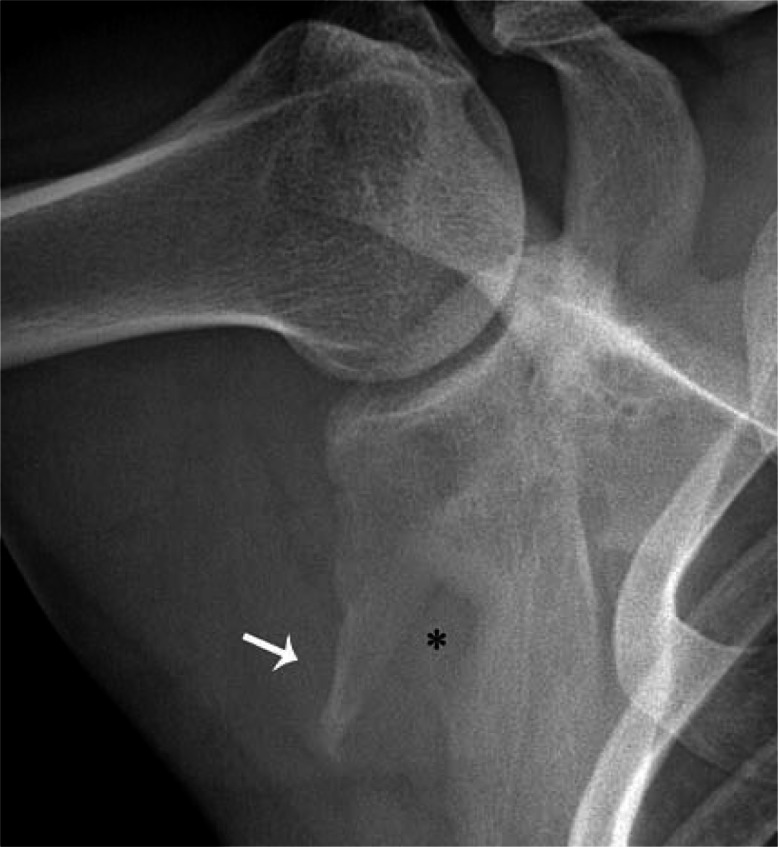
A serial radiograph of the shoulder obtained in abduction and external rotation 6 years after the injury reveals complete healing of the fractures with significant malunion consisting of a 4 cm long triangular subglenoid fragment (white arrow) and a persistent defect in the axillary border of the scapula (*)
Figure 3E:

Clinical photographs obtained 6 years after the initial injury reveal restriction of active internal rotation, extension and adduction of the right shoulder resulting in a 7.5 cm discrepancy between the right and left side on the inferior Apley’s scratch test. Note that the right hand cannot reach as high as the left.
Acknowledgments
The authors thank Leslie M. Stoklosa, DC, MS and Karol A. Donaubauer, BA, DC, CCSP for sharing their case material and Christopher J. Herrington, BS for his technical assistance with images and literature acquisition.
Footnotes
Commercial associations and conflicts of interest: None
Sources of support: None
References
- 1.Ada JR, Miller ME. Scapular fractures: Analysis of 113 cases. Clin Orthop Relat Res. 1991;269:174–180. [PubMed] [Google Scholar]
- 2.Armstrong CP, Van der Spuy J. The fractured scapula: Importance and management based on a series of 62 patients. Injury. 1984;15(5):324–9. doi: 10.1016/0020-1383(84)90056-1. [DOI] [PubMed] [Google Scholar]
- 3.McGahan JP, Rab GT, Dublin A. Fractures of the scapula. J Trauma. 1980;20(10):880–3. doi: 10.1097/00005373-198010000-00011. [DOI] [PubMed] [Google Scholar]
- 4.McGinnis M, Denton JR. Fractures of the scapula: A retrospective study of 40 fractured scapulae. J Trauma. 1989;29(11):1488–93. [PubMed] [Google Scholar]
- 5.Zlowodzki M, Bhandari M, Zelle BA, Kregor PJ, Cole PA. Treatment of scapula fractures: Systematic review of 520 fractures in 22 case series. J Orthop Trauma. 2006;20(3):230–3. doi: 10.1097/00005131-200603000-00013. [DOI] [PubMed] [Google Scholar]
- 6.Desault PJ. A treatise on fractures, luxations and other affections of the bones. Philadelphia: Fry and Kammerer; 1805. pp. 57–67. [Google Scholar]
- 7.Wilson PD. Experience of the management of fractures and dislocations (Based on an analysis of 4390 cases) by Staff of the Fracture Service MGH, Boston. Philiadelphia: JB Lippincott; 1938. [Google Scholar]
- 8.Cole PA. Scapula fractures. Orthop Clin North Am. 2002;33(1):1–18. vii. doi: 10.1016/s0030-5898(03)00069-5. [DOI] [PubMed] [Google Scholar]
- 9.Goss TP. Scapular fractures and dislocations: Diagnosis and treatment. J Am Acad Orthop Surg. 1995;3(1):22–33. doi: 10.5435/00124635-199501000-00004. [DOI] [PubMed] [Google Scholar]
- 10.Thompson DA, Flynn TC, Miller PW, Fischer RP. The significance of scapular fractures. J Trauma. 1985;25(10):974–7. doi: 10.1097/00005373-198510000-00008. [DOI] [PubMed] [Google Scholar]
- 11.Veysi VT, Mittal R, Agarwal S, Dosani A, Giannoudis PV. Multiple trauma and scapula fractures: So what? J Trauma. 2003;55(6):1145–7. doi: 10.1097/01.TA.0000044499.76736.9D. [DOI] [PubMed] [Google Scholar]
- 12.Gosens T, Speigner B, Minekus J. Fracture of the scapular body: Functional outcome after conservative treatment. J Shoulder Elbow Surg. 2009;18(3):443–8. doi: 10.1016/j.jse.2009.01.030. [DOI] [PubMed] [Google Scholar]
- 13.Salimi J, Khaji A, Karbakhsh M, Saadat S, Eftekhar B. Scapular fracture: Lower severity and mortality. Sao Paulo Med J. 2008;126(3):186–9. doi: 10.1590/S1516-31802008000300009. [DOI] [PMC free article] [PubMed] [Google Scholar]
- 14.Hudak P, Amadio PC, Bombardier C, the Upper Extremity Collaborative Group Development of an Upper Extremity Outcome Measure: The DASH (Disabilities of the Arm, Shoulder, and Hand) Am J Ind Med. 1996;29:602–608. doi: 10.1002/(SICI)1097-0274(199606)29:6<602::AID-AJIM4>3.0.CO;2-L. [DOI] [PubMed] [Google Scholar]
- 15.Tadros AM, Lunsjo K, Czechowski J, Abu-Zidan FM. Causes of delayed diagnosis of scapular fractures. Injury. 2008;39(3):314–8. doi: 10.1016/j.injury.2007.10.014. [DOI] [PubMed] [Google Scholar]
- 16.Imatani RJ. Fractures of the scapula: A review of 53 fractures. J Trauma. 1975;15(6):473–8. doi: 10.1097/00005373-197506000-00002. [DOI] [PubMed] [Google Scholar]
- 17.Scavenius M, Sloth C. Fractures of the scapula. Acta Orthop Belg. 1996;62(3):129–32. [PubMed] [Google Scholar]
- 18.Dimitroulias A, Molinero KG, Krenk DE, Muffly MT, Altman DT, Altman GT. Outcomes of nonoperatively treated displaced scapular body fractures. Clin Orthop Relat Res. 2011;469(5):1459–65. doi: 10.1007/s11999-010-1670-4. [DOI] [PMC free article] [PubMed] [Google Scholar]
- 19.Guttentag IJ, Rechtine GR. Fractures of the Scapula: A Review of the Literature. Orthop Rev. 1988;17(2):147–58. [PubMed] [Google Scholar]
- 20.Ferraz IC, Papadimitriou NG, Sotereanos DG. Scapular body nonunion: A case report. J Shoulder Elbow Surg. 2002;11(1):98–100. doi: 10.1067/mse.2002.118479. [DOI] [PubMed] [Google Scholar]
- 21.Gupta R, Sher J, Williams GR, Jr, Iannotti JP. Non-Union of the scapular body. A case report. J Bone Joint Surg Am. 1998;80:428–30. doi: 10.2106/00004623-199803000-00017. [DOI] [PubMed] [Google Scholar]
- 22.Deltoff MN, Bressler HB. Atypical scapular fracture: A case report. Am J Sports Med. 1989;17(2):292–5. doi: 10.1177/036354658901700225. [DOI] [PubMed] [Google Scholar]