Abstract
The master pacemaker in the suprachiasmatic nucleus (SCN) controls daily rhythms of behavior in mammals. C57BL/6J mice lacking Period1 (Per1−/−) are an anomaly because their SCN molecular rhythm is weak or absent in vitro even though their locomotor activity rhythm is robust. To resolve the contradiction between the in vitro and in vivo circadian phenotypes of Per1−/− mice, we measured the multi-unit activity (MUA) rhythm of the SCN neuronal population in freely-behaving mice. We found that in vivo Per1−/− SCN have high-amplitude MUA rhythms, demonstrating that the ensemble of neurons is driving robust locomotor activity in Per1−/− mice. Since the Per1−/− SCN electrical activity rhythm is indistinguishable from wild-types, in vivo physiological factors or coupling of the SCN to a known or unidentified circadian clock(s) may compensate for weak endogenous molecular rhythms in Per1−/− SCN. Consistent with the behavioral light responsiveness of Per1 −/− mice, in vivo MUA rhythms in Per1 −/− SCN exhibited large phase shifts in response to light. Since the acute response of the MUA rhythm to light in Per1−/− SCN is equivalent to wild-types, an unknown mechanism mediates enhanced light responsiveness of Per1−/− mice. Thus, Per1−/− mice are a unique model for investigating the component(s) of the in vivo environment that confers robust rhythmicity to the SCN as well as a novel mechanism of enhanced light responsiveness.
Introduction
The suprachiasmatic nucleus (SCN) is the master pacemaker in mammals that controls circadian rhythms in locomotor activity. Lesions of the SCN cause arrhythmicity of circadian rhythms [1]–[3] and transplants of fetal SCN tissue restore behavioral rhythms with the periodicity of the donor SCN [4]–[6]. A small number of rhythmic SCN cells is sufficient to drive behavioral rhythmicity, since the circadian locomotor activity rhythm was restored in hamsters by transplanting only a few SCN neurons [7]. In vitro monitoring of rhythms of neural activity and circadian gene expression has also demonstrated that the periods of rhythmic activity in SCN explants approximate the respective behavioral periodicities of wild-type and mutant rodents [8]–[13].
Together these studies have contributed to the conceptualization that SCN neurons, each having a cell-autonomous rhythm governed by molecular feedback loops, are coupled to each other resulting in an integrated period that drives the periodicity of the locomotor activity rhythm. Recent studies have identified exceptions to this tenet of mammalian circadian organization. For example, some mutant mice (Transgenic R6/2, Fmr1−/−/Fxr2−/−) have rhythmic SCN in vitro, but arrhythmic locomotor activity [14], [15]. In contrast, in C57BL/6J mice lacking the circadian gene, Period1 (Per1−/− mice), circadian gene promoter activity (Period1-luciferase) and circadian gene fusion protein expression (PERIOD2::LUCIFERASE) in Per1−/− SCN explants are arrhythmic or have low-amplitude oscillations in vitro, while the wheel-running activity rhythm of Per1−/− mice is robust and indistinguishable from wild-types [16]. The Per1-luciferase rhythms in individual cells in Per1−/− SCN explants are severely compromised (most cells are arrhythmic), but a small subset of neurons retain rhythmicity. The phenotype of Per1−/− mice is perplexing because if the SCN molecular rhythm is necessary to drive rhythmic wheel-running activity, then how is it possible that Per1−/− mice have robust activity rhythms? One possibility is that those few neurons that retain molecular rhythms in vivo in the Per1−/− SCN are sufficient to drive behavior [7]. Consistent with this hypothesis, Per1−/− mice display large behavioral phase shifts in response to light pulses, suggesting that Per1−/− SCN have low-amplitude molecular rhythms [17]. Alternatively, in vivo physiological factors (which are not present in the in vitro culture) or coupling to a rhythmic oscillator may confer robust rhythmicity to the entire population of neurons in the Per1−/− SCN.
In vivo MUA (multi-unit activity) measures the ensemble electrical activity rhythm from a large population of SCN neurons and thus is ideal for investigating the in vivo phenotype of the Per1−/− SCN. In this study, we sought to resolve the contradiction between the in vitro and in vivo circadian phenotypes of Per1−/− mice by measuring MUA from the SCN of freely-behaving mice.
Materials and Methods
Ethics Statement
All experiments were conducted in accordance with animal protocols approved by the Animal Care and Use Committees at Osaka University (permission#AD-20-042-0) and Vanderbilt University (M/08/096). All surgery was performed under isoflurane anesthesia, and all efforts were made to minimize suffering.
Animals
C57BL/6J (N10 to N11) heterozygous mPer1ldc+/− mice [16] were intercrossed to generate wild-type and mPer1ldc−/− mice that were genotyped as previously described [18]. For experiments measuring MUA, mice were bred and group-housed in the Osaka University Graduate School of Dentistry animal facility in a 12 h-light/12 h-dark cycle (12L∶12D) and provided food and water ad libitum. For experiments assessing the effects of ocular enucleation and activity feedback, mice were bred and group-housed in the Vanderbilt University animal facility in 12L∶12D and provided food and water ad libitum.
Surgery and in vivo multi-unit neural activity recording
The experiments were performed according to the technique developed by Yamazaki et al. [19] for hamsters with minor modifications for mice [20], [21]. Bipolar electrodes were constructed from pairs of epoxy-coated stainless steel wires (bare diameter, 100 µm; tip distance, 75 µm; Unique Medical, Tokyo, Japan) and an uncoated platinum-iridium wire (diameter, 75 µm; A-M Systems, Sequim, WA, USA) was used as a signal ground in the cortex. Wires were connected to a 5-pin receptacle (1.25 mm pitch; Morex, Taipei Hsien, Taiwan) wrapped in insulated copper tape.
Male mice, 24–35 weeks of age and weighing 25–32 g, were anesthetized under isoflurane (1.5%; Abbott, Tokyo, Japan) and placed in a stereotaxic device (Narishige, Tokyo, Japan). The dorsal aspects of the parietal bones were exposed and cleaned. One hole was drilled around the bregma in the parietal bones. The electrodes were inserted into the brain, aimed at the SCN (0.4 mm posterior and 0.2 mm lateral to the bregma, 5.8 mm depth from the skull surface) and attached directly to the skull with orthodontic bond (3M Unitek, Monrovia, CA, USA).
After surgery mice were singly housed in an open-top cage (410 mm width×260 mm length×270 mm height) with a running wheel (170 mm diameter). The light intensity in the recording chamber was ∼100 lux at cage level (white LED, DL18E26D Denryo, Tokyo, Japan; mounted to the top of the light-tight box). The mice were allowed to recover from surgery for at least 1 week. Thereafter, the electrodes were connected to head stage buffer amplifiers (TL082; Texas Instruments, Dallas, TX, USA) located on the head of the mouse. Buffer amplifiers were connected to a slip ring (Biotex, Kyoto, Japan) that allowed free movement of the animal. Output signals were processed by a differential input integration amplifier (INA 101 AM; Burr-Brown, Tucson, AZ, USA; gain, ×10) and then fed into an AC amplifier (band-pass, 500 Hz to 5 kHZ; gain, ×10,000). Spikes were discriminated by amplitude and counted in 1-min bins using a computer-based window discrimination system (KPCI-1801HC; Keithley Instruments, Cleveland, OH, USA). The number of wheel-running revolutions was simultaneously recorded by the same computer-based system.
At the conclusion of the experiment, mice were anesthetized and positive current (1 µA; 60 s) was passed through the recording electrodes. Brains were removed and fixed in 4% paraformaldehyde in 0.1 M phosphate buffer containing 2% potassium ferrocyanide (Sigma-Aldrich, St. Louis, MO, USA). Serial coronal sections (25 µm thick) were stained with neutral-red and blue spots of deposited iron were identified as the recording site. Data were analyzed from mice that had both recording electrodes localized to one side of the SCN with one electrode in the ventral SCN and the other in the dorsal SCN, so that the rhythm was measured from the whole SCN. This electrode localization was confirmed in 4 wild-type mice (from a total of 8 mice) and 4 Per1−/− mice (from a total of 24 mice).
Light responsiveness experiments
The onset of activity [circadian time (CT) 12] was determined for 3 d in constant darkness (DD) and linear regression (ClockLab, Actimetrics, Wilmette, IL, USA) was used to predict the onset of activity (CT12) on the day of the light pulse. At CT15, the mouse received a 15-min light pulse (intensity 100 lux, white LED as described above for recording chamber). To determine the phase shift in locomotor activity, one regression line was fit to the onset of activity for 3 d before the light pulse and the second line was fit to the onset of activity for 4 d following the light pulse (excluding the first cycle immediately after the pulse) and the phase shift was calculated that took into account the change in period that occurred with the light pulse (ClockLab). The phase shifts in the MUA rhythms were analyzed similarly except that acrophase was used as the phase marker for the MUA rhythm (ClockLab). Mean (±SD) MUA responses of the SCN to light pulses were plotted relative to baseline, which was determined from the mean counts 15 min before the light pulse and set to 100% in wild-type and Per1 −/− mice. The number of units recorded varies from mouse to mouse so the absolute baseline is different for each animal. Thus, the relative MUA response must be determined for each animal. The relative MUA response during the light pulse was determined for each mouse by dividing the average relative MUA response (%) during the 15 min light pulse by the average MUA (%) during the 15 min before the light pulse. Then the relative MUA responses during the light pulse were averaged for each genotype.
Ocular enucleation experiments
Male Per1−/− mice (n = 2; 12 weeks of age) were singly housed in cages (140 mm width×330 mm length×170 mm height) with unlimited access to a running wheel (110 mm diameter), food, and water. The cages were placed in light-tight, ventilated boxes in l2L∶12D (light intensity: 200–300 lux). Cages were changed every 3 weeks. Wheel-running activity (recorded every minute by computer) was monitored using ClockLab. Mice were anesthetized under isoflurane anesthesia and Buprenex (1 mg/kg) was administered subcutaneously immediately before bilateral ocular enucleation. After surgery, mice were returned to their cages and wheel-running was recorded in 12L∶12D for 2 days and then in DD. Wheel-running activity data were double-plotted in actograms in 5-minute bins using ClockLab.
Activity feedback experiments
Male Per1−/− mice (n = 5; 8–22 weeks of age) were singly housed in cages (140 mm width×330 mm length×170 mm height) with locked wheels (wheels were present in the cages but they could not rotate), food, and water. The cages were placed in light-tight, ventilated boxes in l2L∶12D (light intensity: 200–300 lux) for 7 days and then the mice were released into DD. Cages were changed every 3 weeks. General activity was monitored every minute with passive infrared sensors [22] and data were double-plotted in actograms in 5-min bins using ClockLab. χ2 periodogram analysis (p<0.001) were performed on days 1–14 in DD to determine if significant rhythmicity was present.
Data analysis
Actograms of wheel-running and multi-unit neural activity were generated using ClockLab analysis software. Cosinor analyses of MUA rhythms were performed using Circadian Physiology software (v2.3; http://www.circadian.org/softwar.html) [23]. Cosine curves were fit to the data (3 cycle in LD, 3 cycles in DD) in 6-min bins (all data fit with p<0.000001) using periods ranging from 22 to 26-h by 0.1-h steps. For the best period, the mesor and amplitude were determined. To determine the phase [in Zeitgeber time (ZT), where ZT0 is lights on and ZT12 is lights off) of the MUA rhythm in LD, data were fit with a cosine curve with a fixed 24-h period. The period of the wheel-running rhythm was determined by linear regression (ClockLab).
Means were statistically compared (SPSS Statistics software, IBM, Armonk, NY, USA) with independent samples t tests (two-tailed), with the following exceptions. If the variances were not homogeneous, then a Welch's t-test was used. If the data were not normally distributed (determined by the Sapiro-Wilk test), then the non-parametric Mann-Whitney U test was used. The detection of statistically different differences is limited by n = 4/group. Significance was ascribed at p<0.05. Results are expressed as the mean ± SD.
Results
In vivo MUA measures the rhythm of the SCN neuronal population
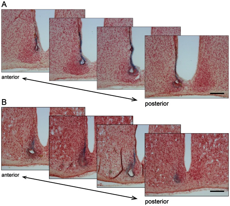
We first measured the rhythm from a large, diverse population of SCN neurons by differential recording from two bipolar electrodes, one electrode implanted in the ventral (core) region and the other electrode in the dorsal (shell) region of wild-type and Per1−/− SCN. Histological examination of each SCN after recording showed that one electrode was placed in the ventral (core) region and the other in the dorsal (shell) region of the SCN in wild-type (Fig. 1A) and Per1−/− mice (Fig. 1B). These data demonstrate that MUA rhythms were measured from the whole SCN in all animals examined.
Figure 1. Histological examination of recording sites in the ventral and dorsal SCN.
Representative serial coronal sections (25 µm, every other section shown) of a wild-type (A) and Per1−/− (B) SCN following MUA recording. Sections were stained with neutral-red and blue spots of deposited iron are the recording sites. Recording sites were present in the ventral and dorsal SCN. The anterior to posterior orientation of the sections is indicated. Scale bar represents 200 µm. The data in A and B correspond to the data shown in Fig. 2A and B, respectively.
In vivo MUA rhythms in Per1−/− SCN are robust
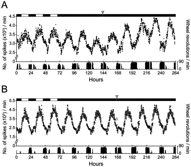
To assess the in vivo phenotype of the Per1−/− SCN, we measured MUA from freely moving wild-type (Fig. 2A) and Per1−/− (Fig. 2B) mice in the light-dark cycle (LD) and in constant darkness (DD). In vivo, the amplitude of neural activity in Per1−/− SCN was indistinguishable from wild-types in LD and DD (Table 1), indicating that the many neurons in the Per1−/− SCN are rhythmic. The phases of the MUA rhythms in LD did not differ between wild-type (ZT: 6.25±0.31) and Per1−/− (ZT: 6.49±0.89) mice (t-test, p = 0.628). The period of the MUA rhythm in DD was slightly, but statistically significantly, longer in Per1−/− SCN compared to wild-type SCN (Table 1).
Figure 2. The in vivo circadian rhythm in the multi-unit neural activity of Per1 −/− SCN is similar to wild-type SCN.
Representative serial-plotted actograms (6-min bins) of multi-unit neural activity (top trace; y-axis: spikes/min) recorded from wild-type (A; black circles) and Per1−/− (B; open circles) SCN in freely-behaving mice. The bottom trace is simultaneously-recorded wheel-running activity (y-axis: wheel revolutions/min). Open bars are light and closed bars are dark (top of figure). A 15-min light pulse (indicated by open inverted triangle; 100 lux) was administered during the early subjective night (CT15). The number of spikes (for MUA) or wheel revolutions were counted every minute and integrated in 6-min bins.
Table 1. Comparison of circadian rhythm parameters in wild-type and Per1−/ − mice.
| Parameter | wild-type | Per1−/− | p-value |
| MUA in LD | |||
| Mesor (spikes/min) | 4339±2351 | 6781±6466 | 0.773### |
| Amplitude (spikes/min) | 1415±1119 | 1258±717 | 0.820# |
| Period (h) | 23.93±0.39 | 24.18±0.56 | 0.491# |
| MUA in DD | |||
| Mesor (spikes/min) | 4171±1968 | 6615±6475 | 0.686# |
| Amplitude (spikes/min) | 1150±825 | 1085±509 | 0.898# |
| Period (h) | 23.60±0.14 | 23.85±0.13 | 0.040# |
| Wheel-running period (h) | 23.80±0.23 | 23.87±0.12 | 0.885### |
| Light pulse at CT15 | |||
| Relative MUA during light pulse (%) | 124±18 | 117±19 | 0.579# |
| Phase shift in wheel-running activity | −1.50±0.84 | −3.10±0.21 | 0.028## |
| Phase shift in MUA | −1.59±0.30 | −3.00±0.42 | 0.002# |
Cosine curves were fit to the multi-unit (MUA) neural activity data recorded from wild-type or Per1−/− SCN in the light-dark cycle (LD; 3 d) or in constant darkness (DD; 3 d) using periods ranging from 22 to 26-h by 0.1-h steps. For the best period, the mesor and amplitude were determined. Fifteen-min light pulses (100 lux) were administered at CT15 (3 h after activity onset). The relative MUA response during the light pulse was determined for each mouse by dividing the average relative MUA response (%) during the 15 min light pulse by the average MUA (%) during the 15 min before the light pulse. Then the mean relative MUA responses during the light pulse were averaged for each genotype. To determine the magnitudes of the phase shifts, one regression line was fit to the onset of activity or acrophase of the MUA rhythm for 3 d before the light pulse and the second line was fit to the 4 d following the light pulse (excluding the first cycle immediately after the pulse) and the phase shift was calculated that took into account the change in period that occurred with the light pulse. Data are the mean ± SD; n = 4 wild-type and n = 4 Per1 −/− mice.
Student's t-test,
Welch's t-test,
Mann-Whitney U test.
Robust circadian locomotor activity rhythms persist in Per1−/− mice after ocular enucleation and removal of activity feedback
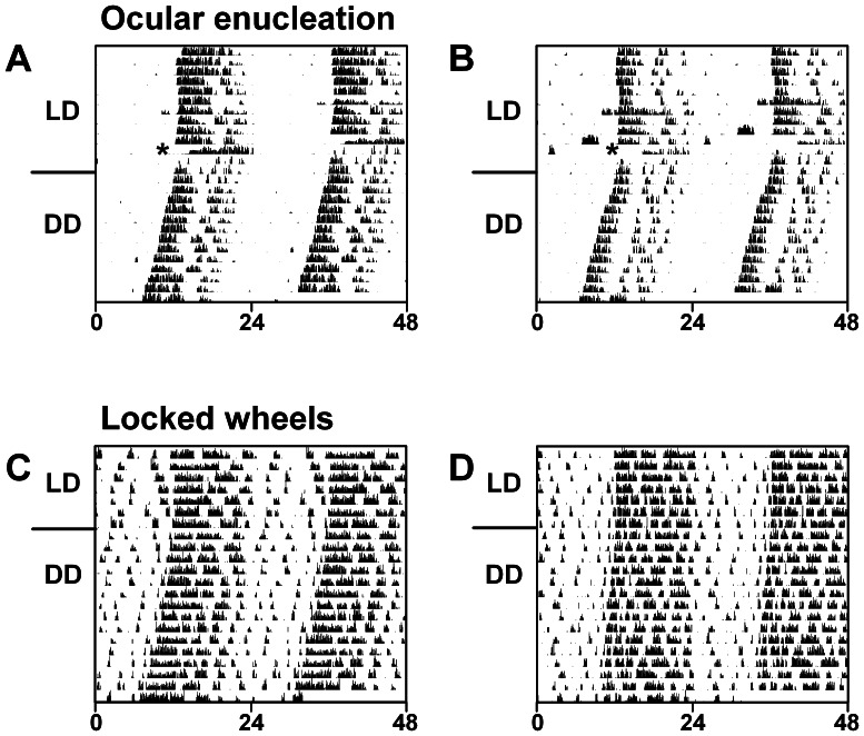
To determine if robust oscillations were conferred to the Per1−/− SCN by coupling of the SCN with the circadian clock in the retina, we recorded wheel-running activity after ocular enucleation of Per1−/− mice (Fig. 3A, B). We found that robust circadian rhythms of wheel-running persisted after ocular enucleation, suggesting that Per1−/− SCN rhythmicity is not conferred by coupling to the retina clock.
Figure 3. Robust locomotor behavior rhythms in Per1−/− mice persist after ocular enucleation and in the absence of wheel-running activity feedback.
Double-plotted actograms (5-min bins) of locomotor activity of male Per1−/− mice (x-axis: time in hours; y-axis: days). A, B. Wheel-running activity was recorded from Per1−/− mice (n = 2) maintained in 12L∶12D (LD) and ocular nucleation was performed (indicated by asterisks). Two days after surgery, mice were released into constant darkness (DD). C, D. Per1−/− mice (n = 5) were housed in cages with locked running wheels (wheels were present but could not rotate), maintained in 12L∶12D and then released into DD. General activity was continuously monitored with passive infrared sensors.
It has previously shown that SCN MUA is altered by running wheel activity [19], [21], [24]. To determine if activity feedback to the SCN contributed to robust SCN rhythms in Per1−/− mice, we analyzed general activity of Per1−/− mice housed with locked running wheels (wheels were present in the cage but could not rotate; Fig. 3C, D). We found that circadian locomotor activity rhythms of Per1−/− mice were robust in LD and DD in the absence of activity feedback (χ2 periodogram detected significant rhythmicity in all Per1−/− mice after ocular enucleation).
Light responsiveness of the SCN in Per1−/− mice
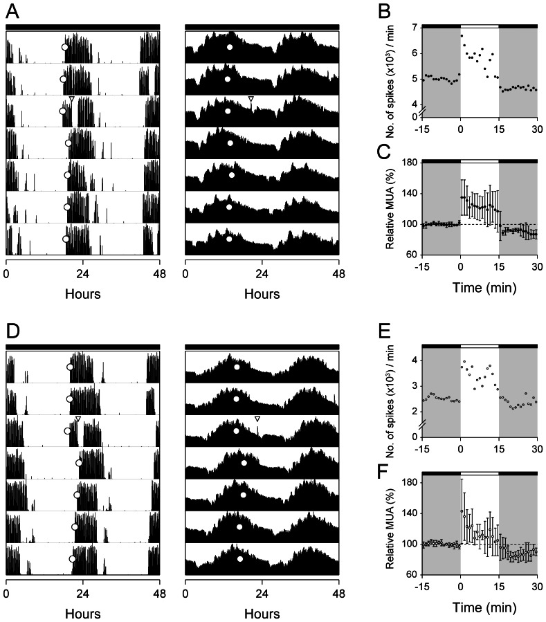
Per1−/− mice exhibit large phase shifts (∼4 h) in behavior in response to light pulses administered during the early subjective night [17]. To assess the response of the Per1−/− SCN to light, we simultaneously measured MUA in the SCN and wheel-running activity in wild-type (Fig. 4A) and Per1−/− (Fig. 4D) mice administered 15-min light pulses at CT15. We found that the acute increase in MUA and the duration of the acute increase in the SCN in response to the light pulse did not differ between wild-type (Fig. 4B, C) and Per1−/− (Fig. 4E, F) mice (Table 1: Mean relative MUA during light pulse). The magnitudes of the phase shifts in the SCN and wheel-running activity rhythms were greater in Per1−/− mice compared to wild-types (Table 1). In both genotypes, the magnitude of the phase shift in the MUA rhythm matched that of the phase shift in behavior.
Figure 4. Behavioral and SCN responses to light.
Representative double-plotted actograms of wheel-running activity (left panels) and multi-unit neural activity in the SCN (right panels) of wild-type (A) and Per1 −/− (D) mice. Each line represents two consecutive days (48 hr), with time in 6-min bins plotted left to right. Consecutive days are aligned vertically. The mice were maintained in constant darkness (indicated by the black bars above actograms) and a 15-min light pulse (100 lux) was administered 3 hr after activity onset (CT15; open inverted triangle). Open circles indicate the respective phase markers: activity onset for wheel-running activity and acrophase for MUA. The magnitudes of phase shifts were determined by measuring the phase differences between least-squares fitted regression lines through the respective phase markers before and after the light pulse [locomotor: −2.20 h, MUA: −1.94 h in (A); locomotor: −3.24 h, MUA:−2.60 h in (D)]. To show the acute response of the SCN to light, MUA data (plotted in 1-min bins) surrounding the light pulse in (A) and (D) are magnified in (B) and (E), respectively. The time of the light pulse is indicated by the open bars at the tops of the figures and dark is indicated by gray shading. Mean (±SD) MUA responses in wild-type (C; n = 4) and Per1−/− (F; n = 4) SCN are shown relative to baseline (baseline determined from the mean counts 15 min before the light pulse and set to 100% in wild-type and Per1 −/− mice).
Discussion
C57BL/6J Per1−/− mice are unique because their circadian behavioral phenotype, which is indistinguishable from wild-types, is not predicted from their arrhythmic (or weakly rhythmic) in vitro SCN phenotype. This discrepancy between the in vivo behavioral and in vitro SCN phenotypes raises intriguing questions regarding the mechanisms whereby the SCN controls circadian behavior in these mice. Is the ensemble of neurons in Per1−/− SCN rhythmic in vivo? Does a physiological factor(s) present in the in vivo environment confer high-amplitude oscillations to the Per1−/− SCN? Is a robust rhythm imparted to the Per1−/− through coupling with an extra-SCN oscillator? To address these questions, we examined the in vivo phenotype of the Per1−/− SCN by simultaneously monitoring MUA in the SCN and wheel-running activity in freely-behaving Per1−/− mice. We found that in vivo Per1−/− SCN had robust, high-amplitude MUA rhythms in the light-dark cycle and in constant darkness that were similar to wild-type mice. The MUA rhythm we measured reflects the neural activity of a large population of SCN neurons because we obtained differential recordings from electrodes implanted in the ventral and dorsal SCN. Thus, the high-amplitude MUA rhythm in the Per1−/− SCN demonstrates that the ensemble of neurons, rather than a few rhythmic neurons, is driving robust locomotor activity in Per1−/− mice. Similar to a previous study demonstrating that the age-related decline in the amplitude of the locomotor activity rhythm was reflected in the amplitude of the in vivo MUA SCN rhythm (but not the rhythm of PER2::LUC expression in ex vivo SCN), this study demonstrates that in vivo MUA is a reliable predictor of circadian behavior [20].
These data suggest that some component of the in vivo environment confers robust rhythmicity to the Per1−/− SCN. Our finding that the in vivo environment may be pro-rhythmic is reminiscent of previous studies that demonstrated differences between the in vitro and in vivo effects of tetrodotoxin (TTX) on the SCN rhythm-inhibition of action potentials with TTX had marked effects on the maintenance of the SCN oscillation in vitro [25] but seemingly not in vivo [26].
Physiological factors, such as temperature and hormones, are candidates for modulating in vivo rhythmicity. Alternatively, a rhythmic circadian clock may exist in Per1−/− mice that couples to the SCN, resulting in high-amplitude SCN MUA rhythms. The retina is one such candidate oscillator, as our previous study suggested that coupling of the retina to the SCN stabilizes the free-running period of locomotor activity in hamsters [27]. However, we found that robust circadian activity rhythms persisted in Per1−/− mice after ocular enucleation, suggesting that coupling to the retina clock does not confer robust rhythmicity to the Per1−/− SCN. Consistent with our result, it was recently demonstrated that Per1−/− retinas are arrhythmic in vitro [28], thus the retina is not a likely candidate as a rhythmic circadian clock in Per1−/− mice.
Since all peripheral tissues in Per1−/− mice that have been examined are arrhythmic in vitro [9], [16], perhaps an unidentified circadian clock, whose rhythm persists without Per1, couples to the SCN [29]. It is also possible that the Per1−/− SCN may be coupled to a rhythmic oscillator that is closely apposed to the SCN. Coronal 300 µm Per1−/− SCN explants are arrhythmic in vitro, but we cannot rule out the possibility that a nucleus just rostral or caudal to the SCN confers robust rhythmicity to the SCN in vivo (a thicker coronal section of the SCN cannot be examined because it will not survive in culture).
Physical activity induced by running wheel availability, forced treadmill running, or drug administration shifts the phase of rodent circadian rhythms and free-running periods of circadian rhythms are shortened in the presence of a running wheel [30]–[34]. We have previously demonstrated that wheel-running activity feeds back to the SCN and decreases its neural activity in hamsters and mice [19], [24] thus it is possible that wheel-running activity drives robust MUA rhythms in the Per1−/− SCN. However, we found that robust locomotor activity rhythms persist in Per1−/− mice housed with locked running wheels, suggesting that wheel-running activity feedback is not responsible for robust SCN rhythms in Per1−/− mice.
Per1−/− mice exhibit large behavioral phase shifts in response to light pulses administered during the subjective night [17]. A previous study demonstrated that the augmented light responsiveness of Clock mutant (Δ19) heterozygous mice is attributed to their low-amplitude SCN rhythms [35]. In contrast to Clock mutant mice, we found that Per1−/− SCN have high-amplitude MUA rhythms, suggesting that the enhanced light responsiveness of Per1−/− mice cannot be attributed to the amplitude of the rhythm in the SCN. However, a recent study demonstrated that coupled limit cycle oscillators may behave differently from single oscillators such that the inverse relationship between MUA amplitude and phase-shifting capacity may not hold true for neuronal networks [36]. It is also possible that the in vivo MUA rhythm does not reflect the amplitude of the molecular timekeeping rhythm, such that the molecular rhythm could be low-amplitude while the MUA rhythm is robust. The relationship between the amplitude of the molecular rhythm and the amplitude of the electrical activity rhythm in the SCN should be further examined.
Large behavioral phase shifts may also occur if the Per1−/− SCN has enhanced sensitivity to light. If so, then the acute MUA response to light pulses would be greater in Per1−/− SCN compared to wild-types. However, we found that the magnitude and duration of the acute MUA responses of the wild-type and Per1−/− SCN to light were identical to each other, suggesting that they have equivalent sensitivities to light. Alternatively, it is possible that the sensitivity of the molecular timekeeping rhythm is altered in Per1−/− SCN, which could then account for enhanced light responsiveness of Per1−/− mice.
Together these data suggest that the high-amplitude rhythm of the Per1−/− SCN is conferred by the in vivo environment, resulting in robust wheel-running activity of Per1−/− mice. Whether a physiological factor or coupling to another oscillator (or both) confers rhythmicity to the Per1−/− SCN is unknown. Furthermore, the enhanced light responsiveness of Per1−/− mice cannot be attributed to either a low-amplitude MUA rhythm in the SCN or to enhanced responsiveness of the MUA rhythm in the SCN to light. Thus, the mechanism of enhanced light responsive in Per1−/− mice is unknown, but does not appear to operate at the level of the MUA rhythm. Future studies of C57BL/6J Per1−/− mice are necessary to elucidate the in vivo physiological factor or unidentified circadian clock(s) that compensates for weak endogenous rhythms in Per1−/− SCN and to identify the mechanism of enhanced responses to light.
Funding Statement
This work was supported by the Japan Science and Technology Agency PRESTO program and the Program to Disseminate Tenure Tracking System from the Ministry of Education, Culture, Sports, Science, and Technology of Japan (to WN), and a Japan Society for Promotion of Science postdoctoral fellowship (to NNT). The funders had no role in study design, data collection and analysis, decision to publish, or preparation of the manuscript.
References
- 1. Moore RY, Eichler VB (1972) Loss of a circadian adrenal corticosterone rhythm following suprachiasmatic lesions in the rat. Brain Res 42: 201–206. [DOI] [PubMed] [Google Scholar]
- 2. Rusak B, Zucker I (1979) Neural regulation of circadian rhythms. Physiol Rev 59: 449–526. [DOI] [PubMed] [Google Scholar]
- 3. Stephan FK, Zucker I (1972) Circadian rhythms in drinking behavior and locomotor activity of rats are eliminated by hypothalamic lesions. Proc Natl Acad Sci U S A 69: 1583–1586. [DOI] [PMC free article] [PubMed] [Google Scholar]
- 4. Lehman MN, Silver R, Gladstone WR, Kahn RM, Gibson M, et al. (1987) Circadian rhythmicity restored by neural transplant. Immunocytochemical characterization of the graft and its integration with the host brain. J Neurosci 7: 1626–1638. [DOI] [PMC free article] [PubMed] [Google Scholar]
- 5. Ralph MR, Foster RG, Davis FC, Menaker M (1990) Transplanted suprachiasmatic nucleus determines circadian period. Science 247: 975–978. [DOI] [PubMed] [Google Scholar]
- 6. Sawaki Y, Nihonmatsu I, Kawamura H (1984) Transplantation of the neonatal suprachiasmatic nuclei into rats with complete bilateral suprachiasmatic lesions. Neurosci Res 1: 67–72. [DOI] [PubMed] [Google Scholar]
- 7. Silver R, Lehman MN, Gibson M, Gladstone WR, Bittman EL (1990) Dispersed cell suspensions of fetal SCN restore circadian rhythmicity in SCN-lesioned adult hamsters. Brain Res 525: 45–58. [DOI] [PubMed] [Google Scholar]
- 8. Herzog ED, Takahashi JS, Block GD (1998) Clock controls circadian period in isolated suprachiasmatic nucleus neurons. Nat Neurosci 1: 708–713. [DOI] [PubMed] [Google Scholar]
- 9. Liu AC, Welsh DK, Ko CH, Tran HG, Zhang EE, et al. (2007) Intercellular coupling confers robustness against mutations in the SCN circadian clock network. Cell 129: 605–616. [DOI] [PMC free article] [PubMed] [Google Scholar]
- 10. Liu C, Weaver DR, Strogatz SH, Reppert SM (1997) Cellular construction of a circadian clock: period determination in the suprachiasmatic nuclei. Cell 91: 855–860. [DOI] [PubMed] [Google Scholar]
- 11. Meng QJ, Logunova L, Maywood ES, Gallego M, Lebiecki J, et al. (2008) Setting clock speed in mammals: the CK1 epsilon tau mutation in mice accelerates circadian pacemakers by selectively destabilizing PERIOD proteins. Neuron 58: 78–88. [DOI] [PMC free article] [PubMed] [Google Scholar]
- 12. Nakamura W, Honma S, Shirakawa T, Honma K (2002) Clock mutation lengthens the circadian period without damping rhythms in individual SCN neurons. Nat Neurosci 5: 399–400. [DOI] [PubMed] [Google Scholar]
- 13. Yoo SH, Ko CH, Lowrey PL, Buhr ED, Song EJ, et al. (2005) A noncanonical E-box enhancer drives mouse Period2 circadian oscillations in vivo. Proc Natl Acad Sci U S A 102: 2608–2613. [DOI] [PMC free article] [PubMed] [Google Scholar]
- 14. Pallier PN, Maywood ES, Zheng Z, Chesham JE, Inyushkin AN, et al. (2007) Pharmacological imposition of sleep slows cognitive decline and reverses dysregulation of circadian gene expression in a transgenic mouse model of Huntington's disease. J Neurosci 27: 7869–7878. [DOI] [PMC free article] [PubMed] [Google Scholar]
- 15. Zhang J, Fang Z, Jud C, Vansteensel MJ, Kaasik K, et al. (2008) Fragile X-related proteins regulate mammalian circadian behavioral rhythms. Am J Hum Genet 83: 43–52. [DOI] [PMC free article] [PubMed] [Google Scholar]
- 16. Pendergast JS, Friday RC, Yamazaki S (2009) Endogenous rhythms in Period1 mutant suprachiasmatic nuclei in vitro do not represent circadian behavior. J Neurosci 29: 14681–14686. [DOI] [PMC free article] [PubMed] [Google Scholar]
- 17. Pendergast JS, Friday RC, Yamazaki S (2010) Photic entrainment of period mutant mice is predicted from their phase response curves. J Neurosci 30: 12179–12184. [DOI] [PMC free article] [PubMed] [Google Scholar]
- 18. Bae K, Jin X, Maywood ES, Hastings MH, Reppert SM, et al. (2001) Differential functions of mPer1, mPer2, and mPer3 in the SCN circadian clock. Neuron 30: 525–536. [DOI] [PubMed] [Google Scholar]
- 19. Yamazaki S, Kerbeshian MC, Hocker CG, Block GD, Menaker M (1998) Rhythmic properties of the hamster suprachiasmatic nucleus in vivo. J Neurosci 18: 10709–10723. [DOI] [PMC free article] [PubMed] [Google Scholar]
- 20. Nakamura TJ, Nakamura W, Yamazaki S, Kudo T, Cutler T, et al. (2011) Age-related decline in circadian output. J Neurosci 31: 10201–10205. [DOI] [PMC free article] [PubMed] [Google Scholar]
- 21. Nakamura W, Yamazaki S, Nakamura TJ, Shirakawa T, Block GD, et al. (2008) In vivo monitoring of circadian timing in freely moving mice. Curr Biol 18: 381–385. [DOI] [PubMed] [Google Scholar]
- 22. Pendergast JS, Branecky KL, Yang W, Ellacott KL, Niswender KD, et al. (2013) High-fat diet acutely affects circadian organisation and eating behavior. Eur J Neurosci [Epub ahead of print]. [DOI] [PMC free article] [PubMed] [Google Scholar]
- 23.Refinetti R (2006) Circadian physiology. Boca Raton: CRC Press/Taylor & Francis Group.
- 24. van Oosterhout F, Lucassen EA, Houben T, vanderLeest HT, Antle MC, et al. (2012) Amplitude of the SCN clock enhanced by the behavioral activity rhythm. PLoS One 7: e39693. [DOI] [PMC free article] [PubMed] [Google Scholar]
- 25. Yamaguchi S, Isejima H, Matsuo T, Okura R, Yagita K, et al. (2003) Synchronization of cellular clocks in the suprachiasmatic nucleus. Science 302: 1408–1412. [DOI] [PubMed] [Google Scholar]
- 26. Schwartz WJ, Gross RA, Morton MT (1987) The suprachiasmatic nuclei contain a tetrodotoxin-resistant circadian pacemaker. Proc Natl Acad Sci U S A 84: 1694–1698. [DOI] [PMC free article] [PubMed] [Google Scholar]
- 27. Yamazaki S, Alones V, Menaker M (2002) Interaction of the retina with suprachiasmatic pacemakers in the control of circadian behavior. J Biol Rhythms 17: 315–329. [DOI] [PubMed] [Google Scholar]
- 28. Ruan GX, Gamble KL, Risner ML, Young LA, McMahon DG (2012) Divergent roles of clock genes in retinal and suprachiasmatic nucleus circadian oscillators. PLoS One 7: e38985. [DOI] [PMC free article] [PubMed] [Google Scholar]
- 29. Pendergast JS, Oda GA, Niswender KD, Yamazaki S (2012) Period determination in the food-entrainable and methamphetamine-sensitive circadian oscillator(s). Proc Natl Acad Sci U S A 109: 14218–14223. [DOI] [PMC free article] [PubMed] [Google Scholar]
- 30. Edgar DM, Kilduff TS, Martin CE, Dement WC (1991) Influence of running wheel activity on free-running sleep/wake and drinking circadian rhythms in mice. Physiol Behav 50: 373–378. [DOI] [PubMed] [Google Scholar]
- 31. Shioiri T, Takahashi K, Yamada N, Takahashi S (1991) Motor activity correlates negatively with free-running period, while positively with serotonin contents in SCN in free-running rats. Physiol Behav 49: 779–786. [DOI] [PubMed] [Google Scholar]
- 32. Turek FW, Losee-Olson S (1986) A benzodiazepine used in the treatment of insomnia phase-shifts the mammalian circadian clock. Nature 321: 167–168. [DOI] [PubMed] [Google Scholar]
- 33. Yamada N, Shimoda K, Ohi K, Takahashi S, Takahashi K (1988) Free-access to a running wheel shortens the period of free-running rhythm in blinded rats. Physiol Behav 42: 87–91. [DOI] [PubMed] [Google Scholar]
- 34. Yamada N, Shimoda K, Takahashi K, Takahashi S (1990) Relationship between free-running period and motor activity in blinded rats. Brain Res Bull 25: 115–119. [DOI] [PubMed] [Google Scholar]
- 35. Vitaterna MH, Ko CH, Chang AM, Buhr ED, Fruechte EM, et al. (2006) The mouse Clock mutation reduces circadian pacemaker amplitude and enhances efficacy of resetting stimuli and phase-response curve amplitude. Proc Natl Acad Sci U S A 103: 9327–9332. [DOI] [PMC free article] [PubMed] [Google Scholar]
- 36. vanderLeest HT, Rohling JH, Michel S, Meijer JH (2009) Phase shifting capacity of the circadian pacemaker determined by the SCN neuronal network organization. PLoS One 4: e4976. [DOI] [PMC free article] [PubMed] [Google Scholar]


