Summary
Medulloblastoma is an aggressively-growing tumour, arising in the cerebellum or medulla/brain stem. It is the most common malignant brain tumour in children, and displays tremendous biological and clinical heterogeneity1. Despite recent treatment advances, approximately 40% of children experience tumour recurrence, and 30% will die from their disease. Those who survive often have a significantly reduced quality of life.
Four tumour subgroups with distinct clinical, biological and genetic profiles are currently discriminated2,3. WNT tumours, displaying activated wingless pathway signalling, carry a favourable prognosis under current treatment regimens4. SHH tumours show hedgehog pathway activation, and have an intermediate prognosis2. Group 3 & 4 tumours are molecularly less well-characterised, and also present the greatest clinical challenges2,3,5. The full repertoire of genetic events driving this distinction, however, remains unclear.
Here we describe an integrative deep-sequencing analysis of 125 tumour-normal pairs. Tetraploidy was identified as a frequent early event in Group 3 & 4 tumours, and a positive correlation between patient age and mutation rate was observed. Several recurrent mutations were identified, both in known medulloblastoma-related genes (CTNNB1, PTCH1, MLL2, SMARCA4) and in genes not previously linked to this tumour (DDX3X, CTDNEP1, KDM6A, TBR1), often in subgroup-specific patterns. RNA-sequencing confirmed these alterations, and revealed the expression of the first medulloblastoma fusion genes. Chromatin modifiers were frequently altered across all subgroups.
These findings enhance our understanding of the genomic complexity and heterogeneity underlying medulloblastoma, and provide several potential targets for new therapeutics, especially for Group 3 & 4 patients.
As a first phase of the International Cancer Genome Consortium (ICGC) PedBrain Tumor Project (www.pedbraintumor.org), we have collected matched tumour and germline samples from 125 medulloblastoma patients aged from 0–17 years (Supplementary Table 1). Whole-genome sequencing (WGS, n=39) and whole-exome sequencing (WES, n=21) were applied to a ‘Discovery’ set, with a custom-capture approach used to sequence 2,734 genes in an additional ‘Replication’ set (n=65). All tumour samples were obtained at primary diagnosis, prior to adjuvant therapy, and the distribution of molecular subgroups was similar across cohorts (Supplementary Figure 1).
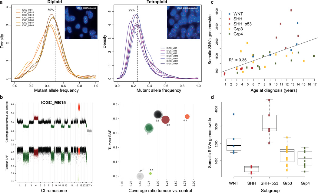
Investigation of genome-wide somatic mutation allele frequencies identified several cases with a clear peak at ~25%, rather than the expected ~50% allele frequency for early, heterozygous events (Figure 1a). Analysis of coverage depth and allele frequencies in regions of copy-number change ruled out stromal contamination, but rather suggested a tetraploid baseline in the tumour genome (Figure 1b). Predicted ploidy status was confirmed by fluorescence in situ hybridisation (FISH) using multiple centromeric probes in 17/18 cases analysed (Figure 1a). The extremely low fraction of mutations at ~50% allele frequency suggests that genome duplication occurred very early during tumourigenesis. Some cases likely went through even higher polyploidy states before reaching a ~4n baseline (e.g. ICGC_MB45, displaying 4n chromosomes with 4:0 or 3:1 allele ratios; Supplementary Figure 2). Across the Discovery set, tetraploidy was most commonly observed in Group 3 (7/13, 54%) and Group 4 tumours (8/20, 40%), followed by SHH (4/14, 29%) and WNT tumours (1/7, 14%). Interestingly, the four tetraploid SHH tumours all harboured TP53 mutations and also displayed chromothripsis6. Tetraploid Group 3 & 4 tumours showed significantly more large-scale copy number alterations compared with diploid cases (median 10 changes per tumour in tetraploid versus 4 per tumour in diploid cases, p=0.008, two-tailed Mann-Whitney U test; Supplementary Figure 3). Thus, tetraploidy followed by genomic instability may be an early driving event in a large proportion of Group 3 & 4 medulloblastomas, which pose a significant clinical challenge due to their dismal prognosis and lack of targeted treatment options. Novel classes of drugs such as mitotic checkpoint kinase or kinesin inhibitors, which target the maintenance of tetraploidy through successive cell divisions, may therefore represent a rational therapeutic strategy in these cases7,8. The value of tetraploidy as a prognostic marker also requires further investigation.
Figure 1. Tetraploidy is a frequent early event in MB tumourigenesis, and mutation rates vary with age and subgroup.
a, Distributions of genome-wide somatic mutation allele frequencies (the proportion of sequence reads supporting a mutation) for diploid tumours (with a peak at ~50% for heterozygous events, n=7) and tetraploid cases (with a peak at ~25%, n=7). Insets show centromeric FISH for chromosomes 1 (red) and 11 (green), confirming the predicted ploidy status.
b, Top left: Rescaled tumour:germline coverage ratio, indicating copy-number gains (red) or losses (green). Bottom left: B-Allele frequency (BAF) in the tumour at SNP positions which are heterozygous in the germline. Right: Genome alteration print (GAP) of segmented copy number and allele frequency profiles. Chromosomes with predicted 3:0/2:1/3:2 allele ratios show a BAF of ~0/0.33/0.4 and coverage ratios of ~0.75/0.75/1.25. Due to random sampling, the 2:2 allele ratio is slightly below 0.5.
c, Genome-wide somatic mutation rates are positively correlated with patient age (n=39).
d, Distribution of somatic mutation rates by tumour subgroup (n=39). p-values are according to a Wilcoxon rank-sum test with Bonferroni correction. SHH-p53: SHH-subgroup tumours harbouring a somatic or germline TP53 mutation.
The average somatic mutation rate in the WGS cohort was 0.52/Mb, with an average of 10.3 non-synonymous coding single nucleotide variants (SNVs) in the Discovery cohort (Supplementary Table 2). This is slightly higher than previously reported for medulloblastoma9, possibly due to improved coverage and technical sensitivity, but considerably lower than in deep-sequenced adult tumours, e.g.10,11. There were significantly fewer transitions in the somatic alterations compared with germline variation (p=4.6×10⁻⁷, Wilcoxon rank-sum test; Supplementary Figure 4). All coding somatic SNVs identified in the combined cohort are listed in Supplementary Table 3.
We identified a positive correlation between genome-wide mutation rate and patient age, as previously reported for coding mutations9 (r2 = 0.35, p=7.8×10⁻⁵ Pearson's product-moment correlation; Figure 1c). Intriguingly, this association was more pronounced in diploid tumours (r2 = 0.52, p=3×10⁻⁵), and virtually absent in tetraploid cases (r2 = 0.04, p=0.5) (Supplementary Figure 5a,b). A similar trend was observed for non-synonymous mutations across the Discovery cohort (Supplementary Figure 5c). Coverage level did not correlate with mutation rate (Supplementary Figure 5d). One explanation may be that all medulloblastomas originate during embryogenesis, with some tumours needing to accumulate more genetic ‘hits’ before becoming symptomatic. Alternatively, tumours arising in older patients may derive from more differentiated cells that require a greater number of alterations to undergo malignant transformation. Investigation of additional tumours from older patients may help to clarify this.
Five SHH tumours harbouring TP53 mutations, including three previously described Li-Fraumeni syndrome (LFS)-associated tumours with germline mutations6, one newly-identified LFS case (ICGC_MB23), and one somatically mutated tumour (ICGC_MB34), had significantly more mutations than the remaining cases, both genome wide (mean 1.1/Mb vs 0.43/Mb, p=4.5×10⁻⁶; two-tailed t-test) and for non-synonymous changes (mean 23 vs 8.8, p=2.6×10⁻⁶). Interestingly, the WNT subgroup, which typically shows a good prognosis and few copy-number changes, had the next highest mutation rate (Figure 1d).
Forty-one somatic, coding, small insertions/deletions (InDels) were identified across the cohort, with an average of 0.4 coding InDels per case in the Discovery set (range 0–2; Supplementary Table 4). Some genes, however, were more commonly affected by InDels than SNVs. For example, frameshift InDels in PTCH1 were detected in 6/125 cases, while only 2 SNVs were observed. Recurrent InDels were also seen in the chromatin modifiers MLL2, KDM6A (3 cases each) and BCOR (2 cases).
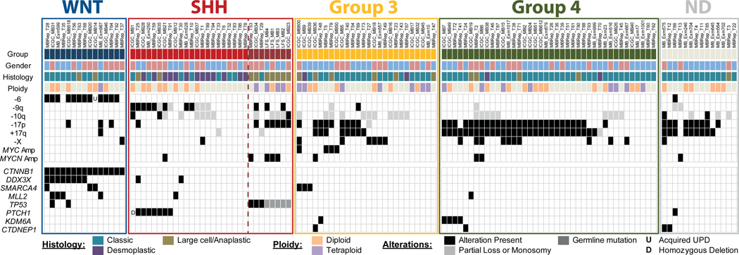
In contrast to another paediatric brain tumour, glioblastoma, in which we recently identified frequently recurrent hotspot mutations12, the majority of mutated genes in this study were unique to a single case (587/760 non-synonymous SNVs in the 125 cases, 77%) - demonstrating the pronounced genetic heterogeneity of medulloblastoma. Twenty-five of these singleton mutations, and 53 SNVs in total, were at positions listed in the COSMIC database of somatic alterations in tumours (available at http://http://www.sanger.ac.uk/genetics/CGP/cosmic/), suggesting a rare but important contribution of many known cancer genes in MB (Supplementary Table 5). Only 8 genes were somatically altered in more than 3% of the whole series: CTNNB1 (15 cases, 12%); DDX3X (10 cases, 8%); PTCH1 (8 cases, 6%), SMARCA4 (6 cases, 5%), MLL2 (6 cases, 5%), TP53 (somatically mutated in 5 cases, 4%), KDM6A (5 cases, 4%) and CTDNEP1 (4 cases, 3%) (Figure 2). These were also the only genes found to be significantly altered upon analysis of the combined cohort with MutSig - an algorithm testing whether the observed mutations in a gene are not simply a consequence of random background mutation processes. It takes into account gene length and composition, silent to non-silent mutation ratios, and other factors (see https://confluence.broadinstitute.org/display/CGATools/MutSig; Supplementary Table 6). Large-scale copy-number changes known to be associated with medulloblastoma, such as formation of an isodicentric 17q and losses of 10q / 9q / X13–15, were more frequently recurrent than SNVs (Supplementary Figure 6a–e).
Figure 2. Subgroup specificity of common genetic alterations.
Summary of clinical data and recurrent alterations in the combined cohort (n=125). Genes which were found to be significantly mutated by MutSig analysis were included. UPD: uniparental disomy, ND: no material available for conclusive molecular subgroup assignment.
Many alterations were enriched in specific medulloblastoma subgroups. For example, all of the WNT tumours (15/15) harboured a mutation in CTNNB1, and 13/15 displayed loss of one copy of chromosome 6 (or acquired uniparental disomy in one case) – alterations which have previously been associated with this subgroup 4,13,15. Mutations in DDX3X were also clearly enriched in WNT tumours (adjusted p=7.06×10⁻⁶, two-tailed Fisher’s exact test with a Bonferroni correction), and these mutations were clustered within the helicase domain (Supplementary Figure 7a). Three were localised at the RNA binding surface of the protein and three were predicted to disrupt the closed (RNA binding) conformation (Supplementary Figure 7b). The remainder were predicted to indirectly disrupt either the positive charge on the RNA binding surface (n=2) or the folding of the closed form (n=2). No truncating mutations were found, suggesting an alteration rather than simply a loss of function. DDX3X has recently been proposed to have an oncogenic role10,11, although its exact function in tumourigenesis remains to be determined.
As anticipated from previous studies 13,16, SHH tumours frequently showed loss of the whole of chromosome arm 9q, as well as alterations in key hedgehog-pathway signalling molecules (e.g. PTCH1, altered in 8 cases; MYCN, amplified in 5 cases, and SMO, mutated in ICGC_MB12).
The most frequently mutated gene in Group 3 tumours was SMARCA4, (3/26 cases). As with DDX3X, these mutations were clustered in the helicase domain (Supplementary Figure 7a). As noted above, tetraploidy was also a common event in this subgroup, and in Group 4 tumours. Recurrent truncating mutations in KDM6A (on chromosome X, which frequently shows copy-number loss in female Group 3 & 4 medulloblastoma patients; also known as UTX), encoding a histone 3 lysine 27 (H3K27) demethylase, were also seen in Group 4 (4/40, 10%), indicating a tumour suppressive role in this subgroup, as previously described for other cancers17. CTDNEP1 (a homologue of the Xenopus gene DULLARD), was also affected by truncating alterations in four tumours. In three of these cases, the mutation was accompanied by loss of the wild-type allele through isodicentric 17q formation. This gene, encoding a nuclear envelope phosphatase, was shown in Xenopus to have roles in BMP signalling and neural development18. In mammalian cells it is involved in the lipin activation pathway, regulating nuclear membrane biogenesis and production of diacylglycerol19,20. Given the high frequency of isodicentric 17q in medulloblastoma, genetic targets on this chromosome have long been sought after. CTDNEP1 may be a good candidate for one of the medulloblastoma tumour suppressors on 17p.
Aside from these subgroup-enriched events, a commonly recurring theme across all medulloblastomas is alterations in genes involved in chromatin modification. Some point mutations and DNA copy number alterations in this pathway have previously been implicated in medulloblastoma9,21. Overall, 45/125 cases (36%) harboured a mutation in a gene categorised under the Gene Ontology term ‘Chromatin Modification’ (GO:0015168, Supplementary Figure 6f,g).
We recently described an enrichment of catastrophic DNA rearrangements (‘chromothripsis’) in TP53-mutated SHH medulloblastomas6. Three new TP53-mutant SHH tumours were identified in this study: ICGC_MB23 (germline mutation), MBRep_T29 and MBRep_T53 (somatic mutations). Two of these, ICGC_MB23 and MBRep_T53, showed complex genomic rearrangements suggestive of the chromothripsis model (Supplementary Figure 8)22.
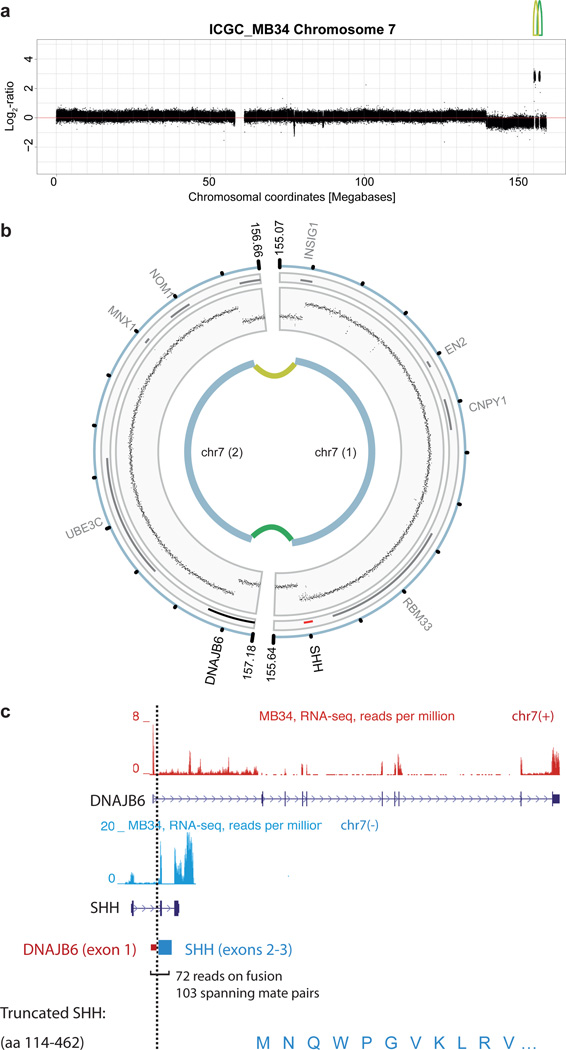
Deep sequencing also allowed fine-mapping of two amplicons on chromosome 7 in ICGC_MB34 (a SHH tumour with a somatic TP53 mutation, relating to MB2034 in6). One amplicon included the entire SHH gene, while the second disrupted DNAJB6, such that its first exon was juxtaposed to SHH (Figure 3a,b). RNA sequencing further revealed a novel fusion transcript, not expected from the DNA data, containing the first exon of DNAJB6 and exons 2 & 3 of SHH. The first exon of SHH was skipped, resulting in a predicted N-terminally truncated SHH protein (Figure 3c). Expression of SHH was extremely high in this case, whilst virtually absent in 301 other medulloblastomas (Supplementary Figure 9a). Predicted DNA and RNA junctions were validated by PCR (Supplementary Figure 9b).
Figure 3. Identification of novel fusion genes in MB.
a, Read-depth plot with log2 tumour:germline coverage ratio showing alterations on chromosome 7 in ICGC_MB34. Lines indicate connected segments.
b, Schematic of the rearrangement.
c, Details of the SHH fusion gene structure and support for its expression, derived from RNA sequencing data.
Several additional in-frame gene fusions were identified by large insert mate-pair sequencing, which gives better resolution for structural variant detection. ICGC_MB18, for example, carried an intrachromosomal translocation resulting in a fusion between LCLAT1 and ERBB4, the latter of which has previously been associated with MB oncogenesis23 (Supplementary Figure 9c–f). In ICGC_MB6, a complex rearrangement of fragments from chromosomes 1 and 17 produced a fusion between MLLT6 and MRPL45, a mitochondrial ribosomal protein, resulting in strong overexpression of the latter (Supplementary Figure 10a–c). These findings indicate that gene fusions involving well-established medulloblastoma oncogenes may play a more important role in MB than previously recognised, and warrant further investigation.
High-coverage, strand-specific RNA sequencing of 28 cases allowed us to determine the proportion of DNA SNVs that were observable in the transcriptome (Supplementary Tables 3 & 4). Overall, 129/268 (48%) non-synonymous mutations in the DNA were also detectable at the RNA level. A further 38% (101/268) resided in genes expressed at extremely low abundance (reads per kilobase of exon model per million mapped reads (RPKM) <1). Thus, the fraction of expressed mutations is even smaller than the already low number of DNA alterations, supporting the hypothesis that very few driving hits are needed to generate this paediatric tumour. It may also be the case that some mutations required for tumour initiation are not essential for later tumour cell maintenance.
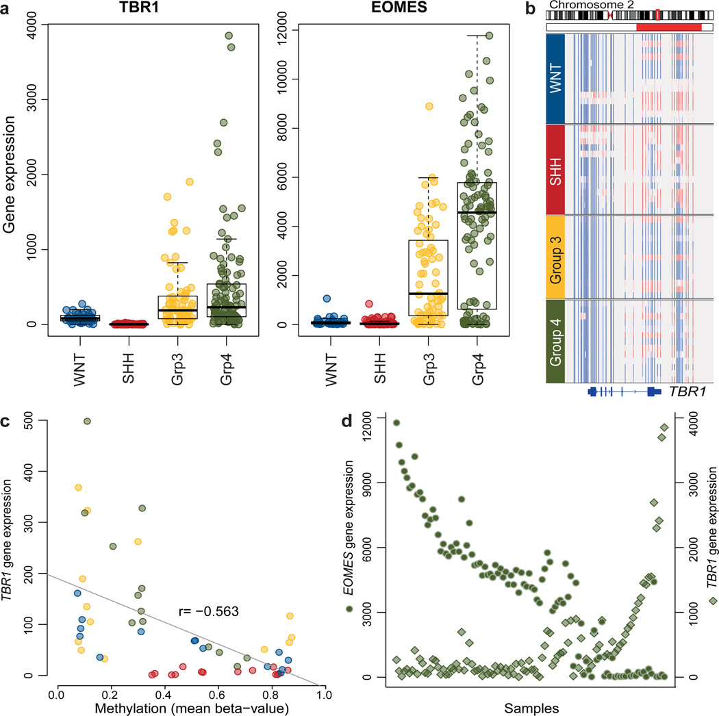
RNA sequencing further revealed monoallelic expression of a heterozygous mutation in TBR1, producing a p.G275C change, which was also seen in a previous study9 (Supplementary Figure 11a). TBR1 encodes a T-box transcription factor involved in brain development24. This gene, and a second family member, EOMES (or TBR2), clearly showed subgroup-specific differential expression (Figure 4a). Sequencing of TBR1 exon 2 in a further 85 medulloblastomas revealed one additional case with an identical mutation. All three mutated tumours were in Group 4. Gene expression was also strongly correlated with DNA methylation for both TBR1 and EOMES (Figure 4b,c, Supplementary Figure 11b,c), and expression of TBR1 and EOMES is inversely correlated in Group 4 tumours (Figure 4d), giving subsets that are either TBR1-methylated and EOMEShi or EOMES-methylated and TBR1hi (Supplementary Figure 11d,e). These two genes are markers for different stages of neuronal lineage commitment, suggesting possible differences in cell-of-origin or differentiation within Group 4 subpopulations25.
Figure 4. Integration of mutation, expression and methylation data shows differential regulation of TBR1 and EOMES in medulloblastoma.
a, Microarray data showing clear differences in TBR1 and EOMES expression between medulloblastoma subgroups (n=301).
b, DNA methylation of TBR1 (n=54), ranging from low (blue) to high (red). Horizontal red bar indicates the region used for correlation analysis in c.
c, Expression of TBR1 is tightly correlated with gene methylation (n=54; Pearson’s correlation values, r). SHH tumours show high methylation and virtually no expression, while WNT, Group 3 and Group 4 tumours display a more varied pattern.
d, Expression levels of TBR1 (diamonds) and EOMES (circles) are inversely related in Group 4 tumours (n=104).
This large, integrative genomics study has provided a detailed insight into new mechanisms contributing to medulloblastoma tumourigenesis and disclose novel targets for therapeutic approaches, especially for Group 3 & 4 patients. The molecular subgroup-related enrichment of many alterations highlights the importance of considering this distinguishing factor in research, trial design and clinical practice.
Methods Summary
All patient material was collected after receiving informed consent according to ICGC guidelines and as approved by the institutional review board of contributing centres. Tumour subgrouping was based on gene expression profiling or immunohistochemical analysis as described by Northcott et al5.
Next generation sequencing was performed using Illumina technologies. Mean DNA sequence coverage was 35-fold for whole-genome cases (range 26–56×), while mean on-target coverage in the whole-exome and replication cohorts was 68-fold (74% of targets above 20× for whole-exome, 66% for the replication cohort). Exome capture was carried out with Agilent SureSelect (Human All Exon 50 Mb and XT Custom Library) in-solution reagents. Sequence data were aligned to the hg19 human reference genome assembly; duplicate and non-uniquely mapping reads were excluded. Tumour ploidy was predicted from sequencing data by a novel approach integrating copy number aberrations with allele frequencies. A subset of sequence variants were validated using PCR and Sanger sequencing. Verification rates were 95% (128/135) for SNVs and 100% (14/14) for InDels (Supplementary Tables 3 and 4). A complete description of the materials and methods is provided in the Supplementary Information.
Supplementary Material
Acknowledgements
We thank GATC Biotech AG for sequencing services. For technical support and expertise we thank: Bettina Haase, Dinko Pavlinic, and Bianka Baying from the EMBL Genomics Core facility; Michael Wahlers and Rupert Lück from the EMBL high-performance computing facility; the DKFZ Genomics and Proteomics Core Facility; Ina Kutschera from the NCT Heidelberg, Karin Schlangen, Macha Metsger, Kerstin Schulz, Asja Nürnberger, Alexander Kovacsovics, and Matthias Linser from the Max Planck Institute for Molecular Genetics, Janet C. Lindsey, Simon Bailey and Danita M. Pearson.
This work was principally supported by the PedBrain Tumor Project contributing to the International Cancer Genome Consortium, funded by German Cancer Aid (109252) and the German Federal Ministry of Education and Research (BMBF, NGFNplus #01GS0883). Additional support came from the German Cancer Research Center – Heidelberg Center for Personalized Oncology (DKFZ-HIPO), the Max Planck Society, the Pediatric Brain Tumor Foundation, the Italian Neuroblastoma Foundation and the Samantha Dickson Brain Tumour Trust. This study included samples provided by the UK Children’s Cancer and Leukaemia Group (CCLG) as part of CCLG-approved biological study BS-2007-04.
Footnotes
Supplementary Information is linked to the online version of the paper at www.nature.com/nature.
Author Contributions
D.T.W.J., M.Su., A.M.S., H-J.W., S.B., S.P., H.C., E.P., L.S., A.W., S.H., T.T., B.R., C.C.B., M.Sch., C.v.K., V.B., R.V., S.Wo., S.Wi., and J.F. performed and/or coordinated experimental work.
N. Jäger, D.T.W.J., M.K., T.Z., B.H., M.Su, T.P., V.Ho., T.R., H-J.W., J.W., M.A., V.Am, M.Z., Q.W., B.L., V.Ast, C.L., J.E., R.K., P.v.S., J.K., D.Sh., M.J.B., R.B.R. and P.A.N. performed data analysis.
Y-J.C., M.Ry., M.Re., S.C., G.P.T., U.S., V.Ha., N.G., Y-J.K., C.M., W.R., A.U., C.H-M., T.M., A.E.K., A.v.D., O.W., E.M., J.R., M.E., M.U.S., M.C.F., M.H., N.Jabado, S.R., A.O.v.B., D.W., S.C.C., M.G.M., V.P.C., W.S., G.R., M.D.T., and A.K. collected data and provided patient materials.
D.T.W.J., N.Jäger, D.St., M.K., V.Ho., H.W., R.E., S.M.P. and P.L. prepared the initial manuscript and figures.
U.D.W., H.L., B.B., G.R., M.M., S.L.P., M-L.Y., J.O.K., R.E., A.K., S.M.P., and P.L. provided project leadership.
All authors contributed to the final manuscript
Author Information Short-read sequencing data have been deposited at the European Genome-phenome Archive (EGA, http://www.ebi.ac.uk/ega/) hosted by the EBI, under accession number EGAS00001000215. Reprints and permissions information is available at www.nature.com/reprints. The authors declare no competing financial interests. Readers are welcome to comment on the online version of this article at www.nature.com/nature.
References
- 1.Louis D, et al. The 2007 WHO Classification of Tumours of the Central Nervous System. Acta Neuropathologica. 2007;114:97–109. doi: 10.1007/s00401-007-0243-4. [DOI] [PMC free article] [PubMed] [Google Scholar]
- 2.Kool M, et al. Molecular subgroups of medulloblastoma: an international meta-analysis of transcriptome, genetic aberrations, and clinical data of WNT, SHH, Group 3, and Group 4 medulloblastomas. Acta Neuropathol. 2012;123:473–484. doi: 10.1007/s00401-012-0958-8. [DOI] [PMC free article] [PubMed] [Google Scholar]
- 3.Taylor MD, et al. Molecular subgroups of medulloblastoma: the current consensus. Acta Neuropathol. 2012;123:465–472. doi: 10.1007/s00401-011-0922-z. [DOI] [PMC free article] [PubMed] [Google Scholar]
- 4.Clifford S, et al. Wnt/Wingless Pathway Activation and Chromosome 6 Loss Characterise a Distinct Molecular Sub-Group of Medulloblastomas Associated with a Favourable Prognosis. Cell Cycle. 2006;5:2666–2670. doi: 10.4161/cc.5.22.3446. [DOI] [PubMed] [Google Scholar]
- 5.Northcott PA, et al. Medulloblastoma comprises four distinct molecular variants. J Clin Oncol. 2011;29:1408–1414. doi: 10.1200/JCO.2009.27.4324. [DOI] [PMC free article] [PubMed] [Google Scholar]
- 6.Rausch T, et al. Genome sequencing of pediatric medulloblastoma links catastrophic DNA rearrangements with TP53 mutations. Cell. 2012;148:59–71. doi: 10.1016/j.cell.2011.12.013. [DOI] [PMC free article] [PubMed] [Google Scholar]
- 7.Rello-Varona S, et al. Preferential killing of tetraploid tumor cells by targeting the mitotic kinesin Eg5. Cell Cycle. 2009;8:1030–1035. doi: 10.4161/cc.8.7.7950. [DOI] [PubMed] [Google Scholar]
- 8.Vitale I, et al. Inhibition of Chk1 kills tetraploid tumor cells through a p53-dependent pathway. PLoS One. 2007;2:e1337. doi: 10.1371/journal.pone.0001337. [DOI] [PMC free article] [PubMed] [Google Scholar]
- 9.Parsons DW, et al. The Genetic Landscape of the Childhood Cancer Medulloblastoma. Science. 2011;331:435–439. doi: 10.1126/science.1198056. [DOI] [PMC free article] [PubMed] [Google Scholar]
- 10.Stransky N, et al. The mutational landscape of head and neck squamous cell carcinoma. Science. 2011;333:1157–1160. doi: 10.1126/science.1208130. [DOI] [PMC free article] [PubMed] [Google Scholar]
- 11.Wang L, et al. SF3B1 and other novel cancer genes in chronic lymphocytic leukemia. N Engl J Med. 2011;365:2497–2506. doi: 10.1056/NEJMoa1109016. [DOI] [PMC free article] [PubMed] [Google Scholar]
- 12.Schwartzentruber J, et al. Driver mutations in histone H3.3 and chromatin remodelling genes in paediatric glioblastoma. Nature. 2012;482:226–231. doi: 10.1038/nature10833. [DOI] [PubMed] [Google Scholar]
- 13.Kool M, et al. Integrated Genomics Identifies Five Medulloblastoma Subtypes with Distinct Genetic Profiles, Pathway Signatures and Clinicopathological Features. PLoS ONE. 2008;3:e3088. doi: 10.1371/journal.pone.0003088. [DOI] [PMC free article] [PubMed] [Google Scholar]
- 14.Pfister S, et al. Outcome prediction in pediatric medulloblastoma based on DNA copynumber aberrations of chromosomes 6q and 17q and the MYC and MYCN loci. J Clin Oncol. 2009;27:1627–1636. doi: 10.1200/JCO.2008.17.9432. [DOI] [PubMed] [Google Scholar]
- 15.Thompson MC, et al. Genomics identifies medulloblastoma subgroups that are enriched for specific genetic alterations. J Clin Oncol. 2006;24:1924–1931. doi: 10.1200/JCO.2005.04.4974. [DOI] [PubMed] [Google Scholar]
- 16.Pietsch T, et al. Medulloblastomas of the Desmoplastic Variant Carry Mutations of the Human Homologue of Drosophila patched. Cancer Res. 1997;57:2085–2088. [PubMed] [Google Scholar]
- 17.van Haaften G, et al. Somatic mutations of the histone H3K27 demethylase gene UTX in human cancer. Nat Genet. 2009;41:521–523. doi: 10.1038/ng.349. [DOI] [PMC free article] [PubMed] [Google Scholar]
- 18.Satow R, Kurisaki A, Chan TC, Hamazaki TS, Asashima M. Dullard promotes degradation and dephosphorylation of BMP receptors and is required for neural induction. Dev Cell. 2006;11:763–774. doi: 10.1016/j.devcel.2006.10.001. [DOI] [PubMed] [Google Scholar]
- 19.Han S, et al. Nuclear envelope phosphatase 1-regulatory subunit 1 (formerly TMEM188) is the metazoan Spo7p ortholog and functions in the lipin activation pathway. J Biol Chem. 2012;287:3123–3137. doi: 10.1074/jbc.M111.324350. [DOI] [PMC free article] [PubMed] [Google Scholar]
- 20.Kim Y, et al. A conserved phosphatase cascade that regulates nuclear membrane biogenesis. Proc Natl Acad Sci U S A. 2007;104:6596–6601. doi: 10.1073/pnas.0702099104. [DOI] [PMC free article] [PubMed] [Google Scholar]
- 21.Northcott PA, et al. Multiple recurrent genetic events converge on control of histone lysine methylation in medulloblastoma. Nat Genet. 2009;41:465–472. doi: 10.1038/ng.336. [DOI] [PMC free article] [PubMed] [Google Scholar]
- 22.Stephens PJ, et al. Massive Genomic Rearrangement Acquired in a Single Catastrophic Event during Cancer Development. Cell. 2011;144:27–40. doi: 10.1016/j.cell.2010.11.055. [DOI] [PMC free article] [PubMed] [Google Scholar]
- 23.Gilbertson RJ, Perry RH, Kelly PJ, Pearson ADJ, Lunec J. Prognostic Significance of HER2 and HER4 Coexpression in Childhood Medulloblastoma. Cancer Res. 1997;57:3272–3280. [PubMed] [Google Scholar]
- 24.Hevner RF, et al. Tbr1 regulates differentiation of the preplate and layer 6. Neuron. 2001;29:353–366. doi: 10.1016/s0896-6273(01)00211-2. [DOI] [PubMed] [Google Scholar]
- 25.Englund C, et al. Pax6, Tbr2, and Tbr1 are expressed sequentially by radial glia, intermediate progenitor cells, and postmitotic neurons in developing neocortex. J Neurosci. 2005;25:247–251. doi: 10.1523/JNEUROSCI.2899-04.2005. [DOI] [PMC free article] [PubMed] [Google Scholar]
Associated Data
This section collects any data citations, data availability statements, or supplementary materials included in this article.


