Abstract
Purpose
To evaluate a simplified method to measure peripapillary choroidal thickness using commercially available, three-dimensional optical coherence tomography (3D-OCT).
Methods
3D-OCT images of normal eyes were consecutively obtained from the 3D-OCT database of Korea University Medical Center On the peripapillary images for retinal nerve fiber layer (RNFL) analysis, choroidal thickness was measured by adjusting the segmentation line for the retinal pigment epithelium to the chorioscleral junction using the modification tool built into the 3D-OCT image viewer program. Variations of choroidal thickness at 12 sectors of the peripapillary area were evaluated.
Results
We were able to measure the peripapillary choroidal thickness in 40 eyes of our 40 participants, who had a mean age of 41.2 years (range, 15 to 84 years). Choroidal thickness measurements had strong inter-observer correlation at each sector (r = 0.901 to 0.991, p < 0.001). The mean choroidal thickness was 191 ± 62 µm. Choroidal thickness was greatest at the temporal quadrant (mean ± SD, 210 ± 78 µm), followed by the superior (202 ± 66 µm), nasal (187 ± 64 µm), and inferior quadrants (152 ± 59 µm).
Conclusions
The measurement of choroidal thickness on peripapillary circle scan images for RNFL analysis using the 3D-OCT viewing program was highly reliable and efficient.
Keywords: Choroid, Optical coherence tomography, Retina, Retinal nerve fiber layer
Advances in spectral domain optical coherence tomography (SD-OCT) and enhanced depth-imaging techniques have allowed for measurement of the deep layers of the retina and choroid [1]. Recently, some studies have been published regarding the configuration of choroidal thickness on the macula and the peripapillary area of normal controls, as well as those of patients with various macular and optic disc diseases [1-6]. However, the method used to measure choroidal thickness has varied among studies and could be dependent on the observers and the location of the scans used to measure choroidal thickness.
In previous optical coherence tomography (OCT) studies, line scanning was used to investigate choroidal thickness around the optic disc [4,5]. In these studies, choroidal thickness was determined between two boundaries set by the observers. Although they found excellent inter-observer agreement for the measurements, variations between observers were still present. Choroidal thickness was determined at a specific point on the line scans of the optic disc, rather than the entire line scan. More time and effort may be needed to manually measure the thickness of the entire area of the line scan. They topographically related choroidal thickness with those of the overlying retina or retinal nerve fiber layer (RNFL). While a relationship between choroidal thickness and overlying RNFL thickness was found, the comparison was not based on the normative database of peripapillary RNFL thickness.
In contrast to measurements taken from line scans of the optic disc, measurement of peripapillary RNFL thickness by OCT circle scan has been widely used for the diagnosis and monitoring of optic disc disorders, such as glaucoma and anterior ischemic optic neuropathy [7-10]. The circle scan mode of current SD-OCT systems provides automatic retinal and RNFL thickness information at 12 sectors around the optic disc. The OCT viewer program provides a normative database for peripapillary RNFL thickness. Measurement of choroidal thickness by circle scan for RNFL thickness could be useful to investigate the relationship between choroidal thickness and the overlying RNFL thickness.
In the current SD-OCT scanning protocol for peripapillary circle scans, each image is scanned several times and averaged to improve the signal-to-noise ratio. In the averaged circle scan image, the choroid could be better delineated than in those images obtained without averaging. In the current study, we evaluated the method used to measure choroidal thickness on peripapillary images for RNFL analysis using an OCT viewer program.
Materials and Methods
The institutional review board at Korea University approved our study protocol. This study followed the tenets of the Declaration of Helsinki. Study cases were consecutively collected from a database of three-dimensional OCT (3D-OCT; 3D-OCT 1000 Mark II, software ver. 4.21, Topcon, Tokyo, Japan) images obtained at Korea University Medical Center in 2010. We excluded eyes with refractive errors greater than -/+ 3.0 diopters (D) spherical equivalent, as well as any retinal, choroidal, or optic nerve head (ONH) pathology or patients with a history of intraocular surgery. We also excluded eyes whose peripapillar atropy involved the OCT scanning ring or whose peripapillary RNFL thickness was outside 95% of the 3D-OCT age-matched normative database. The 3D-OCT used in the current study was an SD-OCT device with a wavelength of 840 nm, a horizontal resolution of ≤20 µm, and an axial resolution of up to 5 µm. Its imaging speed was 27,000 axial scans per second. This device, which includes a camera for color fundus photography, is programmed to automatically capture a color image of the fundus just after obtaining the OCT images. We selected 3D-OCT circle scan images used to capture RNFL images in a 3.4-mm zone around the optic disc. Each circle scan consisted of 1024 A-scans. Each image was scanned four times, and these scans were averaged to improve the signal-to-noise ratio. We excluded images with a Q-factor of less than 45, as suggested by the manufacturer, for image quality assurance.
Choroidal thickness measurements
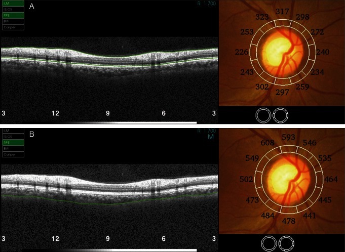
The circle scan mode of 3D-OCT automatically provides two parameters of tissue thickness at 12 sectors around an optic disc: RNFL thickness and retinal thickness. The thickness at each sector represents the mean thickness of the layer(s) within each sector. Retinal thickness was determined between the segmentation lines for the internal limiting membrane (ILM) and the retinal pigment epithelium (RPE). Next, the segmentation line, which is set automatically for the RPE, was modified to the chorioscleral junction by two independent observers (JO and CMY) using the modification tool built into the 3D-OCT image viewer program. In this manner, the chorioretinal thickness between the ILM and chorioscleral junction was obtained (Fig. 1). The chorioscleral junction was defined as a hyperreflective line between the large vessel layer of the choroid and the sclera. In cases where there was no clear chorioscleral junction boundary, observers used the signal-and-noise modulation tool built into the viewer system. Even with signal modulation, the posterior choroid was marked as a smooth line joining the outer limits of the large choroidal vascular space in the poor images. The choroidal thickness was defined as the thickness between the segmentation lines for RPE and for the chorioscleral junction, and was calculated by subtracting the retinal thickness from the chorioretinal thickness.
Fig. 1.
Measurement of choroidal thickness on peripapillary circle scan images of three-dimensional optical coherence tomography (3DOCT) used to capture retinal nerve fiber layer images using the Topcon 3D-OCT viewer program. The segmentation line, which was automatically set for retinal pigment epithelium (RPE, A), was modified to the chorioscleral junction (B). The choroidal thickness was calculated by subtracting the retinal thickness (top right) from the chorioretinal thickness (bottom right). The peripapillary choroidal thickness was the greatest in the temporal quadrant (9), followed by the superior (12) and nasal ones (3). It was thinnest at the inferior quadrant (6). LM = limiting membrane; IS/OS = inner segement / outer segment; RPE = retinal pigemnt epithelium; BM = basement membrane.
Statistical methods
Intraclass correlation coefficients were calculated to evaluate inter-observer reliability for the measurement of chorioretinal thickness, and they were considered to be strong if the correlation coefficient was greater than 0.9. A paired t-test was used to analyze differences between means in chorioretinal thickness by sector. Multiple linear regression analysis was performed to obtain covariate-adjusted partial correlation coefficients among three thickness parameters. We also conducted multivariate regression analyses for choroidal thickness, age, gender, and refractive error; p-values less than 0.05 were considered statistically significant. All statistical analyses were performed using the SPSS ver. 12.0 (SPSS Inc., Chicago, IL, USA).
Results
Forty images from 40 eyes of 40 participants (32 males, 8 females) were included in our analysis. The mean age of the participants was 41.2 years (SD, ± 20.6 years). The mean refractive error was -0.4 D (SD, ± 1.1 D). Reliable measurements were obtained in all eyes in our dataset. Choroidal thickness measurements had strong inter-observer correlation at all 12 sectors (r = 0.907 to 0.982, p < 0.001). The mean difference in the choroidal thickness measurements between the two observers at each sector showed no statistical significance (Table 1).
Table 1.
Inter-observer reliability and mean difference of peripapillary choroidal thickness measurements between two observers

The clock hours were aligned based on right-eye orientation. Nine o'clock corresponded to the temporal region, 12 o'clock to the superior, 3 o'clock to the nasal, and 6 o'clock to the inferior.
p-values less than 0.05 were considered statistically significant.
ICC = intraclass correlation; CI = confidence interval; SD = standard deviation.
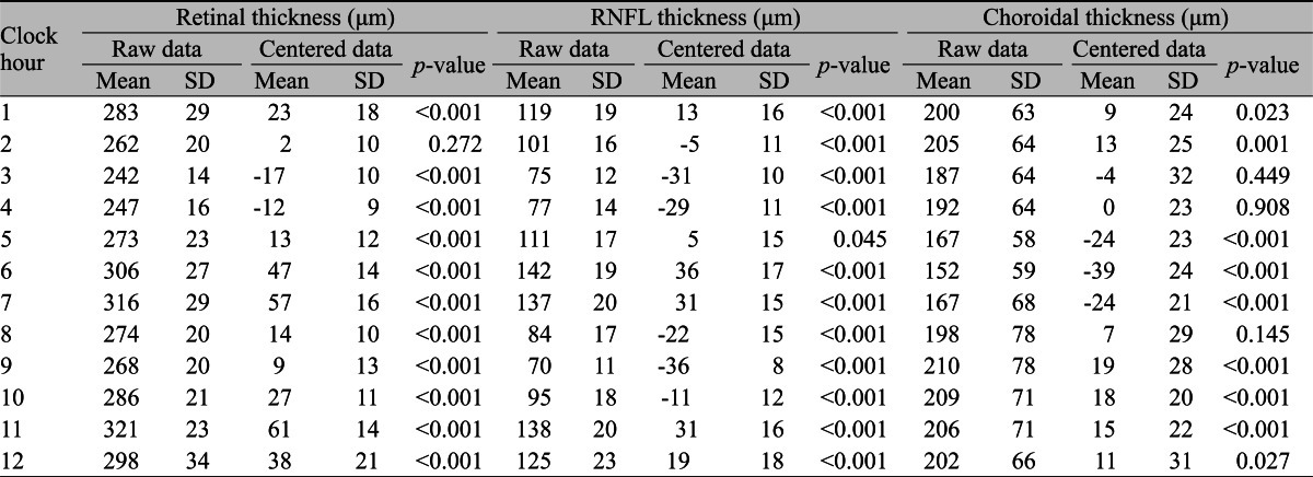
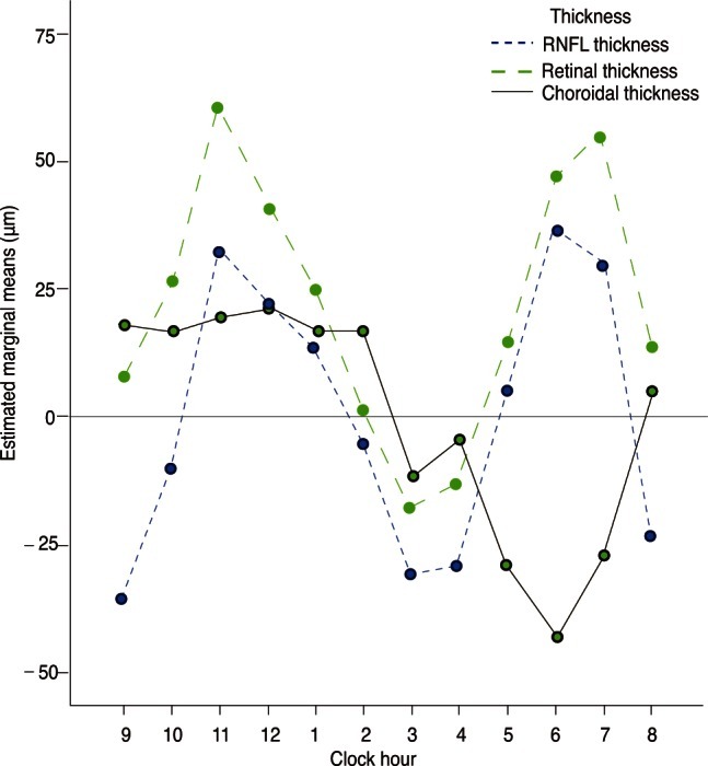
The mean average choroidal thickness was 191 ± 62 µm. Choroidal thickness was greatest at the temporal area (mean ± SD, 210 ± 78 µm), followed by the superior (202 ± 66 µm), nasal (187 ± 64 µm), and inferior areas (152 ± 59 µm) (Table 2). In Fig. 2, each centered value was obtained by subtraction of the overall mean within 12 sectors from the choroidal, retinal, and RNRL thickness measured at each sector. Centered values of choroidal thickness were greater than zero in some sectors (1 to 6, 12) but less than zero in the other sectors. The minimum value was noted in the inferior region (6), and the maximum value was located in the temporal region (9). In contrast, the values of retinal and RNFL thickness centered around the optic disc were greater in the superior and inferior regions.
Table 2.
Peripapillary retinal thickness, RNFL thickness, and choroidal thickness measurements at clock-hour sectors

The clock hours were aligned based on right-eye orientation, as described in Table 1.
p-values less than 0.05 were considered statistically significant for testing of within-subjects contrasts between three types of thicknesses (linear) and sectors (fourth-order).
RNFL = retinal nerve fiber layer; SD = standard deviation.
Fig. 2.

Profile plot of the centered choroidal, retinal, and retinal nerve fiber layer (RNFL) thicknesses. Choroidal thickness around the optic disc had the minimum value in the inferior region, while the retina and RNFL were thicker in the superior and inferior regions.
We evaluated partial correlation coefficients between the means of each thickness from multiple linear regression analysis, adjusting for the effects of age, sex, and refractive error. The partial correlation coefficient between choroidal and retinal thickness was 0.36 (p = 0.047). The same value was 0.77 (p < 0.001) between RNFL thickness and retinal thickness. However, this coefficient was not significant between choroidal and RNFL thickness.
Multivariate regression analyses that included age, gender and refractive error showed that choroidal thickness values were correlated with age (p < 0.001) and decreased an average of 1.97 µm for each year of age (choroidal thickness [µm] = 272 - 1.97 × age; confidence interval for annual decrease, -2.724 to -1.214). Retinal thickness was correlated with age (p < 0.001) and refractive error (p = 0.033), decreasing 0.70 µm for each year of age (retinal thickness [µm] = 290 - 0.70 × age + 4.6 × refractive error; confidence interval for annual decrease, -0.921 to -0.486).
Discussion
In addition to studies of macular choroidal thickness, studies of the measurement of peripapillary choroidal thickness are increasing. Recently, Ehrlich et al. [11] used SD-OCT to examine peripapillary choroidal thickness in 31 patients with primary open-angle glaucoma and in 39 glaucoma suspects. They found excellent inter-observer agreement for choroidal thickness measurement and no correlation between RNFL and choroidal thickness. Maul et al. [4] also assessed peripapillary choroidal thickness using SD-OCT in 38 patients with glaucoma and in 36 glaucoma suspects. They found that age, axial length, central corneal thickness, and diastolic perfusion pressures were correlated with choroidal thickness. Ho et al. [5] measured peripapillary choroidal thickness using two raster scans (a horizontal and a vertical scan) centered at the optic nerve. However, the measurements required additional scans not typically used in clinics. Even though advances in OCT technology have reduced acquisition time, additional time and effort are still needed to obtain the additional scans needed to measure choroidal thickness, which is not always easy in busy clinics. We measured choroidal thickness using the same peripapillary scans obtained to determine RNFL thickness, and we provided an analysis of sectorial configuration of the peripapillary choroid in healthy eyes. Choroidal thickness was calculated between two boundaries, which were determined by observers in previous studies. However, in this study, we used the automatically determined segmentation line. The method used in the current study may reduce the time and effort needed to obtain measurements of choroidal thickness.
Enhanced depth imaging of SD-OCT enables clear detection of the choroid-retina and choroid-scleral interfaces and in vivo measurements of choroidal thickness [1]. Several studies have used enhanced SD-OCT depth imaging to demonstrate that choroidal thickness at the macula was correlated significantly with age [1,2,12]. However, we were able to measure peripapillary choroidal thickness using 3D-OCT without the addition of enhanced depth imaging. A recent study reported that choroidal thickness could be measured without enhanced depth imaging, although reliable measurements of choroidal thickness were obtainable in only 74% of the examined eyes [3]. This study only used the pixel averaging technique to improve the signal-to-noise ratio, thereby obtaining signals from deep layers. Using this technique, we were able to collect reliable measurements of choroidal thickness around the optic disc in all examined eyes. The greater yield of reliable choroidal thickness measurements in our study may be attributed to the fact that the choroid is thinner around the optic disc than in the macula. Using the signal-and-noise modulation tool built into the viewer system may have also contributed to the higher number of reliable measurements. In this study, measurements of choroidal thickness were highly reproducible. We obtained choroidal thickness as a mean thickness value using all A-scans on the circle scan with 12 sectors instead of using an A-scan on a line scan centered at the optic nerve. The measurements taken using all A-scans in each sector may have contributed to our high reproducibility rates.
In this study, the peripapillary choroidal thickness was greatest at the temporal quadrant, followed by the superior, nasal, and inferior areas. Moreover, peripapillary choroidal thickness showed an abrupt decrease only in the inferior region ("single-dip" configuration). The results of the current study are consistent with previous studies [13]. This pattern of choroidal thickness in the peripapillary area has been suggested to reflect the peripapillary choroidal circulation and developmental characteristics of the peripapillary area.
In the current study, choroidal thickness was correlated with the overlying retinal thickness around the optic disc. This finding was inconsistent with previous findings in which choroidal thickness was not correlated with retinal thickness on the macula [11]. Such a discrepancy may be due to differences in the locations at which measurements were taken. The present study also found that the peripapillary RNFL, retinal and choroidal thicknesses showed a negative correlation with age. This finding is in agreement with those of previous SD-OCT studies on macular choroidal thickness [5]. For example, Ikuno et al. [12] showed that age, refractive error, and axial length were correlated with macular choroidal thickness [11]. Our study showed a relatively weaker correlation with refractive error. This difference may be attributable to the narrower range of refractive errors in our study, which excluded eyes exceeding -/+ 3 D.
The present study had some limitations. The small sample size and the hospital-based design of this study may have introduced a selection bias. In this retrospective study, only the thickness of the choroid and adjacent structures was measured and compared; systemic hemodynamic parameters and local blood flow were not checked. Future studies should include vascular parameter measurements to validate our results. Other researchers may also criticize our technique of measuring peripapillary choroidal thickness. Because the total RPE thickness was included in the choroidal thickness when this value was calculated using our method, it is difficult to compare our choroidal thickness measurements with those obtained in other studies. However, we propose that our method may be less prone to observer error because only the chorioscleral junction was chosen manually, and the other border was determined automatically by the built-in software. In previously reported studies of macular choroid thickness, both the outer edges of the hyper-reflective RPE and the chorioscleral junction had to be determined manually using a caliper tool by an observer, which could introduce a higher margin of error. Moreover, the RPE layer is not expected to show significant differences in thickness around the optic disc because it is very thin. Our method also showed excellent inter-observer reproducibility.
In conclusion, we were able to measure choroidal thickness on 3D-OCT circle scan images used to capture RNFL images using a 3D-OCT viewing program. The measurements were highly reliable and efficient.
Acknowledgements
This study was supported by a grant of the Korean Health Technology R&D Project, Ministry for Health, Welfare and Family Affairs, Republic of Korea (A102024).
Footnotes
No potential conflict of interest relevant to this article was reported.
References
- 1.Spaide RF, Koizumi H, Pozzoni MC. Enhanced depth imaging spectral-domain optical coherence tomography. Am J Ophthalmol. 2008;146:496–500. doi: 10.1016/j.ajo.2008.05.032. [DOI] [PubMed] [Google Scholar]
- 2.Fujiwara T, Imamura Y, Margolis R, et al. Enhanced depth imaging optical coherence tomography of the choroid in highly myopic eyes. Am J Ophthalmol. 2009;148:445–450. doi: 10.1016/j.ajo.2009.04.029. [DOI] [PubMed] [Google Scholar]
- 3.Manjunath V, Taha M, Fujimoto JG, Duker JS. Choroidal thickness in normal eyes measured using Cirrus HD optical coherence tomography. Am J Ophthalmol. 2010;150:325–329.e1. doi: 10.1016/j.ajo.2010.04.018. [DOI] [PMC free article] [PubMed] [Google Scholar]
- 4.Maul EA, Friedman DS, Chang DS, et al. Choroidal thickness measured by spectral domain optical coherence tomography: factors affecting thickness in glaucoma patients. Ophthalmology. 2011;118:1571–1579. doi: 10.1016/j.ophtha.2011.01.016. [DOI] [PMC free article] [PubMed] [Google Scholar]
- 5.Ho J, Branchini L, Regatieri C, et al. Analysis of normal peripapillary choroidal thickness via spectral domain optical coherence tomography. Ophthalmology. 2011;118:2001–2007. doi: 10.1016/j.ophtha.2011.02.049. [DOI] [PMC free article] [PubMed] [Google Scholar]
- 6.Shin JW, Shin YU, Lee BR. Choroidal thickness and volume mapping by a six radial scan protocol on spectral-domain optical coherence tomography. Ophthalmology. 2012;119:1017–1023. doi: 10.1016/j.ophtha.2011.10.029. [DOI] [PubMed] [Google Scholar]
- 7.Danesh-Meyer HV, Boland MV, Savino PJ, et al. Optic disc morphology in open-angle glaucoma compared with anterior ischemic optic neuropathies. Invest Ophthalmol Vis Sci. 2010;51:2003–2010. doi: 10.1167/iovs.09-3492. [DOI] [PMC free article] [PubMed] [Google Scholar]
- 8.Hood DC, Anderson SC, Wall M, Kardon RH. Structure versus function in glaucoma: an application of a linear model. Invest Ophthalmol Vis Sci. 2007;48:3662–3668. doi: 10.1167/iovs.06-1401. [DOI] [PubMed] [Google Scholar]
- 9.Contreras I, Rebolleda G, Noval S, Munoz-Negrete FJ. Optic disc evaluation by optical coherence tomography in nonarteritic anterior ischemic optic neuropathy. Invest Ophthalmol Vis Sci. 2007;48:4087–4092. doi: 10.1167/iovs.07-0171. [DOI] [PubMed] [Google Scholar]
- 10.Contreras I, Noval S, Rebolleda G, Munoz-Negrete FJ. Follow-up of nonarteritic anterior ischemic optic neuropathy with optical coherence tomography. Ophthalmology. 2007;114:2338–2344. doi: 10.1016/j.ophtha.2007.05.042. [DOI] [PubMed] [Google Scholar]
- 11.Ehrlich JR, Peterson J, Parlitsis G, et al. Peripapillary choroidal thickness in glaucoma measured with optical coherence tomography. Exp Eye Res. 2011;92:189–194. doi: 10.1016/j.exer.2011.01.002. [DOI] [PubMed] [Google Scholar]
- 12.Ikuno Y, Kawaguchi K, Nouchi T, Yasuno Y. Choroidal thickness in healthy Japanese subjects. Invest Ophthalmol Vis Sci. 2010;51:2173–2176. doi: 10.1167/iovs.09-4383. [DOI] [PubMed] [Google Scholar]
- 13.Tanabe H, Ito Y, Terasaki H. Choroid is thinner in inferior region of optic disks of normal eyes. Retina. 2012;32:134–139. doi: 10.1097/IAE.0b013e318217ff87. [DOI] [PubMed] [Google Scholar]


