

These historic pictures were taken at the time of the operation in 1961 by Donald Ross who gave them to me.1 Photography was routine for cardiac surgery in Guys Hospital at this time as Sir Russell Brock was insistent that appearances were accurately recorded as well as described in operation reports.
Figure 1 shows the classic appearances of calcific aortic valvar stenosis in an adult patient undergoing open aortic valvotomy and decalcification, the treatment for this condition at the time. The comissures are fused reducing the orifice to a slit. There is bicuspidisation with compete fusion forming an anterior raphe pointing to the left lower corner and, a posterior cusp opposite the anterior cusp with a medan raphe.
Figure 1.

Calcific valvar aortic stenosis
There are “mountains” in each cusp where calcium has deposited and thickened and made the valve rigid.
Ross was decalcifying the valve and sucking away the dislodged bits under direct vision. The patient was on cardiopulmonary bypass. Suddenly, the weakened valve tissue went down the sucker.
There was no mechanical valve available in the hospital. Ross remembered when he was working in the dog laboratory, and at suggestion of Sir Russell Brock he had been sterilising homograft aorta to use as tissue supplement rather than synthetic material. He had sterilised a human cadaver aortic valve by a freeze drying process, thinking one day it could be useful. He sent for it from where he had left it - above the sink (figure 2).
Figure 2.

The first aortic homograft used, in a sterile air-tight container.
The homograft was rehydrated and reconstituted and placed in the subcoronary position. The patient lived for several years with a near normal functioning aortic valve (figure 3).
Figure 3.
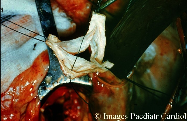
Homograft in subcoronary position.
Figure 4 is a slide from a talk by the author entitled ‘50 years with cardiac surgeons’.
Figure 4.

A summary of Ross operations from a lecture by Professor Jane Somerville
References
- 1.Ross DN. Replacement of aortic and mitral valves with a pulmonary autograft. Lancet. 1967;2:956–8. doi: 10.1016/s0140-6736(67)90794-5. [DOI] [PubMed] [Google Scholar]