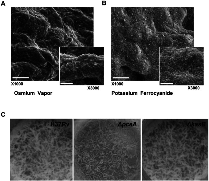
FIG 1 .
M. tuberculosis pellicle is an organized structure. SEM of M. tuberculosis pellicle using osmium tetroxide vapor (A) or potassium ferrocyanide (B) processing shows an organized bacterial community. Scale bars represent 10 µm in ×1,000 and 5 µm in ×3,000 magnification. (C) Pellicle phenotype is distinct from cording. ΔpcaA and ΔkasB mutants (both defective in cording) were inoculated into Sauton’s medium without Tween 80 and incubated for 3 weeks under pellicle-promoting conditions. Both ΔpcaA and ΔkasB mutants are capable of growing as a pellicle, indicating that the ability to cord is not a prerequisite for pellicle growth.

