Figure 1.
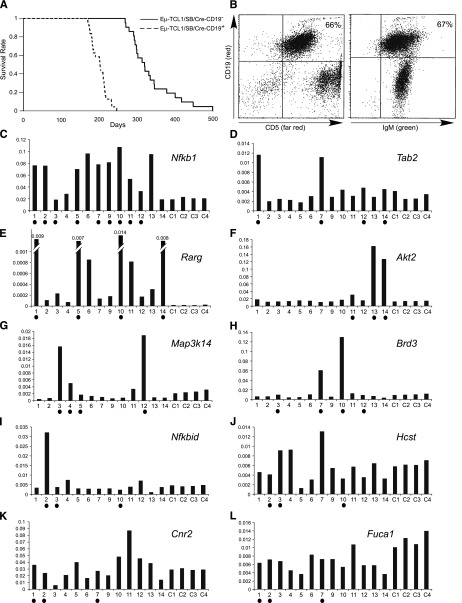
Analysis of Eμ−TCL1/SB mice. (A) Kaplan-Meier survival plot of Eμ−TCL1/SB and Eμ−TCL1 control mice. (B) Fluorescence-activated cell sorter analysis of a representative Eμ−TCL1/SB CLL case (the T cells are green because in this system cells that do not undergo Cre recombination are green fluorescent protein–positive12). (C-L) Real-time RT-PCR analysis of genes affected by CISs. Shown values are relative to actin expression. Samples containing insertions near corresponding genes are marked with solid dots. The following Applied Biosystems assays were used: Hcst (Mm01172975_m1), Nfkbid (Mm00549082_m1), Map3k14 (Mm00444166_m1), Rarg (Mm00441091_m1), Brd3 (Mm00469733_m1), Tab2 (Mm00663112_m1), Fuca1 (Mm00502778_m1), Nfkb1 (Mm00476361_m1), Akt2 (Mm02026778_g1) and Cnr2 (Mm02620087_s1), and Actin (Mm00607939_s1).

