

Abstract
Conventional composite sol-gel method has been modified to enhance the piezoelectric performance of ceramic thick films. Lead zirconate titanate (PZT) and lead magnesium niobate–lead titanate (PMN-PT) thick films were fabricated using the modified sol-gel method for ultrasonic transducer applications. In this work, piezoresponse force microscopy was employed to evaluate the piezoelectric characteristics of PZT and PMN-PT composite sol-gel thick films. The images of the piezoelectric response and the strain-electric field hysteresis loop behavior were measured. The effective piezoelectric coefficient (d33,eff) of the films was determined from the measured loop data. It was found that the effective local piezoelectric coefficient of both PZT and PMN-PT composite films is comparable to that of their bulk ceramics. The promising results suggest that the modified composite sol-gel method is a promising way to prepare the high-quality, crack-free ceramic thick films.
INTRODUCTION
In order to enhance the spatial resolution in imaging applications1, 2 and maximize sensitivity in acoustic tweezers applications,3 ultrasonic transducers at ultrahigh operating frequency (>100 MHz) are highly desired. Consequently, development of piezoelectric thick films has been attracted much interest for high frequency transducer applications.4 Nevertheless, the preparation of the piezoelectric element is still one of the technical challenges in the fabrication of ultrahigh frequency transducers. To achieve such high operating frequency, a few micron thick film is required. Conventional thin film technologies such as sol–gel, pulsed-laser deposition, sputtering, and metal organic chemical vapor deposition are not suitable to produce high quality film in this thickness range due to the slow deposition rate and high level of stress generated on the film itself during the processing. On the other hand, other thick film techniques such as vapor deposition methods,5 screen-printing,6, 7 tape-casting,8 etc. have also been studied to fabricate piezoelectric thick films. However, it still has been difficult to fabricate crack-free thick films with high density and good performance.
In recent years, due to its low production cost and good control of film stoichiometry, a composite sol-gel9 has emerged as a promising technique for the development of dense ceramic thick films.10 It is a modified sol-gel technique in which the composite sol-gel slurry is prepared by mixing a desired concentration of ceramic powder with a chemical sol. The film is fabricated by performing heat treatments on the resultant sol-gel spun on the substrate. Because of a strongly bonded network between the sol–gel and ceramic particles with the adhered substrate, crack-free thick films can be fabricated with the composite sol-gel technique.11 Among the materials used for ultrasonic transducer applications, the most common one is the lead zirconate titanate (Pb(Zr,Ti)O3, PZT) family, which is well-known to have high piezoelectric strain constant, dielectric constant, and electromechanical coupling coefficients.12, 13 Lead magnesium niobate–lead titanate (Pb(Mg1/3Nb2/3)O3–PbTiO3, PMN-PT) system has also been attracted much attention due to their much higher piezoelectric and dielectric constants.14, 15 Previously, a variety of techniques including double-beam interferometer16 and scanning laser Doppler vibrometer17 have been utilized to measure the piezoelectric properties of the composite sol-gel thick film, where the reported piezoelectric coefficients of the films are small (30–100 pm/V).
In order to enhance the piezoelectric characteristics of the film, the porosity was proposed to be reduced by performing sol-infiltration and adding sintering aids on each layer of the as-prepared film.18 As higher piezoelectric coefficient of the transducer element would produce higher sensitivity of the ultrasonic transducer, both PZT and PMN-PT thick films were fabricated using the modified composite sol-gel method in our previous work.19, 20 It was found that these two types of thick film yield good performance of the high-frequency ultrasonic transducers.
For these thick films fabricated using the modified composite sol-gel method to be useful transducer, they must exhibit outstanding piezoelectricity. Piezoresponse force microscopy (PFM) is a powerful tool for probing local piezoelectric and ferroelectric properties of materials at nanoscale,21 which can be employed to examine the piezoelectric characteristics of these films. With the PFM technique, the displacement induced on the thick film can be measured for determining the piezoelectric coefficient, as well as providing the vector PFM image of the amplitude.22, 23, 24 In this work, the PFM technique was employed to image the piezoelectric response and the corresponding polarization state of the composite sol-gel thick films. The local piezoelectric characteristics of both the PZT and PMN-PT composite sol-gel thick films were studied. The strain-electric field hysteresis loop and 3-D images of amplitude and phase were also evaluated.
METHODS
Composite sol-gel thick films
In this work, both PZT and PMN-PT thick films were prepared by the modified composite sol-gel method. Besides using different precursors and heat treatment conditions, the preparation processes of PZT and PMN-PT films were very similar including slurry preparation from mixing ultrafine commercial ceramic powder and home-made sol-gel solution, film spinning on a Pt(111)/Ti/SiO2/Si(100) substrate, low-temperature pyrolysis (450 °C), and high-temperature annealing (750 °C). During spinning a mixture of ultrafine ceramic powder and sol-gel precursor solution on the substrate, a strongly bonded network formed (between solution, powder, and substrate) could minimize the cracks in the resultant composite thick film. In the modified composite sol-gel method, the significant step is the post treatment of each as-prepared composite layer. The sol-gel precursor solution was infiltrated into the as-prepared composite layers in vacuum so as to further enhance the density and piezoelectric performance of the film as well. The specific parameters used in the PZT and PMN-PT films preparation can be found in Refs. 25, 26, respectively. For the PFM measurements, these 10 μm-thick composite sol-gel films were without any metallic top electrode.
PFM measurements
PFM is based on the detection of local piezoelectric deformation of a ferroelectric sample induced by an external electric field. In a typical PFM measurement, an AC driving voltage is applied to the specimen through the conductive cantilever tip, which induces a surface vibration of the specimen due to its piezoelectricity. The deflection of the probe cantilever is detected by a standard photodiode detector method and then demodulated using a lock-in amplifier. Consequently, topography and ferroelectric domains of the specimen can be imaged simultaneously with high resolution.
In the present work, the PFM characterizations were carried out using a commercial scanning probe microscope (SPM) (Asylum Research MFP-3D) with a high voltage PFM module. To evaluate local piezoelectric responses, vertical and lateral PFM images were characterized to study the out-of-plane and in-plane polarization domains of the composite sol-gel films by applying an AC driving voltage ranging from 1 to 10 V, through the conductive tip (Ultrasharp NSC18/Pt/AlBS, MicroMasch) while scanning the surface. To confirm that the fabricated thick films do have good ferroelectricity and piezoresponse, a sequence of DC bias imposed by a 2 V AC driving voltage was applied on top of the film and the corresponding PFM phase and amplitude were measured. The corresponding piezoelectric vibration signal was detected so as to acquire the strain-electric field hysteresis loop, butterfly loop, and further evaluate the effective piezoelectric coefficient (d33, eff) of the film. In addition, the nanolithography mode was used to write ferroelectric domain patterns on the composite sol-gel film, where DC voltages of −30 and 30 V were applied to the conductive tip according to the predetermined pattern, which locally poled the sample into different polar domains. The polarization is reoriented beneath the tip when the electric field exceeds the coercive field of the sample.
RESULTS AND DISCUSSION
In vertical PFM measurement, if the polarization direction is parallel to the applied electric field, the sample deformation Aout-plane = d33Vac is positive (expansion) based on its thickness mode piezoelectric d33 performance and the piezoresponse signal is in phase with Vac. On the contrary, if the applied electric field is antiparallel to the spontaneous polarization, the sample contracts and results in the consequent lowering of the cantilever, and thus the electric field and the piezoresponse signal are shifted in phase by 180°. Generally, the amplitude image represents the strength of the spontaneous polarization at various locations, and the phase signal reflects the direction of the spontaneous polarization. Besides the vertical PFM measurement, the in-plane polarization of the films was also characterized by the lateral PFM measurement in which the mechanism is based on the shear mode piezoelectric d15 performance of the sample. When the applied electric field induces a shear deformation of the domain, the concerned fiction force is transferred to the torsional movement of the conductive cantilever tip. The shear piezoresponse signal of the film is demodulated from the tip movement so that the in-plane polarization direction can be determined by the lateral phase images. Similar to the vertical PFM, the amplitude of the in-plane polarization response can be considered as Ain-plane = d15Vac.
As shown in the images of amplitude overlaid on 3D topography (Figures 1a, 1c), the color is virtually uniform inside most of the grains, suggesting that they exist in a single domain state, while polarization orientation changes between the grains. In the phase images (Figures 1b, 1d), different color contrast corresponds to different polarization domains. It is clearly shown that when crossing the oppositely polarized domains, amplitude values are similar to each other. The lateral PFM images of the PZT and PMN-PT composite sol-gel thick films are shown in Figure 2. Both films exhibit strong lateral amplitude as shown in Figures 2a, 2c, which suggests the predominantly in-plane orientation of the polarization vector. From the PFM amplitude image of PZT in Figure 1, it is observed that the average vertical PFM amplitude is approximately 446 pm under an AC modulation voltage of 10 V while the average lateral PFM amplitude is found to be approximately 143 pm under an AC modulation voltage of only 3 V as shown in Figure 2. With the strong piezoresponse response of both the PZT and PMN-PT films, the images present the domain structure clearly along the thickness and lateral directions of the thick films.
Figure 1.

Vertical PFM images of 1.6 × 1.6 μm2 PZT thick film: (a) amplitude, (b) phase, and PMN-PT thick film: (c) amplitude, (d) phase mappings overlaid on 3D topography image.
Figure 2.

Lateral PFM images of 1.6 × 1.6 μm2 PZT thick film: (a) amplitude, (b) phase, and PMN-PT thick film: (c) amplitude, (d) phase mappings overlaid on 3D topography image.
Switching PFM mode was exploited to verify the ferroelectricity of PZT and PMN-PT thick films. A sequence of DC bias voltage up to 40 V was applied through the conductive AFM tip onto the films, which effectively polarized the films underneath the AFM tip. The resulted piezoresponse was measured using an AC modulation voltage of 2 V and 1 V for PZT and PMN-PT films, respectively. The PFM phase-voltage hysteresis loop and amplitude-voltage butterfly loop of PZT and PMN-PT films were then acquired as shown in Figures 34, respectively. The measurements were taken on three different locations of the films, with differences among these three points rather small. Furthermore, the effective piezoelectric coefficient of the thick film was evaluated by , as shown in Figures 3c, 4c, respectively, where the amplitude, A, and phase, ϕ, are combined into a single term, and V is the amplitude of the AC modulation. A large piezoelectric coefficient of up to 600 pm/V was observed. The effective piezoelectric coefficient of the PZT and PMN-PT films was determined to be ∼600 and ∼500 pm/V (in average), respectively. The results are comparable to the piezoelectric coefficient of the PZT (∼300–600 pC/N (Ref. 27)) and PMN-PT bulk ceramics (∼450 pC/N (Ref. 28)). Since the electric field in PFM measurement is not uniform and the material is clamped on the substrate, the reported effective piezoelectric coefficient cannot be considered as the real piezoelectric coefficients of the material. Nevertheless, the results are still very impressive especially while comparing to the performance of the films reported in the literatures. The significant reason should be attributed to the improved processing science of the modified composite sol-gel method. With performing the sol-gel infiltration process, the porosity of the resultant film would be greatly reduced. The increment of film density would lead to the enhancement of film performance. The outstanding piezoresponse of both the PZT and PMN-PT thick films reported in the present work proves that the modified composite sol-gel method can develop high-quality ceramic thick films.
Figure 3.

(a) Phase-voltage hysteresis loop, (b) amplitude-voltage butterfly loop, and (c) piezoelectric hysteresis loop of the PZT thick film.
Figure 4.

(a) Phase-voltage hysteresis loop, (b) amplitude-voltage butterfly loop, and (c) piezoelectric hysteresis loop of the PMN-PT thick film.
Using the nanolithography mode of the PFM, a ferroelectric domain pattern of characters “USC” is written on a PZT film as shown in Figure 5. The observed PFM phase image reflects the original written pattern very well, which indicates that the local polarization of the film can be induced by an external electric field. It can be seen that the interface of the pattern is clear in which the phase contrast approaches 180°, demonstrating good stability of domains of the PZT composite film.
Figure 5.
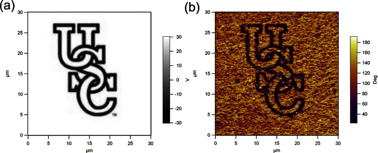
(a) A written pattern of characters “USC,” (b) PFM phase image of the PZT composite film polarized with the written pattern.
CONCLUSIONS
The piezoelectric response of the PZT and PMN-PT composite sol-gel thick films has been characterized using PFM technique. Both the local switching and piezoelectric responses show that the films exhibit superior piezoelectric properties. The effective piezoelectric coefficient of both the PZT and PMN-PT composite films was determined to be 600 and 500 pm/V, respectively, and these effective local values are comparable to that of their corresponding bulk ceramics. The results suggest that the modified composite sol-gel method is capable of preparing high-quality ceramic thick films that have found many applications in biomedical ultrasonics.
ACKNOWLEDGMENTS
This work was financially supported by NIH Grant No. P41-EB2182.
References
- Silverman R. H., Kruse D. E., Coleman D. J., and Ferrara K. W., “ High-resolution ultrasonic imaging of blood flow in the anterior segment of the eye,” Invest. Ophthalmol. Vis. Sci. 40, 1373–1381 (1999). [PubMed] [Google Scholar]
- Rallan D. and Harland C. C., “ Ultrasound in dermatology—Basic principles and applications,” Clin. Exp. Dermatol. 28(6 ), 632–638 (2003). 10.1046/j.1365-2230.2003.01405.x [DOI] [PubMed] [Google Scholar]
- Lee J., Lee C., Kim H. H., Jakob A., Lemor R., Teh S. Y., Lee A., and Shung K. K., “ Targeted cell immobilization by ultrasound microbeam,” Biotechnol. Bioeng. 108, 1643–1650 (2011). 10.1002/bit.23073 [DOI] [PMC free article] [PubMed] [Google Scholar]
- Zhou Q., Lau S., Wu D., and Shung K. K., “ Piezoelectric films for high frequency ultrasonic transducers in biomedical applications,” Prog. Mater. Sci. 56, 139–174 (2011). 10.1016/j.pmatsci.2010.09.001 [DOI] [PMC free article] [PubMed] [Google Scholar]
- Yokoyama S., Honda Y., Morioka H., Okamoto S., Funakubo H., Iijima T., Matsuda T. H., Saito K., Yamamoto T., Okino H., Sakata O., and Kimura S., “ Dependence of electrical properties of epitaxial Pb(Zr,Ti)O3 thick films on crystal orientation and Zr/(Zr+Ti) ratio,” J. Appl. Phys. 98, 094106 (2005). 10.1063/1.2126156 [DOI] [Google Scholar]
- Thiele E. S., Damjanovic D., and Setter N., “ Processing and properties of screen-printed lead zirconate titanate piezoelectric thick films on electroded silicon,” J. Am. Ceram. Soc. 84, 2863–2868 (2001). 10.1111/j.1151-2916.2001.tb01106.x [DOI] [Google Scholar]
- Yao K., He X. J., Xu Y., and Chen M. M., “ Screen-printed piezoelectric ceramic thick films with sintering additives introduced through a liquid-phase approach,” Sens. Actuator A-Phys. 118, 342–348 (2005). 10.1016/j.sna.2004.08.022 [DOI] [Google Scholar]
- Navarro A., Alcock J. R., and Whatmore R. W., “ Aqueous colloidal processing and green sheet properties of lead zirconate titanate (PZT) ceramics made by tape casting,” J. Eur. Ceram. Soc. 24, 1073–1076 (2004). 10.1016/S0955-2219(03)00460-6 [DOI] [Google Scholar]
- Corker D. L., Zhang Q., Whatmore R. W., and Perrin C., “ PZT ‘composite’ ferroelectric thick films,” J. Eur. Ceram. Soc. 22(3 ), 383–390 (2002). 10.1016/S0955-2219(01)00260-6 [DOI] [Google Scholar]
- Wu D. W., Zhou Q. F., Shung K. K., Bharadwaja S. N., Zhang D. S., and Zheng H. X., “ Dielectric and piezoelectric properties of PZT composite thick films with variable solution to powder ratios,” J. Am. Ceram. Soc. 92(6 ), 1276–1279 (2009). 10.1111/j.1551-2916.2009.03065.x [DOI] [PMC free article] [PubMed] [Google Scholar]
- Barrow D. A., Petroff T. E., Tandon R. P., and Sayer M., “ Characterization of thick lead zirconate titanate films fabricated using a new sol-gel based process,” J. Appl. Phy. 81, 876–881 (1997). 10.1063/1.364172 [DOI] [Google Scholar]
- Jaffe B., Roth R. S., and Marzullo S., “ Properties of piezoelectric ceramics,” J. Res. Natl. Bur. Standard 55, 239–254 (1955). 10.6028/jres.055.028 [DOI] [Google Scholar]
- Damjanovic D., “ Ferroelectric, dielectric and piezoelectric properties of ferroelectric thin films and ceramics,” Rep. Prog. Phys. 61, 1267–1324 (1998). 10.1088/0034-4885/61/9/002 [DOI] [Google Scholar]
- Zhao J., Zhang Q. M., Kim N., and Shrout T. R., “ Electromechanical properties of relaxor ferroelectric lead magnesium niobate-lead titanate ceramics,” Jpn. J. Appl. Phys. 34, 5658–5663 (1995). 10.1143/JJAP.34.5658 [DOI] [Google Scholar]
- Lam K. H., Li K., and Chan H. L. W., “ Lead magnesium niobate-lead titanate fibres by a modified sol-gel method,” Mater. Res. Bull. 40, 1955–1967 (2005). 10.1016/j.materresbull.2005.05.024 [DOI] [Google Scholar]
- Huang Z., Zhang Q., Corkovic S., Dorey R. A., Duval F., Leighton G., Wright R., Kirby P., and Whatmore R. W., “ Piezoelectric PZT films for MEMS and their characterization by interferometry,” J. Electroceram. 17(17 ), 549–556 (2006). 10.1007/s10832-006-6707-4 [DOI] [Google Scholar]
- Wang Z., Zhu W., Chao C., Zhao C., and Chen X., “ Characterization of composite piezoelectric thick film for MEMS application,” Surf. Coat. Technol. 198, 384–388 (2005). 10.1016/j.surfcoat.2004.10.104 [DOI] [Google Scholar]
- Dorey R. A., Stringfellow S. B., and Whatmore R. W., “ Effect of sintering aid and repeated sol infiltrations on the dielectric and piezoelectric properties of a PZT composite thick film,” J. Eur. Ceram. Soc. 22, 2921–2926 (2002). 10.1016/S0955-2219(02)00062-6 [DOI] [Google Scholar]
- Zhou Q. F., Shung K. K., and Huang Y., “ Improvement electrical properties of sol–gel derived lead zirconate titanate thick films for ultrasonic transducer application,” J. Mater. Sci. 42, 4480–4484 (2007). 10.1007/s10853-006-1414-8 [DOI] [Google Scholar]
- Zhu B., Han J., Shi J., Shung K. K., Wei Q., Huang Y., Kosec M., and Zhou Q., “ Lift-off PMN-PT thick film for high-frequency ultrasonic biomicroscopy,” J. Am. Ceram. Soc. 93(10 ), 2929–2931 (2010). 10.1111/j.1551-2916.2010.03873.x [DOI] [PMC free article] [PubMed] [Google Scholar]
- Kalinin S. V., Rar A., and Jesse S., “ A decade of piezoresponse force microscopy: Progress, challenges, and opportunities,” IEEE Trans. Ultrason., Ferroelect., Freq. Control 58(1 ), 249–254 (2011). [DOI] [PubMed] [Google Scholar]
- Hong S., Woo J., Shin H., Jeon J. U., Pak Y. E., Colla E. L., Setter N., Kim E., and No K., “ Principle of ferroelectric domain imaging using atomic force microscope,” Appl. Phys. Lett. 89(2 ), 1377–1386 (2001). [Google Scholar]
- Nath R., Hong S., Klug J. A., Imre A., Bedzyk M. J., Katiyar R. S., and Auciello O., “ Effects of cantilever buckling on vector piezoresponse force microscopy imaging of ferroelectric domains in BiFeO3 nanostructures,” Appl. Phys. Lett. 96, 163101 (2010). 10.1063/1.3327831 [DOI] [Google Scholar]
- Park M., Hong S., Klug J. A., Bedzyk M. J., Auciello O., No K., Petford-Long A., “ Three-dimensional ferroelectric domain imaging of epitaxial BiFeO3 thin films using angle-resolved piezoresponse force microscopy,” Appl. Phys. Lett. 97, 112907 (2010). 10.1063/1.3487933 [DOI] [Google Scholar]
- Zhou Q. F., Chan H. L. W., and Choy C. L., “ PZT ceramic-ceramic 0-3 nanocomposite films for ultrasonic transducer applications,” Thin Solid Films 375, 95–99 (2000). 10.1016/S0040-6090(00)01232-3 [DOI] [Google Scholar]
- Zhou Q., Zhang Q., Xu B., and Trolier-McKinstry S., “ In-plane polarized 0.7Pb(Mg1/3Nb2/3)O3-0.3PbTiO3 thin films,” J. Am. Ceram. Soc. 85, 1997–2000 (2002). 10.1111/j.1151-2916.2002.tb00394.x [DOI] [Google Scholar]
- Morgan Electro Ceramics, Transducer Products Division, “ Excellence in piezoelectric technology,” Typical Values of Lead Zirconate Titanate Materials (1999), Table 3, pp. 10.
- Kelly J., Leonard M., Tantigate C., and Safari A., “ Effect of composition on the electromechanical properties of (1-x)Pb(Mg1/3Nb2/3)O3-xPbTiO3,” J. Am. Ceram. Soc. 80, 957–964 (1997). 10.1111/j.1151-2916.1997.tb02927.x [DOI] [Google Scholar]