Abstract
Juvenile Paget’s disease (JPD) is a rare heritable osteopathy characterized biochemically by markedly increased serum alkaline phosphatase (ALP) activity emanating from generalized acceleration of skeletal turnover. Affected infants and children typically suffer bone pain and fractures and deformities, become deaf, and have macrocranium. Some who survive to young adult life develop blindness from retinopathy engendered by vascular microcalcification. Most cases of JPD are caused by osteoprotegerin (OPG) deficiency due to homozygous loss-of-function mutations within the TNFRSF11B gene that encodes OPG. We report a 3-year-old Iranian girl with JPD and craniosynostosis who had vitamin D deficiency in infancy. She presented with fractures during the first year-of-life followed by bone deformities, delayed development, failure-to-thrive, and pneumonias. At 1 year-of-age, biochemical studies of serum revealed marked hyperphosphatasemia together with low-normal calcium and low inorganic phosphate and 25-hydroxyvitamin D levels. Several family members in previous generations of this consanguineous kindred may also have had JPD and vitamin D deficiency. Mutation analysis showed homozygosity for a unique missense change (c.130T>C, p.Cys44Arg) in TNFRSF11B that would compromise the cysteine-rich domain of OPG that binds receptor activator of NF-κB ligand (RANKL). Both parents were heterozygous for this mutation. The patient’s serum OPG level was extremely low and RANKL level markedly elevated. She responded well to rapid oral vitamin D repletion followed by pamidronate treatment given intravenously. Our patient is the first Iranian reported with JPD. Her novel mutation in TNFRSF11B plus vitamin D deficiency in infancy was associated with severe JPD uniquely complicated by craniosynostosis. Pamidronate treatment with vitamin D sufficiency can be effective treatment for the skeletal disease caused by the OPG deficiency form of JPD.
Keywords: alkaline phosphatase, bone remodeling, craniosynostosis, deafness, hyperphosphatasemia, hyperphosphatasia, osteoclast, osteolysis, osteoprotegerin, pamidronate, retinopathy, rickets, tumor necrosis factor, vascular calcification, vitamin D deficiency
II) Introduction
Juvenile Paget’s disease (JPD: #239000 in Mendelian Inheritance in Man)(1) is a rare heritable osteopathy that markedly increases serum alkaline phosphatase (ALP) activity (hyperphosphatasemia) from greatly accelerated turnover of osseous tissue throughout the skeleton.(2) JPD was first called “fragile bones with macrocranium” when characterized in 1956 by Bakwin and Eiger in young sisters of Puerto Rican heritage living in New York City.(3) Approximately 60 cases have been reported worldwide.(3–11) Affected infants and children suffer bone pain, fractures, and skeletal deformities and most become deaf by early childhood.(4,6,7) Those who live longer may acquire diffuse vascular microcalcification (VMC)(8) that causes retinopathy and blindness(5,10) and perhaps intracranial aneurysms.(11) JPD can be fatal in childhood or by young adult life unless antiresorptive therapy is given.(4,8)
Beginning in 2002,(4,7) the molecular basis for most but not all cases of JPD became understood as autosomal recessive transmission of loss-of-function mutations within TNFRSF11B that encodes osteoprotegerin (OPG).(12) OPG is a receptor member of the tumor necrosis factor (TNF) superfamily, and slows osteoclastogenesis and osteoclast (OC) action by “decoying” receptor activator of nuclear factor-κB (RANK) ligand (RANKL) away from its cognate receptor RANK.(2) Thus, the prevalent form of JPD can now be called “OPG deficiency”.(2)
Here, we describe a 3-year-old Iranian girl with the OPG deficiency form of JPD. The additional burden of vitamin D deficiency documented during infancy likely exacerbated her skeletal disease and may have caused craniosynostosis. A great-aunt in her consanguineous kindred was probably similarly affected. In keeping with most cases of JPD, we found that our patient was homozygous for a unique “founder” mutation in TNFRSF11B.
III) Materials and Methods
A) Case Report
This 3-year-old Iranian girl appeared well until age 3 months when she developed irritability with excessive crying that was explained by a clavicle fracture. The bone break was treated conservatively. At age 1 year, she sustained a pelvic fracture and had wide wrists and ankles and a “rachitic rosary”. Our review of the radiographs of her lower extremities showed bowing, osteopenia, and wide medullary cavities with indistinct inner margins, but the physeal lines were well-defined and without the fraying associated with rickets. Examination of her skull showed a closed fontanel. Serum calcium (Ca) was 8.5 mg/dl (Nl 8.5 – 10.8), inorganic phosphate (Pi) 3.0 mg/dl (Nl 3.8 – 6.5), 25-hydroxyvitamin D [25(OH)D] 10 ng/ml (Nl 12 – 40), and ALP 1897 U/L (Nl 145 – 420). Serum 1,25-dihydroxyviamin D [1,25(OH)2D] was not measured. Vitamin D deficiency was diagnosed and she was given 600,000 units of vitamin D3 orally once (“stoss therapy”) and then 600 units orally each day as maintenance therapy. Her serum 25(OH)D and Pi levels were normal after ten days of this treatment, but her serum ALP level increased further, radiographic changes were not improved, and the skeletal deformities and irritability from bone pain were no better.
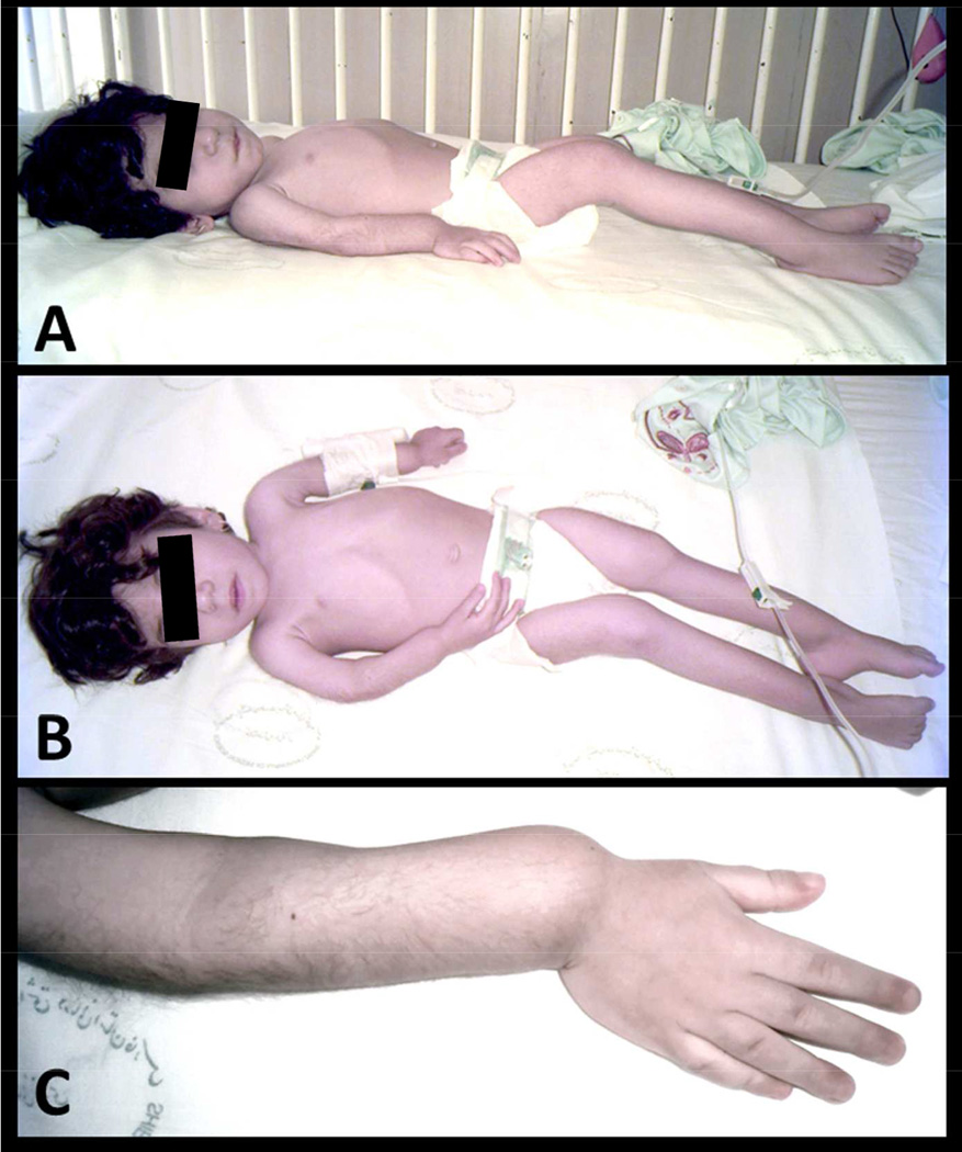
She was referred to us at 2.5 years-of-age with failure-to-thrive. Biochemical testing during the previous year had shown normal serum Ca, Pi, 25(OH)D, and 1,25(OH)2D levels, yet there was biochemical evidence of continued rapid bone turnover with serum ALP activity of 2920 – 5010 U/L (Nl 145 – 420) and urinary deoxypyridinoline of 178 nmol per millimole of creatinine (Nl 2 – 41 for children). She was irritable and appeared chronically ill (Figure 1). Her weight (7500 g), length (74 cm), and head circumference (44 cm) were all below the 3rd percentile for age and gender. Both the anterior and posterior fontanels were closed. Chest deformity included widening of the costochondral junctions giving the appearance of a “rachitic rosary”. Her humeri were short, femurs laterally bossed, tibias curved anteriorly, and wrists and ankles wide. Developmental milestones were delayed. She could speak only a few words (e.g., “papa” and “mama”), was unable to stand, and had markedly delayed gross motor skills. There was poor muscle tone. Her lungs and heart were unremarkable, and she had no hepatosplenomegaly. An umbilical hernia was present. Hearing assessment (pure tone audiometry and oto-acoustic emission) was normal. An ophthalmologist reported a normal eye examination, including the retina. Radiographic studies were consistent with JPD (see Radiographic Findings).
Figure 1. Patient At Referral.
At 2.5 years-of-age (panel A, B) our patient had not walked. There is bowing deformity of all limbs with muscle wasting. Note the flaring of the wrists (panel C) that had been attributed to vitamin D deficiency rickets.
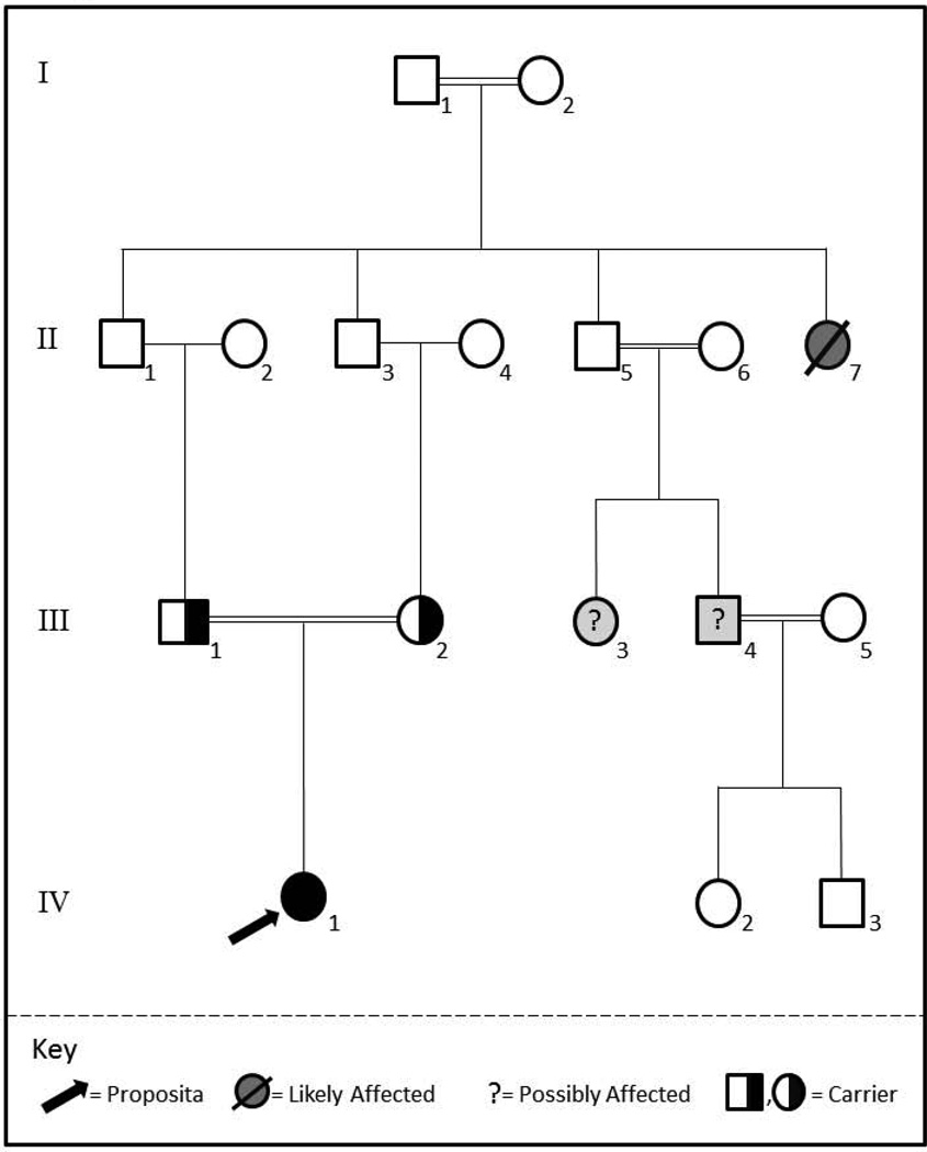
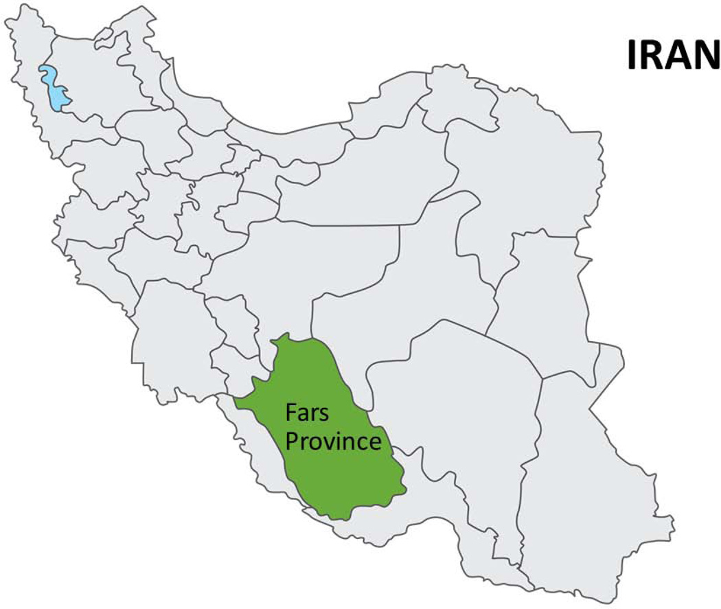
Her healthy appearing parents were first cousins (Figure 2) from Fars Province in the south of Iran (Figure 3). They had normal serum ALP activity and unremarkable findings on skeletal radiographs. However, the kindred’s medical history revealed a great-aunt also with consanguineous parents (Figure 2) who had similar skeletal problems. According to a grandfather, she suffered bone fractures and deformities, poor weight gain, short stature, delayed development, and mental retardation, but could hear and speak. She too had been treated for nutritional rickets, but further evaluation was not undertaken. Subsequently, she required hospitalizations for respiratory failure and died from pneumonia at age 12 years. Her medical records could not be located. Our patient’s father had two cousins, also from consanguineous parents (Figure 2), with bone deformities and severe short stature. One is a 27-year-old man without mental, visual, or vascular problems who has two healthy children. The other is a 34-year-old woman who is blind, mentally retarded, and childless. We have been unable to study these two individuals.
Figure 2. Family Pedigree.
Four generations reveal that our patient is consanguineous, because her parents are first cousins. Her maternal great-aunt was likely affected by JPD and also consanguineous, giving a pseudo-dominant appearance of transmission of their JPD.
Figure 3. Geographical Region of Patient’s OPG Mutation.
Our patient lives in Fars Province, Iran where the population is largely of Persian heritage and where the prevalence of JPD is unknown.
B) Mutation Analysis
After informed written consent, peripheral blood from the patient and her parents was sent to our research laboratory in St. Louis, MO, USA. DNA was isolated from the leukocytes using the Gentra Puregene DNA extraction kit (Invitrogen, Carlsbad, CA, USA). All five coding exons of TNFRSF11B were amplified by PCR and sequenced using an Applied Biosystems 3130 (Foster City, CA, USA) as previously described.(4) The TNFRSF11B mutation (see Results) was identified by visual examination of electropherograms and alignment of the DNA sequence to a control sequence using AlignX/VectorNTI software (Invitrogen, Carlsbad, CA, USA).
C) Serum OPG and RANKL
Enzyme immunoassay kits (Bio Net, Southbridge, MA, USA) were used to quantitate soluble OPG and RANKL levels in serum. In two separate dedicated assay runs, performed as specified by the manufacturer (Biomedica Medizinprodukte GmbH & Co, KG), the serum OPG and RANKL levels in our patient and her parents were contrasted to levels in sera from five healthy children and five healthy adults chosen randomly as age- and ethnicity-matched controls.
IV) Results
A) Radiographic Findings
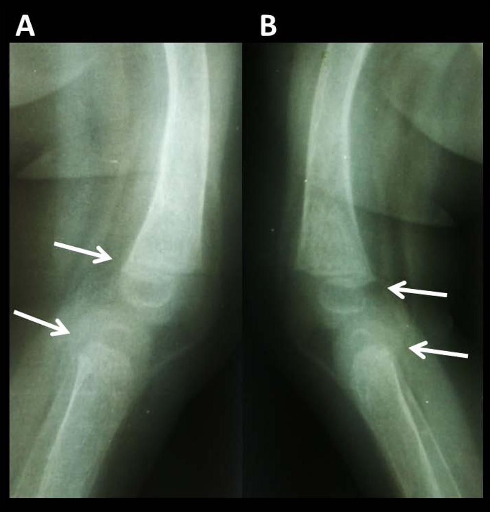
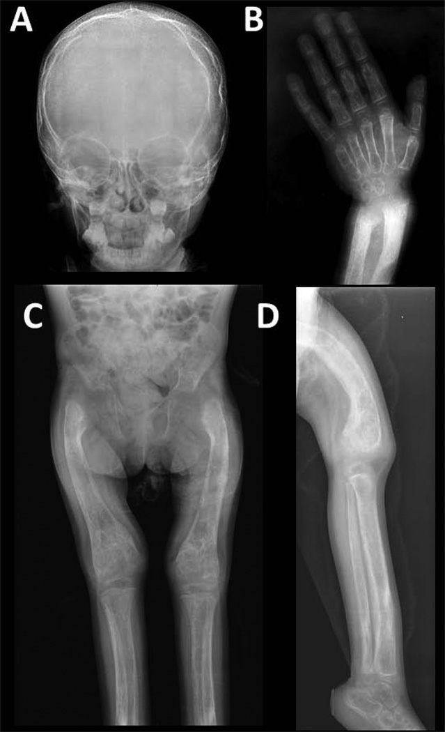
Our review of the patient’s radiographs at 8 months-of-age revealed changes most consistent with JPD, but without superimposed vitamin D deficiency rickets (Figure 4). At 2.5 years-of-age, her findings were again typical for JPD(3–11) and included widened osteopenic long bones with coarse trabeculae and indistinct corticomedullary junctions (Figure 5). The diploic space of the skull was wide with coarse vertical trabeculae giving a “hair-on-end” appearance, and the outer table of the skull was thin and indistinct. However, the sutures were prematurely closed. The chest was deformed with widening of costochondral junctions similar to a rachitic rosary. There were short bowed arms, wide wrists and ankles, coxa vara, bowed thighs and legs, osteopenic widened long tubular bones with irregular cortical thickenings primarily on their inner surfaces causing indistinct corticomedullary junctions, irregular focal areas of sclerosis and lucencies, modeling errors, and cystic changes in the distal femurs.
Figure 4. Radiographic Findings at 8 Months-of-Age.
Lateral radiographs (panel A = right, panel B = left) of bones at the knees at 8 months-of-age show bowing, osteopenia, wide medullary cavities with indistinct inner margins (especially in the tibias) that all can be seen in rickets, but the physeal lines (arrows) are distinct and without fraying expected with rickets.
Figure 5. Radiographic Findings at Referral at 2.5 Years-of-Age.
A) The skull has a wide diploic space with coarse vertically oriented trabeculae and an indistinct thin outer table. The sagittal suture is closed.
B) Radiographs of the hand and wrist shows severe osteopenia. The short tubular bones are wide, especially the phalanges, and there are thin cortices. The radius and ulna show metaphyseal and metadiaphyseal sclerosis, loss of corticomedullary definition, thin cortices, distal flaring, and slight fraying of the provisional zones of calcification. Soft tissue swelling is apparent.
C) The femurs and tibias are osteopenic, wide, and have irregular cortical thickening, indistinct corticomedullary junctions, focal areas of sclerosis and lucencies, marked coxa vara, failure of modeling (especially in the distal femora), and lateral femoral bowing. There is mild acetabular protrusion.
D) This leg and distal thigh demonstrate tibial and femoral bowing, wide medullary cavities with indistinct margins, irregular cortical thickening, and interference with modeling. Coarse and thick trabeculae and focal areas of sclerosis and lucencies are present.
B) Mutational Analysis
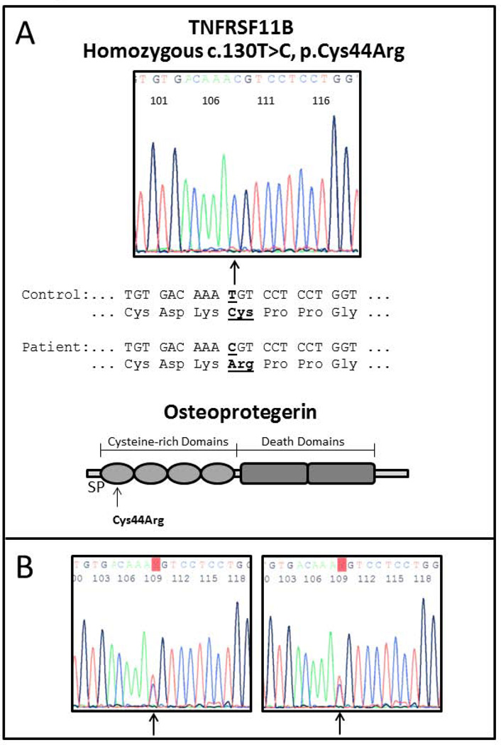
Leukocyte DNA from our patient revealed a homozygous missense change (c.130T>C, p.Cys44Arg) in exon 2 of TNFRSF11B (Figure 6). Her parents were heterozygous for this finding. This missense alteration was likely a mutation that causes JPD because it was not listed in dBSNP as a single nucleotide polymorphism, and was not found after sequencing TNFRSF11B in > 100 Americans.(4) Also, it was not a conservative amino acid change. However, it had not been encountered in other cases of JPD, either ours(5) or published reports. The observation was instead consistent with our experience that various geographic/ethnic groups worldwide with JPD typically carry unique homozygous TNFRSF11B mutations.(13) The exceptional patient, from the United Kingdom, was compound heterozygous for a single amino acid deletion and a nonsense mutation.(13)
Figure 6. TNFRSF11B Mutation Analysis.
A) Sequencing of our patient’s TNFRFS11B (top) shows a homozygous missense mutation c.130T>C, p.Cys44Arg located within the cysteine-rich domain of OPG (bottom) where loss-of-function defects cause severe JPD.(15) (SP = signal peptide).
B) Sequencing of the parents’ TNFRSF11B shows the same heterozygous mutation.
C) Serum Levels of OPG and RANKL
Our patient’s serum OPG level was very low compared to age-matched controls at 0.29 pmol/L (Nl 2.93 ± 1.36) whereas the values were 1.05 pmol/L, 2.51 pmol/L, and 2.11 – 3.97 pmol/L in her father, mother, and five healthy adult controls, respectively. Our patient’s serum RANKL level was markedly elevated (39 pmol/L) in contrast to undetectable in her parents and all controls.
D) Treatment
Our patient was given pamidronate (PMD: Novartis Pharmaceuticals Corporation, East Hanover, NJ, USA) intravenously beginning at age 2.5 years at a dose of 1 mg/kg/day for 3 days, every 3 months, while oral vitamin D3 supplementation was continued at 600 U/day. Serum ALP levels had been increasing during her second year of life from 2156 to 2920, and then to 4653 U/L. Subsequently, irritability was significantly less and her muscle power increased so that she moved her extremities easily and could stand with help. She can now sit, stand, and walk with assistance without pain. Serum calcium levels were measured monthly and have been normal, whereas ALP decreased from 5010 U/L at referral at age 2.5 years to 2187 U/L at age 3 years (i.e.; after 6 months of PMD therapy), and is currently 1126 U/L (Nl 145 – 420) at 38/12 years-of-age.
V) Discussion
Our patient represents the first report of JPD in Iran. We found that she has the OPG deficiency form of this disorder, that for her is caused by a unique homozygous missense mutation (c.130T>C, p.Cys44Arg) within TNFRSF11B.(4,5,7,14) Her healthy appearing consanguineous parents are heterozygous carriers for this defect, but several other individuals in this kindred of Persian heritage may also have suffered OPG deficiency. Hence, it will now be important to: i) understand whether JPD is prevalent in this geographical region, ii) assure that the disorder is recognized and correctly diagnosed, and iii) treat promptly any additional burden of vitamin D deficiency before antiresorptive therapy is given.
Our patient’s clinical course seemed relatively severe for OPG deficiency.(5,7) She presented in infancy with fractures followed by skeletal deformity, delayed developmental milestones, failure-to-thrive, and recurrent pneumonias. Her great-aunt may have succumbed at 12 years-of-age to JPD. However, the JPD in these individuals was likely exacerbated during infancy by vitamin D deficiency with secondary hyperparathyroidism further accelerating osteoclastogenesis and bone turnover. Perhaps vitamin D deficiency also caused our patient’s craniosynostosis because premature suture closure is a consequence of various types of rickets, including nutritional rickets,(15) and our literature review revealed no reports of this complication in JPD. Our patient had small head size, rather than the macrocranium typical of JPD.(3–11) Although she lived in a tropical area of southern Iran, her sunlight exposure was probably limited by her disabilities that kept her indoors in a city. Furthermore, she was breast fed during infancy without receiving supplemental vitamin D. Her mother’s exposure to sunlight was likely also limited because she wore the burka customary for women in this part of Iran. This tradition has been reported to cause low serum levels of 25(OH)D.(16–20) Accordingly, quantitation of serum 25(OH)D and correction of any vitamin D deficiency as early as possible for JPD patients may be important to avoid related complications. Indeed, Polyzos and colleagues(21) reported in 2010 profound hypocalcemia after zoledronic acid treatment given to an adult with JPD.
Our patient responded well to PMD treatment after vitamin D deficiency had been rapidly corrected by “stoss” (German – “push” or “shove”) therapy. She was able to safely begin the PMD infusions that have been helpful for JPD.(6) However, anti-resorptive treatment may now be necessary lifelong to control her skeletal disease.(5) Fortunately, she did not have deafness, and it will be important to know if her hearing will be preserved by anti-resorptive therapy to block the destruction of middle ear bones characteristic of OPG deficiency.(2) Even so, without further advances in the medical management of OPG deficiency, she may become blind in adult life from VMC and retinopathy because this complication is not expected to respond to bisphosphonate (or perhaps to the anti-RANKL monoclonal antibody, denosumab) therapy.(2) In 2007, Whyte et al(5) reported a 60-year-old Greek man with OPG deficiency who was homozygous for the "Balkan" TNFRSF11B frame-shift (966_969delTGACinsCTT) mutation(11) and suffered progressive deafness and blindness although skeletal deformity seemed controlled by bisphosphonate therapy that began at age 32 years. His deafness and blindness occurred although the Balkan TNFRSF11B mutation retains some OPG function, and is regarded as the mildest OPG mutation.(5,15,22)
In health, OPG acts as a homodimer with 401 amino acids per monomer,(23) including a series of cysteine-rich domains at the amino-terminal characteristic of TNF receptor superfamily members (Figure 6). These domains form an extended rod-like structure that binds RANKL.(24) Our patient’s homozygous mutation located at c.130T>C, p.Cys44Arg in exon 2 of TNFRSF11B predicts a transition missense mutation. The affected cysteine residue with thiol side chain would be mutated to arginine with a 3-carbon aliphatic straight chain, and the distal end would be capped by a complex guanidinium group. The mutant OPG amino acid residue is larger, positively charged, and less hydrophobic than the wild type OPG residue located in the core of OPG. Receptors in the TNF superfamily have two to six cysteine-rich domains in the extracellular ligand-binding region; OPG has four (Figure 6).(25) These cysteines form disulfide bonds that are critical for the structural integrity and RANKL binding property of OPG. Our patient’s homozygous Cys44Arg OPG is located in the first cysteine-rich domain (Figure 6) and could compromise folding of the mutant protein, or cause loss of hydrophobic interaction within the core of the protein.(26) Accordingly, her loss of a specific disulfide bond would probably destabilize the three-dimensional structure of OPG.
With homozygous selective and complete deletion of TNFRSF11B in Navajos with JPD, serum immunoreactive OPG levels are undetectable and soluble immunoreactive RANKL levels are extremely elevated.(4) Similar findings were present in our current patient. The Navajo patients have OPG deficiency that has been (if untreated with anti-resorptive therapy) lethal in childhood or young adult life.(4) In contrast, some JPD patients with small structural mutations within TNFRSF11B biosynthesize OPG that is detectable in serum using current assays.(5) Our findings,(13) and those of Chong et al,(14) indicated that homozygous mutation of TNFRSF11B accounts for most patients with JPD. Chong et al(14) concluded that the six different TNFRSF11B mutations studied correlated with the clinical severity of JPD. Our patient carries a homozygous missense mutation in TNFRSF11B which could lead to milder disease than mutations that delete or disrupt the coding region for OPG. Nevertheless, her delayed development, repeated fractures, recurrent pneumonias, and failure-to-thrive seemed more severe than with other missense mutations in TNFRSF11B.(14) We wonder if her additional burden of vitamin D deficiency during infancy together with a missense mutation that caused significant structural disruption of OPG is the explanation for her severe JPD. In fact, two other missense mutations that would affect a cysteine residue in the same region of OPG caused severe JPD.(14)
Hence, homozygosity for a unique (c.130T>C, p.Cys44Arg) missense mutation of TNFRSF11B encoding OPG caused a severe and potentially lethal form of JPD in a consanguineous kindred in Iran. Vitamin D deficiency was likely an exacerbating, but treatable, coincidental problem. Distinguishing OPG deficiency from “nutritional” rickets and repletion of any concomitant lack of vitamin D prior to anti-resorptive therapy will be important for the safe and effective medical management of this rare genetic skeletal disease.
Acknowledgements
We are grateful to Ms. Sharon McKenzie and Ms. Vivienne McKenzie for helping to prepare the manuscript. Ms. Margaret Huskey performed the TNFRSF11B mutation analyses. Ms. Angela Nenninger and Ms. Jessica Richards assisted with our review of the JPD literature.
Supported by the Shiraz University of Medical Sciences, Shriners Hospitals for Children, The Clark and Mildred Cox Inherited Metabolic Bone Disease Research Fund, The Hypophosphatasia Research Fund, The Barnes-Jewish Hospital Foundation, The Frederick S. Upton Foundation, and National Institutes of Health (DK067145).
Acronyms and Abbreviations
- ALP
alkaline phosphatase
- JPD
juvenile Paget’s disease
- NF-κB
nuclear factor-kappa B
- Nl
normal
- OPG
osteoprotegerin
- PMD
pamidronate
- RANK
receptor activator of NF-κB
- RANKL
RANK ligand
- TNF
tumor necrosis factor
- TNFRSF11B
gene that encodes OPG
- VMC
vascular microcalcification
- 25(OH)D
25-hydroxyvitamin D
Footnotes
Disclosures: The authors have no conflict of interest.
Contributor Information
Forough Saki, Email: sakeif@sums.ac.ir.
Zohreh Karamizadeh, Email: Zkaramizadeh@yahoo.com.
Shiva Nasirabadi, Email: shiva.nasirabadi@gmail.com.
Steven Mumm, Email: smumm@dom.wustl.edu.
William H. McAlister, Email: mcalisterw@wustl.edu.
Michael P. Whyte, Email: mwhyte@shrinenet.org.
References
- 1.Baltimore, MD: Johns Hopkins University; Online Mendelian Inheritance in Man, OMIM®. MIM Number: 23900. Date last edited: September 4, 2012. World Wide Web URL: http://omim.org/ [Google Scholar]
- 2.Whyte MP. Mendelian Disorders of RANKL/OPG/RANK Signaling, Chapter 20. In: Thakker RV, Whyte MP, Eisman J, Igarashi T, editors. Genetics of Bone Biology and Skeletal Disease. San Diego, CA: Elsevier (Academic Press); 2013. pp. 309–323. [Google Scholar]
- 3.Bakwin H, Eiger MS. Fragile bones and macrocranium. J Pediatr. 1956;49:558–564. doi: 10.1016/s0022-3476(56)80143-1. [DOI] [PubMed] [Google Scholar]
- 4.Whyte MP, Obrecht SE, Finnegan PM, Jones JL, Podgornik MN, McAlister WH, Mumm S. Osteoprotegerin deficiency and juvenile Paget's disease. New Engl J Med. 2002;347:175–184. doi: 10.1056/NEJMoa013096. [DOI] [PubMed] [Google Scholar]
- 5.Whyte MP, Singhellakis PN, Petersen MB, Davies M, Totty WG, Mumm S. Juvenile Paget's disease: The second reported, oldest patient is homozygous for the TNFRSF11B "Balkan" mutation (966_969deITGACinsCTT), which elevates circulating immunoreactive osteoprotegerin levels. J Bone Miner Res. 2007;22:938–946. doi: 10.1359/jbmr.070307. [DOI] [PubMed] [Google Scholar]
- 6.Tau C, Mautalen C, Casco C, Alvarez V, Rubinstein M. Chronic idiopathic hyperphosphatasia: Normalization of bone turnover with cyclical intravenous pamidronate therapy. Bone. 2004;35:210–216. doi: 10.1016/j.bone.2004.03.013. [DOI] [PubMed] [Google Scholar]
- 7.Cundy T, Hegde M, Naot D, Chong B, King A, Wallace R, Mulley J, Love DR, Seidel J, Fawkner M, Banovic T, Callon KE, Grey AB, Reid IR, Middleton-Hardie CA, Cornish J. A mutation in the gene TNFRSF11B encoding osteoprotegerin causes an idiopathic hyperphosphatasia phenotype. Hum Mol Genet. 2002;11:2119–2127. doi: 10.1093/hmg/11.18.2119. [DOI] [PubMed] [Google Scholar]
- 8.Mitsudo SM. Chronic idiopathic hyperphosphatasia associated with pseudoxanthoma elasticum. J Bone Joint Surg Am. 1971;53:303–314. [PubMed] [Google Scholar]
- 9.Fretzin DF. Pseudoxanthoma elasticum in hyperphosphatasia. Arch Dermatol. 1975;111:271–272. doi: 10.1001/archderm.111.2.271. [DOI] [PubMed] [Google Scholar]
- 10.Sharif KW, Doig WM, Kinsella FP. Visual impairment in a case of juvenile Paget's disease with pseudoxanthoma elasticum: an eleven year follow up. J Pediatr Ophthalmol Strabismus. 1989;26:299–302. doi: 10.3928/0191-3913-19891101-13. [DOI] [PubMed] [Google Scholar]
- 11.Choremis C, Yannakos D, Papadatos C, Baroutsou E. Osteitis deformans (Paget's disease) in an 11 year old boy. Helv Paediatr Acta. 1958;13:185–188. [PubMed] [Google Scholar]
- 12.Morinaga T, Nakagawa N, Yasuda H, Tsuda E, Higashio K. Cloning and characterization of the gene encoding human osteoprotegerin/osteoclastogenesis-inhibitory factor. Eur J Biochem. 1998;254:685–691. doi: 10.1046/j.1432-1327.1998.2540685.x. [DOI] [PubMed] [Google Scholar]
- 13.Mumm S, Banze S, Pettifor J, Tau C, Schmitt K, Ahmed A, Whyte MP. Juvenile paget's disease: Molecular analysis of TNFRSF11B encoding osteoprotegerin indicates homozygous deactivating mutations from consanguinity as the predominant etiology. J Bone Miner Res. 2003;18(Suppl 2):S388. [Google Scholar]
- 14.Chong B, Hegde M, Fawkner M, Simonet S, Cassinelli H, Coker M, et al. Idiopathic hyperphosphatasia and TNFRSF11B mutations: Relationships between phenotype and genotype. J Bone Miner Res. 2003;18:2095–2104. doi: 10.1359/jbmr.2003.18.12.2095. [DOI] [PubMed] [Google Scholar]
- 15.Reilly BJ, Leeming JM, Fraser D. Craniosynostosis in the rachitic spectrum. 1964;64:396–405. doi: 10.1016/s0022-3476(64)80192-x. [DOI] [PubMed] [Google Scholar]
- 16.Mishal AA. Effects of different dress styles on vitamin D levels in healthy young jordanian women. Osteoporosis Int. 2001;12:931–935. doi: 10.1007/s001980170021. [DOI] [PubMed] [Google Scholar]
- 17.Allali F, El Aichaoui S, Khazani H, Benyahia B, Saoud B, El Kabbaj S, Bahiri R, Abouqal R, Hajjaj-Hassouni N. High prevalence of hypovitaminosis D in Morocco: relationship to lifestyle, physical performance, bone markers, and bone mineral density. Semin Arthritis Rheum. 2009;38:444–451. doi: 10.1016/j.semarthrit.2008.01.009. [DOI] [PubMed] [Google Scholar]
- 18.Nichols EK, Khatib IM, Aburto NJ, Sullivan KM, Scanlon KS, Wirth JP, Serdula MK. Vitamin D status and determinants of deficiency among non-pregnant Jordanian women of reproductive age. Eur J Clin Nutr. 2012;66:751–756. doi: 10.1038/ejcn.2012.25. [DOI] [PMC free article] [PubMed] [Google Scholar]
- 19.Gannagé-Yared MH, Chemali R, Sfeir C, Maalouf G, Halaby G. Dietary calcium and vitamin D intake in an adult Middle Eastern population: food sources and relation to lifestyle and PTH. Int J Vitam Nutr Res. 2005;75:281–289. doi: 10.1024/0300-9831.75.4.281. [DOI] [PubMed] [Google Scholar]
- 20.Musaiger AO, Hassan AS, Obeid O. The paradox of nutrition-related diseases in the Arab countries: the need for action. Int J Environ Res Public Health. 2011;8:3637–3671. doi: 10.3390/ijerph8093637. [DOI] [PMC free article] [PubMed] [Google Scholar]
- 21.Polyzos SA, Anastasilakis AD, Litsas I, Efstathiadou Z, Kita M, Arsos G, Moralidis E, Papatheodorou A, Terpos E. Profound hypocalcemia following effective response to zoledronic acid treatment in a patient with juvenile Paget's disease. J Bone Miner Metab. 2010;28:706–712. doi: 10.1007/s00774-010-0198-8. [DOI] [PubMed] [Google Scholar]
- 22.Janssens K, de Vernejoul MC, de Freitas F, Vanhoenacker F, Van Hul W. An intermediate form of juvenile Paget’s disease caused by a truncating TNFRSF11B mutation. Bone. 2005;36:542–548. doi: 10.1016/j.bone.2004.12.004. [DOI] [PubMed] [Google Scholar]
- 23.Simonet WS, Lacey DL, Dunstan CR, Kelley M, Chang MS, Lüthy R, Nguyen HQ, Wooden S, Bennett L, Boone T, Shimamoto G, DeRose M, Elliott R, Colombero A, Tan HL, Trail G, Sullivan J, Davy E, Bucay N, Renshaw-Gegg L, Hughes TM, Hill D, Pattison W, Campbell P, Sander S, Van G, Tarpley J, Derby P, Lee R, Boyle WJ. Osteoprotegerin: A novel secreted protein involved in the regulation of bone density. Cell. 1997;89:309–319. doi: 10.1016/s0092-8674(00)80209-3. [DOI] [PubMed] [Google Scholar]
- 24.Banner DW, D'Arcy A, Janes W, Gentz R, Schoenfeld HJ, Broger C, Loetscher H, Lesslauer W. Crystal structure of the soluble human 55 kd TNF receptor-human TNF beta complex: implications for TNF receptor activation. Cell. 1993;73:431–445. doi: 10.1016/0092-8674(93)90132-a. [DOI] [PubMed] [Google Scholar]
- 25.Yamaguchi K, Kinosaki M, Goto M, Kobayashi F, Tsuda E, Morisaga T, Higashio K. Characterization of structural domains of human osteoclastogenesis inhibitory factor. J Biol Chem. 1998;273:5117–5123. doi: 10.1074/jbc.273.9.5117. [DOI] [PubMed] [Google Scholar]
- 26.Venselaar H, Te Beek TA, Kuipers RK, Hekkelman ML, Vriend G. Protein structure analysis of mutations causing inheritable diseases. An e-Science approach with life scientist friendly interfaces. BMC Bioinformatics. 2010;11:548. doi: 10.1186/1471-2105-11-548. [DOI] [PMC free article] [PubMed] [Google Scholar]


