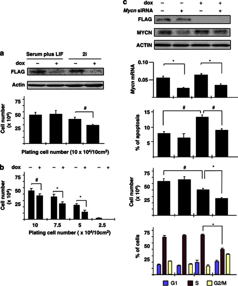
Figure 3.
Arid3b protects ES cells from Mycn-induced apoptosis. Arid3b null ES cells carrying a construct containing FLAG-tagged Arid3b driven by a dox-repressable promoter were established and used to analyze the effect of Arid3b expression in ES cells. Error bars represent s.d. from three independent replicates. Statistical significance was calculated by Student's t test. #P<0.05; *P<0.01. (a) Dox-induced repression of the Arid3b-transgene in ES culture conditions. Western blots of whole-cell extracts using antibodies against FLAG (Arid3b) and β-actin (loading control). Detection of flag-tagged Arid3b protein is lost after 2 days exposure of dox (upper panel). The proliferation of the same ES cell line was compared with or without repression of Arid3b under either serum plus LIF or 2i conditions. ES cells were plated at a density of 104 cells/cm2, and the total cell number determined after 2 days (lower panel). (b) Effect of cell density on the proliferation of ES cells. Cell growth was evaluated (as in a) at different starting cell densities under 2i conditions. (c) Function of Arid3b and Mycn in ES cell proliferation under 2i conditions. The expression of Arid3b and Mycn were repressed by dox or siRNA, respectively. All assays were performed 2 days after transfection and or application of dox. Western blots using antibodies against FLAG (Arid3b), Mycn and β-actin (loading control) shows each depleted level (top). Mycn mRNA levels were also determined by quantative reverse transcription–PCR analysis relative to ubiquitin levels (second panel). The proportion of apoptotic cells was determined by flow cytometric analysis (third panel). Annexin+ and 7-AAD− cells were considered to be apoptotic. Cell numbers were determined (fourth panel) and analyzed for bromodeoxyuridine incorporation by flow cytometry to determine the proportions of cells at different stages of the cycle (bottom panel).

