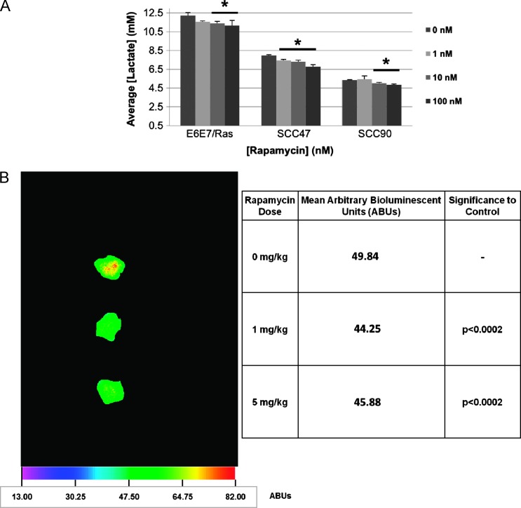
Figure 5.
Rapamycin attenuates tumor cell lactate production. (A) In vitro lactate assay. E6/E7/Ras MOEs or HPV+ SCCs plated to 100% confluence were incubated for 4 hours in the presence of the indicated doses of rapamycin. The media was then subject to a commercially available colorimetric lactate assay, the concentration determined through standard curve, and the results plotted as average lactate concentration per dosage group (n = 3). Rapamycin significantly decreased lactate production in both HPV+ SCC (SCC47: 1 nM, P < .01; 10 nM, P < .05; 100 nM, P < .02; SCC90: 10 nM, P < .02; 100 nM, P < .02) and E6/E7/Ras MOE (10 nM, P < .02; 100 nM, P < .05) cell lines. (B) Quantitative lactate bioluminescence. Lactate levels were visualized in end-point tumor sections of E6/E7/Ras tumors from daily vehicle or rapamycintreated C57Bl/6 mice using a lactate-dependent luciferase-containing buffer system as described by Broggini-Tenzer et al. [34]. Intensities of bioluminescent images of six (n = 6) tumor sections from multiple mice at each dosage group were averaged. A representative image is shown alongside tabulated mean bioluminescent intensity and P values compared to control per dosage group (1 to 5 mg/kg, NS).

