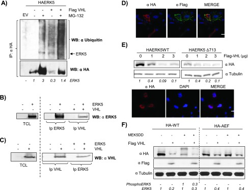
Figure 2.

pVHL promotes ERK5 degradation. (A) Cos7 cells were transfected with 0.5 µg of HA-ERK5 and with 3 µg of Flag-VHL. Thirty-six hours after transfection, cells were incubated in the presence or absence of 20 µM MG-132 for 5 hours. Cells were immunoprecipitated against HA and blotted against ubiquitin. Lower panel showed reblotting of the membrane against HA. HA-ERK5 fold variation observed in this experiment is shown at the bottom. (B) 293T cells were transfected with 5 µg of HA-ERK5 and 5 µg of Flag-VHL. Samples were immunoprecipitated and immunoblotted with indicated antibodies. As positive controls, TCLs overexpressing HA-ERK5 were blotted against HA. (C) Same as B. As positive controls, TCLs overexpressing Flag-VHL were blotted against Flag. (D) Cos7 cells were transfected with 0.25 µg of HA-ERK5 and 0.25 µg of Flag-VHL, and subcellular distributions of both proteins were evaluated by immunofluorescence. Image shows a representative field of five. The scale bar represents 10 µm. (E) Upper panel: Cos7 cells were transfected with 0.5 µg of HA-ERK5 WT or HA-ERK5Δ713 with increasing amounts of Flag-VHL. Thirty-six hours later, 30 µg of TCLs were blotted against HA or tubulin. Fold variation of this experiment is shown at the bottom. Lower panel: Cos7 cells were transfected with 0.25 µg of HA-ERK5Δ713 and processed as in D. (F) Cos7 cells were transfected with 0.5 µg of HA-ERK5 or HA-ERK5-AEF, 1.5 µg of MEK5DD, and 3 µg of Flag-VHL at the indicated combinations. TCLs were processed as in E. Fold variation for both proteins in this experiment is shown at the bottom.
