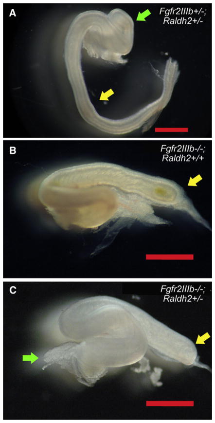
Fig 4.
Morphology of the colon in fibroblast growth factor receptor 2IIIb/retinaldehyde dehydrogenase 2 (Fgfr2IIIb−/−; Raldh2+/−) mutants at embryonic day 18.5. (A) Fgfr2IIIb+/−; Raldh2+/− control littermates have normal morphology of the colon. Both Fgfr2IIIb−/−; Raldh2+/+ (B) and Fgfr2IIIb−/−; Raldh2+/− mutants (C) develop a colonic atresia. Yellow arrow in (A) indicates colon and the atretic end of the proximal colon in (B) and (C). Green arrow in (A) indicates the cecum and the atretic cecum in (C).

