Abstract
Background
Assaying for the parasitic lactate dehydrogenase (pLDH) is widely used as a rapid diagnostic test (RDT), but the efficacy of its serological effectiveness in diagnosis, that is antibody detection ability, is not known. The genetic variation of Korean isolates was analysed, and recombinant protein pLDH was evaluated as a serodiagnostic antigen for the detection of Plasmodium vivax malaria.
Methods
Genomic DNA was purified, and the pLDH gene of P. vivax was amplified from blood samples from 20 patients. The samples came from five epidemic areas: Bucheon-si, Gimpo-si, and Paju-si of Gyeonggi Province, Gangwha-gun of Incheon metropolitan city, and Cheorwon-gun of Gangwon Province, South Korea, from 2010 to 2011. The antigenicity of the recombinant protein pLDH was tested by western blot and enzyme-linked immunosorbent assay (ELISA).
Results
Sequence analysis of 20 Korean isolates of P. vivax showed that the open reading frame (ORF) of 951 nucleotides encoded a deduced protein of 316 amino acids (aa). This ORF showed 100% identity with the P. vivax Belem strain (DQ060151) and P. vivax Hainan strain (FJ527750), 89.6% homology with Plasmodium falciparum FCC1_HN (DQ825436), 90.2% homology with Plasmodium berghei (AY437808), 96.8% homology with Plasmodium knowlesi (JF958130), and 90.2% homology with Plasmodium reichenowi (AB122147). A single-nucleotide polymorphism (SNP) at nucleotide 456 (T to C) was also observed in the isolate from Bucheon, but it did not change in the amino acid sequence. The expressed recombinant protein had a molecular weight of approximately 32 kDa, as analysed by sodium dodecyl sulphate-polyacrylamide gel electrophoresis (SDS-PAGE) analysis. Of the 40 P. vivax patients, 34 (85.0%) were positive by ELISA.
Conclusions
The pLDH genes of 19 isolates of P. vivax were identical, except one for SNP at nucleotide 456. This observation indicates that this gene is relatively stable. Based on these results, the relationship between antibody production against pLDH and the pattern of disease onset should be investigated further before using pLDH for serodiagnosis.
Background
Global figures for deaths caused by malaria range from 1.5 to 2.7 million each year, most of which are children under five years of age and pregnant women. Most of the deaths are caused by Plasmodium falciparum[1,2]. The clinical diagnosis of malaria still relies upon the identification of malaria parasites in Giemsa-stained blood smears of peripheral blood. Therefore, microscopic observation of the Plasmodium species is regarded as the “gold standard” for malaria diagnosis. Despite the simplicity and low cost, such a diagnostic technique is not always available [3]. Rapid diagnostic tests (RDTs) have been introduced to overcome time constraints, a lack of trained personnel in remote or isolated areas, and the low sensitivity when diagnosing malaria infections with a low level of parasitaemia [4]. These lateral-flow immunochromatographic tests detect specific antigens that are produced by malaria parasites and are rapid and simple to carry out without electricity, specific equipment or intensive training [5-8]. To detect Plasmodium, monoclonal antibodies against parasite lactate dehydrogenase (pLDH), histidine-rich protein-2 (HRP-2), and aldolase are widely used. The genetic diversity of HRP-2 is known to partly influence the sensitivity of RDT. pLDH catalyzes the inter-conversion of lactate into pyruvate. Therefore, this enzyme is essential for energy production in Plasmodium species. The level of pLDH in the blood has been directly linked to the level of parasitaemia [9-12]. pLDH (L-lactate: NAD + −oxidoreductase, EC 1.1.1.27) is the one of the first malaria parasite enzymes that was shown to be electrophoretically and kinetically distinct from a human enzyme [13,14]. Glucose utilization in P. falciparum-infected erythrocytes is as much as 100 times the rate observed in uninfected erythrocytes [15]. pLDH plays an important role in regulating glycolysis and in balancing the reduced/oxidized state of the malaria parasites [16,17]. Using RDTs of monoclonal antibodies against the pLDH antigen, sensitivity was over 90%, with high parasite density of P. falciparum. However, the sensitivity decreased to under 70% in parasitaemia of less than 50 parasites/μl [6,18,19].
The creation of diagnostic tools and methods for asymptomatic and low parasitaemia patients has been attempted by the malaria team of the Korean National Institute of Health (KNIH). To accomplish this task, genetic variations of P. vivax pLDH were investigated to identify the typical strain of Korean isolates, and its recombinant protein was evaluated as an antibody detection tool whether it could compensate for the missing cases by antigen detection with RDTs which showing low antigen detection ability in low parasite density.
Methods
Blood sample collection
Patients with clinically suspected malaria attending the Public Health Centers in Gangwha-gun, Gimpo-si, Bucheon-si, and Paju-si of Gyeonggi Province and Cheorwon-gun of Gangwon Province, South Korea from 2010 to 2011 were examined for malaria parasites. Approximately 3 ml of blood was collected from each symptomatic patient. Thin and thick blood smears were prepared for microscopic examination. Blood samples were transported to the Korean National Institute of Health (KNIH), where sera were separated and stored at −20°C for future analysis. Informed consent was obtained from all patients, and all samples were collected under human use protocols that have been reviewed and approved by the Human Ethics Committee of the National Institute of Health (Osong, Korea).
Amplification of pLDH
For the purpose of the expression of the pLDH gene, genomic DNA was extracted from the whole blood of a malaria patient using a QIAamp Blood Kit (Qiagen, Hilden, Germany). PCRs were performed using AccuPower PCR Premix (Bioneer, Taejeon, Korea), 50 ng of purified genomic DNA, and 40 pmoles each of forward (pLDH-F1; 5′-GGA TCC GCT ACT CAG AGG GAG GTG CTC GTC GAA ATC-3′) and reverse primers (pLDH-R1; 5′-GCA TGC GAG GCA GTA CTC TCC GCA GTC CGG ATC AGT-3′), and the total volume was adjusted to 20 ml with distilled water. The thermocycler conditions were as follows: denaturation at 94°C for 5 min; 35 cycles of 1 min at 94°C, 1 min at 58°C and 2 min at 72°C; and incubation at 72°C for 5 min. All of the PCR products were analysed on a 1.0% agarose gel, confirmed under a UV transilluminator and purified with a Qiagen plasmid mini kit (Qiagen). The purified PCR products were ligated into a pCR2.1 cloning vector (Invitrogen, Carlsbad, CA, USA) and then transformed into Escherichia coli Top10 according to Invitrogen’s procedures.
DNA sequencing and analysis
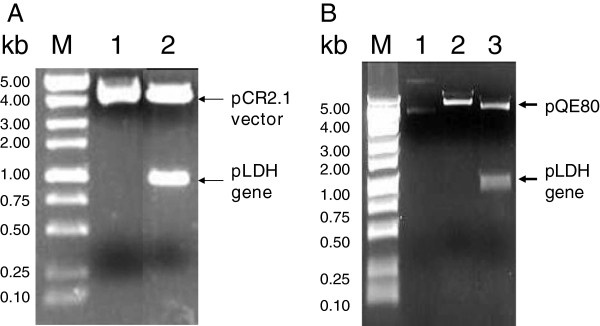
The PCR product inserted into E. coli Top10 was selected for on ampicillin- and 5-bromo-4-chloro-indolyl-β-D-galactopyranoside (X-gal)-containing medium. To confirm transformants, gel electrophoresis was performed after EcoRI digestion (Figure 1A) of a plasmid prepared with a Qiagen plasmid mini kit, according to the protocol supplied by the manufacturer. The pLDH gene sequence was determined using an ABI PRISM dye terminator cycle sequencing ready reaction kit FS (Perkin Elmer, Cambridge, MA, USA) according to the supplied manual. M13 reverse and M13 forward (−20) primers were used in sequencing. Nucleotide and deduced amino acid sequences were analysed using EditSeq and Clustal in the MegAlign program, a multiple alignment program in the DNASTAR package (DNASTAR, Madison, WI, USA). The internet-based BLAST search program of the National Center for Biotechnology Information (NCBI) was used to search protein databases. The gene sequences of pLDH from the Korean isolates were deposited in GenBank (Accession No. JX865768-JX865780, JX872274-JX872280).
Figure 1.

A) The conformation of cloned pCR-pLDH containing the pLDH gene by digestion with EcoRI. M, Molecular size marker; lane 1, non-inserted clone; lane 2, pLDH gene inserted clone (pVpLDH). B) Confirmation of pLDH gene in Eschelichia coli DH5α by restriction enzyme digestion with EcoRI and HindIII. M, Molecular size marker; lane 1, Undigested Plasmid; lane 2, BamHI digested Plasmid; lane 3, EcoRI and HindIII digested plasmid.
Construction of the pLDH expression vector
For the expression of the pLDH gene of the PvKtype19 type strain in E. coli, the gene fragment was amplified from the DNA of blood samples that were confirmed to be infected with P. vivax as described above and which had BamHI and SphI sites on their 5’ ends. Amplified PCR products were digested with BamHI and SphI, purified with a Qiagen Gel Extraction Kit after running on an agarose gel and integrated into the BamHI and SphI cleavage sites of a pQE80 expression vector (Qiagen). The resulting plasmid was subsequently used for the expression of the pLDH recombinant protein in E. coli DH5α. Transformants were confirmed by gel electrophoresis of plasmid DNA after restriction enzyme digestion with EcoRI and HindIII (Figure 1B).
Expression and purification of recombinant pLDH protein
Expression of the pLDH recombinant protein was induced in E. coli with isopropyl-1-thio-β-D-galactopyranoside (IPTG). A total of 1 mM IPTG was added to cultures of E. coli DH5α (pVKtype19) grown to the logarithmic phase in liquid Luria Betani (LB) medium containing 100 μg/ml ampicillin to induce expression of the target protein. Purification of pLDH recombinant protein was carried out using immobilized metal ion affinity chromatography [20]. The purification was performed under native conditions according to the manufacturer’s protocol (Qiagen). Proteins were analysed by sodium dodecyl sulphate-polyacrylamide gel electrophoresis (SDS-PAGE) after each purification step.
Western blot analysis
Recombinant pLDH protein was separated on a 12% SDS-PAGE gel and was then transferred to a nitrocellulose membrane. After the transfer, the membrane was cut into strips and blocked for nonspecific binding with 5% skim milk for 12 hours at 4°C. The membrane was then washed in PBS with 0.15% Tween 20 for 3 × 10 min. The strips were allowed to react with the sera from malaria patients or from uninfected people (diluted 1:100, vol/vol) for four hours and then washed using the procedure described above. The membrane was subsequently incubated with diluted peroxidase-conjugated goat anti-human IgG secondary antibody (1:1,000, v/v) (Sigma) for three hours at room temperature. For colour development, a solution containing 0.2% diaminobenzidine and 0.02% H2O2/PBS was applied to each well [21].
Enzyme-linked immunosorbent assay
Sera from patients infected with P. vivax were analysed for the presence of antigen-specific antibodies using 96-well plates coated with 0.5 mg/ml purified recombinant protein that had been expressed in E. coli and dissolved in phosphate-buffered saline (PBS, pH 7.4) overnight at 4°C. Malaria patient serum was diluted 1:100 (v/v) in blocking buffer (0.25% PBS-Tween 20 with 1% bovine serum albumin, pH 7.4) and incubated for one hour, followed by incubation with peroxidase-conjugated goat anti-human IgG secondary antibody at a 1:1,000 dilution (v/v, Sigma). Optical density was measured with a spectrophotometer at 405 nm (Molecular Devices, Sunnyvale, CA, USA) [22]. Samples were regarded as positive when sera were over the cut-off value, which was calculated as the mean + 2 X the standard deviation (SD) of the negative controls.
Results
Sequence variation of pLDH genes from Plasmodium vivax Korean isolates
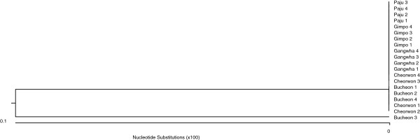
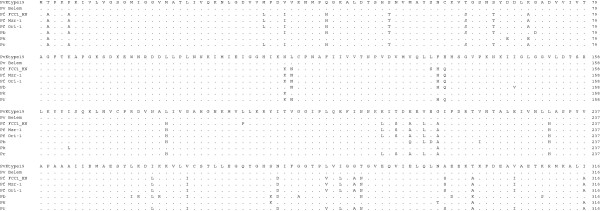
The geographical locations of blood sample collection were Gangwha (37.31 N 125.33E) of the Incheon metropolitan city, Gimpo-si (37.33 N 126.48E), Bucheon (37.29 N 126.46E), Paju (37.88 N 126.76E) of Gyeonggi Province, and Cheorwon (38.10 N 127.30E) of Gangwon Province. Four blood samples infected with indigenous P. vivax were collected from each city during 2010–2011. The pLDH gene was amplified from the genomic DNA of 20 P. vivax Korean isolates. Amplification of the pLDH gene yielded a product of approximately 950 base pairs. After purification, the DNA fragment was ligated into the pCR 2.1 cloning vector (3.9 kb). The plasmid containing the PCR product was named pVpLDH (5.0 kb) and was used for DNA sequence analysis (Figure 1). Based on DNA sequencing, the cloned pLDH gene was 951 bp and consisted of 316 amino acids that were deduced by DNASIS. One single-nucleotide polymorphism (SNP) was detected at base pair 456 (n = 1), from T to C, in the Bucheon 3 isolate (isolated on Sep. 14th 2010) designated as PvKvar (Figures 2 and 3), but it did not change in the amino acid sequence. Therefore, of the 20 Korean isolates of pLDH, 19 isolates had the same DNA and amino acid sequences as P. vivax Belem (DQ060151). These isolates were designated as type strain PvKtype19. When the amino acid sequence of PvKtype19 was compared with several Plasmodium species, PvKtype19 showed 89.5% identity with P. falciparum FCC1_HN (DQ825436), 90.2% with P. falciparum Mzr-1 (JN54719), 90.2% with P. falciparum Ori-1 (JN547218), 90.2% with P. berghei (AY437808), 96.8% with Plasmodium knowlesi (JF958130), and 90.2% with Plasmodium reichenowi (AB122147) (Figures 4 and 5). When the amino acid sequence of PvKtype19 was compared with several P. vivax strains, PvKtype19 showed 100% identity with P. vivax Hainan (FJ527750), 97.8% with P. vivax Ori-1 (JN547221), 99.7% with P. vivax Krt-1 (JN547225), and 98.4% with P. vivax Goa-1 (JN547223) (Figures 6 and 7).
Figure 2.

Comparing the single-nucleotide polymorphism (SNP) of the pLDH gene among 20 Plasmodium vivax Korean isolates. All amino acid sequences were deposited in GenBank (http://WWW.ncbi.nlm.nih.gov/nuccore, Accession No. JX865768-JX865780, JX872274-JX872280).
Figure 3.

Nucleotide sequence differences in pLDH genes from Plasmodium vivax Korean isolates. The nucleotide sequences of 20 P. vivax Korean isolates were aligned. Computer analysis was performed using the multiple sequences alignment tool of MegAlign. All nucleotide sequences were deposited in GenBank BLAST (http://WWW.ncbi.nlm.nih.gov/nuccore, Accession No. JX865768-JX865780, JX872274-JX872280).
Figure 4.

Multiple amino acid sequence alignment of pLDH among Plasmodium species. The deduced amino acid sequence of the PvKtype19 type strain in P. vivax Korean isolates was aligned with those from other Plasmodium species. Computer analysis was performed using the multiple sequences alignment of MegAlign. All amino acid sequences were obtained from GenBank BLAST (http://WWW.ncbi.nlm.nih.gov/nuccore). Pv Belem (P. vivax, Accession; DQ060151), Pf FCC1_HN (P. falciparum, DQ825436), Pf Mzr-1 (P. falciparum, JN54719), Pf Ori-1 (P. falciparum, JN547218), Pb (P. berghei, AY437808), Pk (P. knowlesi strain H, JF958130), and Pr (P. reichenowi, AB122147).
Figure 5.

Amino acid sequence differences in pLDH among Plasmodium species. The deduced amino acid sequence of the PvKtype19 type strain in P. vivax Korean isolates was aligned with those from other Plasmodium species. Computer analysis was performed using the multiple sequences alignment tool of MegAlign. All amino acid sequences were obtained from GenBank BLAST (http://WWW.ncbi.nlm.nih.gov/nuccore). Pv Belem (P. vivax, Accession; DQ060151), Pf FCC1_HN (P. falciparum, DQ825436), Pf Mzr-1 (P. falciparum, JN54719), Pf Ori-1 (P. falciparum, JN547218), Pb (P. berghei, AY437808), Pk (P. knowlesi strain H, JF958130), and Pr (P. reichenowi, AB122147).
Figure 6.

Multiple amino acid sequence alignment of pLDH among Plasmodium vivax. The deduced amino acid sequence of the PvKtype19 type strain in P. vivax Korean isolates was aligned with those from other Plasmodium species. Computer analysis was performed using the multiple sequences alignment tool of MegAlign. All amino acid sequences were obtained from GenBank BLAST (http://WWW.ncbi.nlm.nih.gov/nuccore). PvKtype19 (Korean type strain), PvKvar (Variant form of Korean isolate), Pv Belem (P. vivax, Accession; DQ060151), and Pv Hainan (P. vivax, FJ527750).
Figure 7.

Phylogenetic relationships among the pLDH of several strains of Plasmodium vivax. Computer analysis was performed using the multiple sequences alignment tool of MegAlign. All amino acid sequences were obtained from GenBank BLAST (http://WWW.ncbi.nlm.nih.gov/nuccore). PvKtype19 (Korean type strain), PvKvar (Variant form of Korean isolate), Pv Belem (P. vivax, Accession; DQ060151), Pv Hainan (P. vivax, FJ527750), Ori-1 (P. vivax, JN547221), Krt-1 (P. vivax, JN547225), and Goa-1 (P. vivax, JN547223).
Expression of pLDH inEscherichiacoli
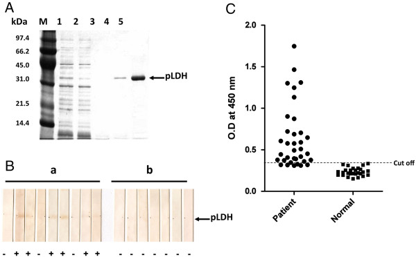
The resulting plasmid pVKtype19 contained a pLDH gene fused to a (His) 6-tag based on pQE80 (Figure 1B). The recombinant plasmid pVKtype19 was then transferred into E. coli DH5α. As analysed by SDS-PAGE followed by Coomassie blue staining, the pLDH recombinant protein was 32 kDa under native purification conditions (Figure 8A).
Figure 8.

A) Purification of pLDH with Ni-NTA agarose affinity chromatography. Lane M, molecular weight protein marker; lane 1, induced E. coli DH5α cell lysate with IPTG; lane 2, flow-through; lane 3, wash; lane 4–5, elutes. B) Western blot analysis of pLDH recombinant protein. a, Malaria patients; b, Healthy individuals. C) Scattergram of absorbances measured by ELISA using pLDH recombinant protein. Sera from healthy individuals and malaria patients infected with P. vivax were used. Cut off showed the mean + 2 X SD of negative controls.
Antigenicity of the pLDH recombinant protein
To determine the antigenicity of the pLDH recombinant protein by western blot and ELISA, the sera of malaria patients that had been collected between 2009 and 2010, which confirmed by microscopic examination but did not count the parasites (parasitaemia), and kept by the KNIH were analysed. Negative sera were collected from staff volunteers from the KNIH.
The sera of six of nine malaria patients exhibited a positive reaction by western blot, while the sera from the normal control group (n = 7), who had never been exposed to malaria, tested negative (Figure 8B). After the number of malaria patients was increased, the antigenicity of the recombinant pLDH protein was evaluated by ELISA. Thirty-four of the 40 sera (85.0%) from malaria patients, as confirmed by microscopic analysis, reacted with the pLDH recombinant protein. In addition, all of the 26 samples from the normal control group failed to react with the pLDH recombinant protein (Figure 8C).
Discussion
pLDH is one of the target antigens that is widely used in developing the monoclonal antibodies that are part of the RDT that comprises the non-microscopic immunochromatographic assay. Interestingly, the level of pLDH has been shown to decline in parallel with the clearance of asexual parasitaemia; therefore, it has been suggested that the disappearance of the parasite-specific enzyme pLDH after anti-malarial drugs may be useful in predicting treatment failure [23]. These characteristics of pLDH led us to investigate the sequence variability of pLDH in Korean isolates. PvKtype19, which was the predominant form of pLDH in Korean isolates, exhibited higher identity with P. knowlesi (96.8%, JF958130) than with P. falciparum Ori-1 (JN547218) (Figures 4 and 5). However, PvKtype19 showed 97.8-100% identity with other subspecies of P. vivax (Figures 6 and 7). Only one synonymous SNP was found in 20 Korean isolates, at base pair 456 (n = 1) (Figures 2 and 3).
Plasmodium vivax has presumably been prevalent in Korea for a long time. However, as a result of a national malaria eradication programme and with help from the World Health Organization (WHO), the incidence of vivax malaria has rapidly decreased [24,25]. After the latest report of two malaria patients in 1985 [26], there were no additional reported cases until one case was reported in 1993 [27] and two indigenous cases were reported in 1994 [28]. Malaria cases then rapidly increased until approximately 2000 [29,30]. After that, the reported malaria cases decreased for several years due to efforts to limit the incidence of malaria. However, malaria has not been thoroughly eradicated in the Korean peninsula because 2 to 3% of patients experience failed drug treatment every year, and many travellers and workers come from malaria-prevalent areas, including North Korea [31]. For these reasons, serological diagnostic tools are needed to support both traditional microscopic diagnosis and antibody testing on a population level, to get a proxy about exposure to malaria in Korea. Currently, IFAT (Immunofluorescence antibody test) is used as the standard serological diagnostic method due to its high sensitivity in this context. However, the sensitivity might be affected by the training and ability of examiners. Therefore, a new antigen is needed for serodiagnosis. Several recombinant proteins cloned from Korean isolates of P. vivax have been tested for use as antigens for serodiagnosis, including circumsporozoite protein (CSP), subtypes Pv210 [32] and Pv247 [33], merozoite surface protein (MSP) [34], and CSP and MSP chimeric proteins [35,36]. None of these antigens were capable of replacing the IFAT method because their sensitivity was less than that of IFAT. Therefore, it was decided to focus on pLDH. Monoclonal antibodies against pLDH have been used in several RDTs and exhibit a relatively high sensitivity for the detection of malaria parasites. However, the ELISA detected only 85.0% (34/40) of microscopic positive samples, even though it was cloned from a Korean vivax malaria strain (pVKtype19, Figure 5). Therefore, antibody detection using the pLDH recombinant protein is not sufficient to compensate the disadvantage of antigen detection using its monoclonal antibody. However, it should be investigated whether pLDH recombinant protein can detect asymptomatic patients or symptomatic patients who have low parasitaemia (under 50/μl) using by antibody detection methods, for example, ELISA or Western blot. Therefore, when using the RDT in the field, it is likely better to use both antigen and antibody detection RDTs to compensate for their individual limitation.
Conclusions
The pLDH gene from P. vivax Korean isolates has an SNP at position 456 (T to C). New information from the geographic mapping of pLDH at the national or regional scale would provide a valuable aid for developing and updating the national anti-malarial policy guidelines in Korea. Additionally, more information is needed before using pLDH as a serological diagnostic antigen.
Competing interests
The authors declare that they have no competing interests.
Authors’ contributions
YJK, YS, HIS and HWL conceived and designed the study and contributed to the execution of the research. HWL wrote the manuscript. JYK, HIS and WJL collected the blood samples in the field. JYK and HIS performed the preparation of DNA samples for DNA sequencing. SWL, who had been working at the University of Florida as a volunteer, (Eastside High School) expressed recombinant pLDH and performed the western blot and ELISA. All authors have read and approved the final manuscript.
Contributor Information
Hyun-Il Shin, Email: shi@nih.go.kr.
Jung-Yeon Kim, Email: creative-kim@hanmail.net.
Won-Ja Lee, Email: wonja@nih.go.kr.
Youngjoo Sohn, Email: youngjoos@khu.ac.kr.
Sang-Wook Lee, Email: swe4444@gmail.com.
Yoon-Joong Kang, Email: yjkang@jwu.ac.kr.
Hyeong-Woo Lee, Email: rainlee67@naver.com.
Acknowledgments
We are grateful to all of the blood donors and the staff of the Public Health Centers in Gangwha-gun, Gimpo-si, Paju-si, Bucheon-si, and Cheorwon-gun. This work was supported by an internal research grants (4847-302 and 4845-300-210-13) from the Korean National Institute of Health, Republic of Korea.
References
- Breman JG. The ears of the hippopotamus: manifestations, determinants and estimates of the malaria burden. Am J Trop Med Hyg. 2001;64:1–11. doi: 10.4269/ajtmh.2001.64.1. [DOI] [PubMed] [Google Scholar]
- Phillips RS. Current status of malaria and potential for control. Clinic Microbiol Rev. 2001;14:208–226. doi: 10.1128/CMR.14.1.208-226.2001. [DOI] [PMC free article] [PubMed] [Google Scholar]
- Reyburn H, Mbatia R, Drakeley C, Carneiro I, Mwakasungula E, Mwerinde O, Saganda K, Shao J, Kitua A, Olomi R, Greenwood BM, Whitty CJ. Overdiagnosis of malaria in patients with severe febrile illness in Tanzania: a prospective study. BMJ. 2004;329:1212. doi: 10.1136/bmj.38251.658229.55. [DOI] [PMC free article] [PubMed] [Google Scholar]
- Iqbal J, Siddique A, Jameel M, Hira PR. Persistent histidine-rich protein 2, parasite lactate dehydrogenase, and panmalarial antigen reactivity after clearance of Plasmodium falciparum monoinfection. J Clin Microbiol. 2004;42:4237–4241. doi: 10.1128/JCM.42.9.4237-4241.2004. [DOI] [PMC free article] [PubMed] [Google Scholar]
- Wongsrichanalai C. Rapid diagnostic techniques for malaria control. Trends Parasitol. 2001;17:307–309. doi: 10.1016/S1471-4922(01)01925-0. [DOI] [PubMed] [Google Scholar]
- Moody A. Rapid diagnostic tests for malaria parasites. Clin Microbiol Rev. 2002;15:66–78. doi: 10.1128/CMR.15.1.66-78.2002. [DOI] [PMC free article] [PubMed] [Google Scholar]
- Bell D, Peeling RW. Evaluation of rapid diagnostic tests: malaria. Nat Rev Microbiol. 2006;4:S34–S38. doi: 10.1038/nrmicro1524. [DOI] [PubMed] [Google Scholar]
- Singh N, Saxena A, Valecha N. Field evaluation of the ICT malaria P.f/P.v immunochromatographic test for diagnosis of Plasmodium falciparum and P. vivax infection in forest villages of Chhindwara, central India. Trop Med Int Health. 2000;5:765–770. doi: 10.1046/j.1365-3156.2000.00645.x. [DOI] [PubMed] [Google Scholar]
- Cho CH, Nam MH, Kim JS, Han ET, Lee WJ, Oh JS, An SS, Lim CS. Genetic variability in Plasmodium vivax aldolase gene in Korean isolates and the sensitivity of the Binax Now malaria test. Trop Med Int Health. 2011;16:223–226. doi: 10.1111/j.1365-3156.2010.02691.x. [DOI] [PubMed] [Google Scholar]
- Baker J, McCarthy J, Gatton M, Kyle DE, Belizario V, Luchavez J, Bell D, Cheng Q. Genetic diversity of Plasmodium falciparum histidine-rich protein 2 (PfHRP2) and its effect on the performance of PfHRP2-based rapid diagnostic tests. J Infect Dis. 2005;192:870–877. doi: 10.1086/432010. [DOI] [PubMed] [Google Scholar]
- Brown WM, Yowell CA, Hoard A, Vader Jagt TA, Hunsaker LA, Deck LM, Royer RE, Piper RC, Dame JB, Makler MT, Vanderjagt DL. Comparative structural analysis and kinetic properties of lactate dehydrogenases from the four species of human malarial parasites. Biochem. 2004;43:6219–6229. doi: 10.1021/bi049892w. [DOI] [PubMed] [Google Scholar]
- Piper R, Lebras J, Wentworth L, Hunt-Cooke A, Houze S, Chiodini P, Marker M. Immunocapture diagnostic assays for malaria using Plasmodium lactate dehydrogenase (pLDH) Am J Trop Med Hyg. 1999;60:109–114. doi: 10.4269/ajtmh.1999.60.109. [DOI] [PubMed] [Google Scholar]
- Sherman IW. Molecular heterogeneity of lactic dehydrogenase in avian malaria (Plasmodium lophurae) J Exp Med. 1961;114:1049–1062. doi: 10.1084/jem.114.6.1049. [DOI] [PMC free article] [PubMed] [Google Scholar]
- Sherman IW. Heterogeneity of lactic dehydrogenase in intraerythrocytic parasites. Trans NY Acad Sci Ser. 1962;2(24):944–953. doi: 10.1111/j.2164-0947.1962.tb01454.x. [DOI] [PubMed] [Google Scholar]
- Rose EF Jr, Raventos S, Perkins M, Nagel RL. Glutathione stability and oxidative stress in P. falciparum infection in vitro: response of normal and G6PD deficient cells. Biochem Biophys Res Commun. 1982;109:355. doi: 10.1016/0006-291X(82)91728-4. [DOI] [PubMed] [Google Scholar]
- Sherman IW. In: Antimalarial drugs I. Biological background, experimental methods and drug resistance. Peters W, Richards WHG, editor. Heidelberg: Springer Verlag; 1984. Metabolism; pp. 31–81. [Google Scholar]
- Fairlamb AH. Novel biochemical pathways in parasitic protozoa. Parasitol. 1989;99:S93–S112. doi: 10.1017/S003118200008344X. [DOI] [PubMed] [Google Scholar]
- Farcas GA, Zhong KJ, Lovegrove FE, Graham CM, Kain KC. Evaluation of the Binax Now ICT test versus polymerase chain reaction and microscopy for the detection of malaria in returning travellers. Am J Trop Med Hyg. 2003;69:589–592. [PubMed] [Google Scholar]
- World Health Organization. A rapid dipstick antigen capture assay for the diagnosis of falciparum malaria: WHO informal consultation on recent advances in diagnostic techniques and vaccines for malaria. Bull World Health Organ. 1996;74:47–54. [PMC free article] [PubMed] [Google Scholar]
- Lee HW, Lee WJ, Lee JS, Lee HS. DNA sequencing and expression of the circumsporozoite protein of Plasmodium vivax Korean isolate in Escherichia coli. Kor J Microbiol. 1999;37:234–242. [Google Scholar]
- Tsang VCW, Peralta JM, Simons AR. Enzyme-linked immunoelectrotransfer blot techniques (EITB) for studying the specificities of antigens and antibodies separated by gel electrophoresis. Meth Enzymol. 1983;92:377–391. doi: 10.1016/0076-6879(83)92032-3. [DOI] [PubMed] [Google Scholar]
- Gao YH, Li HL, Lu Y, Gao FM, Lin YH, Zhou HC, Zhang LH, Wang H. Identification of a vaccine candidate antigen, PfMAg-1, from Plasmodium falciparum with monoclonal antibody M26-32. Parasitol Res. 2009;105:1723–1732. doi: 10.1007/s00436-009-1617-4. [DOI] [PubMed] [Google Scholar]
- Nyunt MH, Kyaw MP, Win KK, Myint KM, Nyunt KM. Field evaluation of HRP2 and pan pLDH-based immunochromatographic assay in therapeutic monitoring of uncomplicated falciparum malaria in Myanmar. Malar J. 2013;12:123. doi: 10.1186/1475-2875-12-123. [DOI] [PMC free article] [PubMed] [Google Scholar]
- National Malaria Eradication Service. Malaria pre-eradication program in Korea. Progress Report 1961–1965. Ministry of Health and Social Affairs. Republic of Korea. 1966;44:70. [Google Scholar]
- Paik YH, Ree HI, Shim JC. Malaria in Korea. Jap J Exp Med. 1988;58:55–66. [PubMed] [Google Scholar]
- Soh JT, Lee KT, Im KI, Min DY, Ahn MH, Kim JJ, Yong TS. Current status of malaria in Korea. Yonsei Rep Trop Med. 1985;16:11–18. [Google Scholar]
- Chai IH, Lim GI, Yoon SN, Oh WI, Kim SJ, Chai JY. Occurrence of tertian malaria in a male patient who has never been abroad. Kor J Parasitol. 1994;32:195–200. doi: 10.3347/kjp.1994.32.3.195. [DOI] [PubMed] [Google Scholar]
- Cho SY, Kong Y, Park SM, Lee JS, Lim YA, Chae SL, Kho WG, Lee JS, Shim JC, Shin HK. Two vivax malaria cases detected in Korea. Kor J Parasitol. 1994;32:281–284. doi: 10.3347/kjp.1994.32.4.281. [DOI] [PubMed] [Google Scholar]
- Lee JS, Kho WG, Lee HW, Seo M, Lee WJ. Current status of vivax malaria among civilians in Korea. Kor J Parasitol. 1998;36:241–248. doi: 10.3347/kjp.1998.36.4.241. [DOI] [PMC free article] [PubMed] [Google Scholar]
- Park JW, Klein TA, Lee HC, Pacha LA, Ryu SH, Yeom JS, Moon SH, Kim TS, Chai JY, Oh MD, Choe KW. Vivax malaria: a continuing health threat to the Republic of Korea. Am J Trop Med Hyg . 2003;69:159–167. [PubMed] [Google Scholar]
- Lee WJ, Kim HH, Choi YK, Choi KM, Kim MA, Kim JY, Sattabongkot J, Sohn Y, Kim H, Lee JK, Park HS, Lee HW. Analysis of the dihydrofolate reductase-thymidylate synthase gene sequences in Plasmodium vivax field isolates that failed chloroquine treatment. Malar J. 2010;9:331. doi: 10.1186/1475-2875-9-331. [DOI] [PMC free article] [PubMed] [Google Scholar]
- Lee HW, Lee JS, Lee WJ, Cho SH, Lee HS. The evaluation of recombinant circumsporozoite protein in malaria diagnosis. Kor J Microbiol. 2000;36:142–149. [Google Scholar]
- Kim TS, Kim HH, Lee SS, Oh CM, Choi KM, Lin K, Kim JY, Na BK, Han ET, Sohn Y, Kim H, Lee HW. Molecular cloning and expression of the VK247 circumsporozoite protein for serodiagnosis of variant form Plasmodium vivax. Parasitol Res. 2011;108:1275–1282. doi: 10.1007/s00436-010-2177-3. [DOI] [PubMed] [Google Scholar]
- Kwon MH, Kim HH, Lee HS, Kim TS, Oh CM, Ahn YJ, Hwang SK, Sohn Y, Kim H, Lee HW. Plasmodium vivax: comparison of the immune responses between oral and parenteral immunization of rPv54 in BALB/c mice. Exp Parasitol. 2010;126:217–223. doi: 10.1016/j.exppara.2010.05.001. [DOI] [PubMed] [Google Scholar]
- Lee C, Chung KW, Kim TS, Choi KM, Choi YK, Chung NJ, Rhie HG, Lee HS, Lee SJ, Lee HW. Trials for the co-expression of the merozoite surface protein-1 and circumsporozoite protein genes of Plasmodium vivax. Exp Parasitol. 2011;129:227–233. doi: 10.1016/j.exppara.2011.08.014. [DOI] [PubMed] [Google Scholar]
- Lee C, Kim HH, Choi KM, Chung KW, Choi YK, Jang MJ, Kim TS, Chung NJ, Rhie HG, Lee HS, Sohn Y, Kim H, Lee SJ, Lee HW. Murine immune responses to a Plasmodium vivax-derived chimeric recombinant protein expressed in Brassica napus. Malar J. 2011;29:106. doi: 10.1186/1475-2875-10-106. [DOI] [PMC free article] [PubMed] [Google Scholar]


