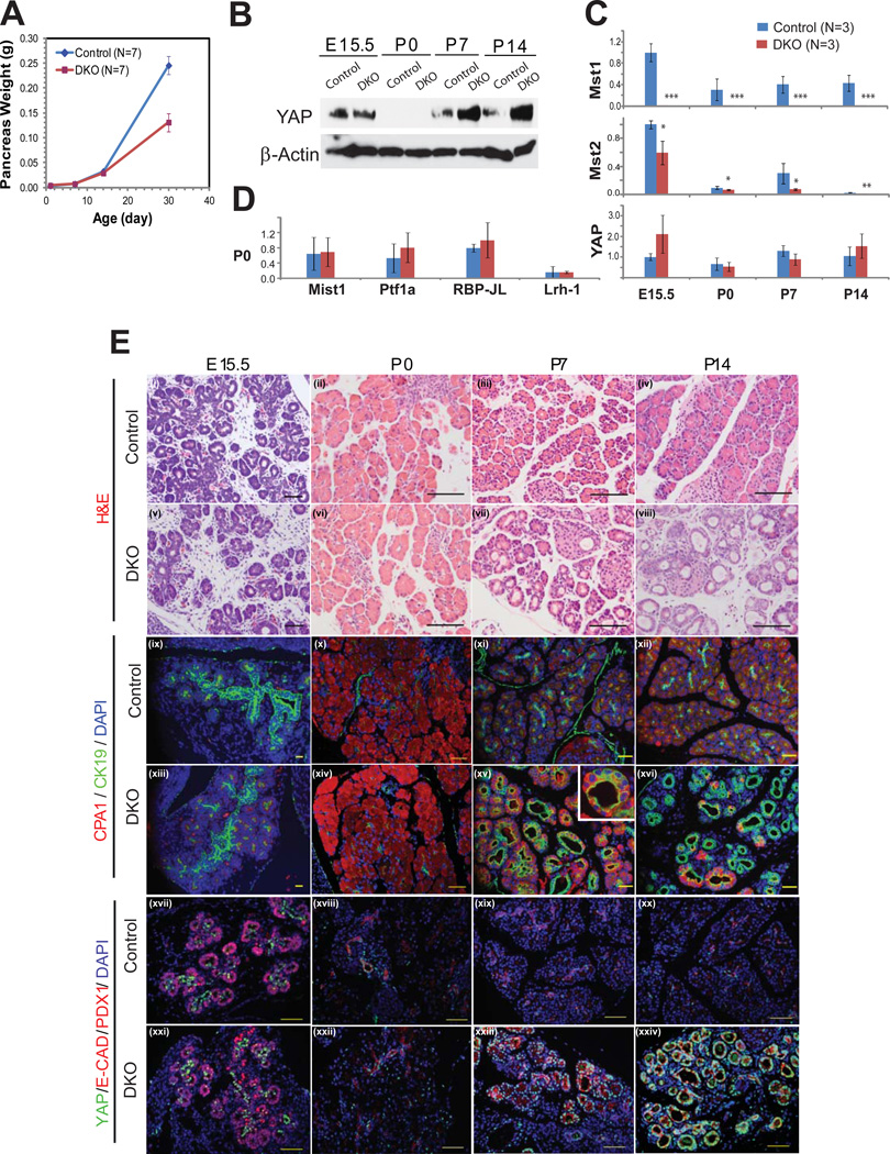
Figure 3. Mst1 and Mst2 are required for postnatal maintenance of the exocrine pancreas.
(A) Total pancreas mass of DKO animal was compared to that of littermate controls at P0, P7, P14, and P30 (N=7 per group). A significant difference of body weight was observed on P30 (p=0.0013).
(B) Western blot assay of pancreas lysates at different time points, starting from E15 through P14, was performed using antibodies against YAP, and β−actin. 30 µg of pancreas lysate was loaded per well..
(C) qPCR was performed for Mst1, Mst2, and YAP at each of 4 time-points comparing control and DKO pancreata (n=3 animals in each group per time point). (mean +/− SD; *, P<0.05; **, P<0.01; ***, P<0.001).
(D) qPCR for the acinar transcription factors Mist1, Ptf1a, RBP-JL, and Lrh-1 were performed at P0 comparing control and DKO pancreata (n=3 animals in each group per time point).
(E) Pancreas tissues of DKO and littermate control were examined by H&E staining (i–viii), immunofluorescence for CK19 and CPA1 (ix–xvi), YAP and PDX1 (xvii,xxi) or YAP and E-Cadherin (xviii–xx, xxii–xxiv) at the indicated developmental stage. Note the strong nuclear YAP staining in the “trunk” regions of both control and DKO embryos at E15.5 and in the duct-like structures of DKO animals at P7 and P14. Scale bar: 100µM.

