Abstract
We examined the temperature dependence of muscle and non-muscle myosin (heavy meromyosin, HMM) with in vitro motility and actin-activated ATPase assays. Our results indicate that myosin V (MV) has a temperature dependence that is similar in both ATPase and motility assays. We demonstrate that skeletal muscle myosin (SK), smooth muscle myosin (SM), and non-muscle myosin IIA (NM) have a different temperature dependence in ATPase compared to in vitro motility assays. In the class II myosins we examined (SK, SM, and NM) the rate-limiting step in ATPase assays is thought to be attachment to actin or phosphate release, while for in vitro motility assays it is controversial. In myosin V the rate-limiting step for both in vitro motility and ATPase assays is known to be ADP release. Consequently, in MV the temperature dependence of the ADP release rate constant is similar to the temperature dependence of in vitro motility. Interestingly, the temperature dependence of the ADP release rate constant of SM and NM was shifted toward the in vitro motility temperature dependence. Our results suggest that the rate-limiting step in SK, SM, and NM may shift from attachment-limited in solution to detachment-limited in the in vitro motility assay. Internal strain within the myosin molecule or by neighboring myosin motors may slow ADP release which becomes rate-limiting in the in vitro motility assay. Within this small subset of myosins examined, the in vitro sliding velocity correlates reasonably well with actin-activated ATPase activity, which was suggested by the original study by Barany et al. (Barany 1967).
Keywords: myosin, muscle contraction, enzymatic activity, motility
Introduction
Muscle contraction is driven by the motor activity of myosin, which uses the energy of ATP hydrolysis to generate force through a cyclic interaction with actin filaments. A highly cited study by Michael Barany published in 1967 (Barany 1967) measured the unloaded shortening velocity of a particular muscle and the ATPase activity of the myosin isolated from that muscle. This landmark study compared a large number of different muscles from a variety of species which had unloaded shortening velocities varying by a factor of 240. The study had implications for understanding the actomyosin contractile mechanism. Specifically, this work implied that the speed of unloaded shortening of a muscle fiber was limited by how fast myosin crossbridges in that muscle could complete their ATPase cycles. Little was known about the kinetic cycle of myosin at the time this study was conducted. A few years later, Lymn and Taylor (Lymn and Taylor 1971) showed that myosin cycles between states that bind strongly to actin and states that bind weakly during a single ATP hydrolysis cycle and speculated on the rate-limiting step for muscle myosin. Many studies have since elaborated on the kinetic cycle of muscle myosins and that of other members of the myosin superfamily (Bloemink and Geeves 2011; De La Cruz and Ostap 2004). A question of particular interest is which step or steps in the kinetic cycle limit(s) the shortening velocity in muscle or the rate at which processive myosins move along actin filaments (Siemankowski et al. 1985; Nyitrai et al. 2006; Weiss et al. 2000; Marston and Taylor 1980; Weiss et al. 2001). An in vitro correlate of the unloaded shortening velocity of muscle is the sliding actin in vitro motility assay developed initially by Kron and Spudich (Kron et al. 1991). In this assay fluorescently-labeled actin filaments are observed to move over a lawn of myosin molecules bound to the coverslip surface using a microscope equipped with optics to detect fluorescence.
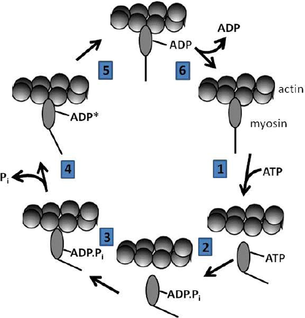
Biochemical studies of myosins have converged on an ATPase mechanism common to most myosins characterized to date (Malnasi-Csizmadia and Kovacs 2010; Sweeney and Houdusse 2010) (Scheme 1). The binding of ATP to actomyosin dramatically weakens the affinity of myosin for actin causing rapid dissociation of the complex (step 1). In addition, ATP binding primes myosin for force generation and the subsequent hydrolysis step stabilizes the lever arm bent (primed) conformation (step 2) (Urbanke and Wray 2001; Malnasi-Csizmadia et al. 2001). The release of phosphate is accelerated when myosin with the hydrolyzed products in the active site (ADP and phosphate) binds to actin (steps 3 & 4). The myosin.ADP complex that follows binds to actin with high affinity. The power stroke is thought to occur when the lever arm straightens during the actin binding and phosphate release steps (step 4), while this point is still debated in the literature (Sweeney and Houdusse 2010). Myosin remains attached to actin until ADP is released and ATP binding detaches myosin from actin. An isomerization between actomyosin.ADP states (step 5) has been found to occur prior to the release of ADP (step 6) (Jacobs et al. 2011; Rosenfeld et al. 2005; Hannemann et al. 2005). Therefore, the period of time after the power stroke that myosin remains attached to actin with high affinity is mediated by the net rate constant of ADP release (influenced by that of the preceding isomerization between actomyosin.ADP states) and/or the rate constant of ATP binding. The proportion of time that myosin spends strongly attached to actin during an ATPase cycle is termed the “duty ratio”. There is tremendous variation in the maximal actin-activated MgATPase (Vmax) among myosins ranging from values as high as 20 s−1 for fast skeletal muscle myosin to rates as slow as 0.1 s−1 for non-muscle myosin IIB at 25 °C in solution (Sellers 1999). In addition to variations in the Vmax, there are also major differences in which steps are rate limiting in different myosins. Therefore, the motor properties of each myosin are precisely tuned for performing their specific cellular functions (De La Cruz and Ostap 2004; Sellers 2000)
Scheme 1.
Contractile velocity (V) is classically interpreted in terms of the relationship between the size of the working stroke or unitary displacement (d) and the period of time that myosin remains attached to actin (ton) (Siemankowski et al. 1985).
Single molecule measurements have determined that d is determined by the length of the lever arm or light chain binding region (Tyska and Warshaw 2002; Tyska and Mooseker 2003; Brenner 2006). For example, single molecule displacements measured with myosin II, which has a lever arm that consists of two light chain binding IQ motifs, are reported in the range of 5–10 nm (Tyska and Warshaw 2002) while myosin V, which has six calmodulins bound to its lever, has a displacement of around 25 nm (Mehta 2001; Moore et al. 2001; Sakamoto et al. 2005; Sakamoto et al. 2003; Veigel et al. 2002). In addition, altering the length of the lever arm in myosin V (between 2–8 IQ motifs) shifts the unitary displacement and the velocity without significantly altering the attachment time (Sakamoto et al. 2003). In muscle myosins, variability in the unloaded shortening velocity was found to be mainly determined by ton (Weiss et al. 2001; Siemankowski et al. 1985; Nyitrai et al. 2006). These studies and others suggest that both the length of the lever arm and the attachment time are important determinants of unloaded actomyosin-based motility.
In the current study we investigated the temperature dependence of the rate of actin filament sliding in the in vitro motility assay using muscle and non-muscle myosins and compared these results with the temperature dependence of actin-activated ATPase activity and the rate constant for ADP release measured by stopped-flow spectrofluorimetry to determine which parameter best correlated with the rate of actin filament sliding. Our results provide support for the idea that the rate constant for ADP release, and hence detachment, limits the velocity of actomyosin based motility under unloaded conditions for both muscle and non-muscle myosins. Interestingly, there is a general scaling of the actin-activated ATPase rate and the rate of in vitro motility in that fast moving myosins typically have faster ATPase activities.
Materials and Methods
All reagents were the highest purity commercially available. ATP and ADP were prepared fresh from powder. N-Methylanthraniloyl (mant)-labeled 2’-deoxy-ADP (dmantADP) was purchased from Jena Scientific. The dmantADP concentration was determined from absorbance measurements at 255 nm using (ε255 = 23,300 M−1·cm−1). ATP and ADP concentrations were determined by absorbance at 259 nm (ε259 = 15,400 M−1·cm−1).
Protein Expression and Purification
A chicken myosin V heavy meromyosin (HMM) construct containing a GCN4 leucine zipper and C-terminal YFP (MV) (Rock et al. 2001), which had the chicken coiled-coil sequence replaced with the mouse coiled-coil (amino acids 926–1103), was obtained from Dr. Lee Sweeney (University of Pennsylvania). The baculovirus system was used to co-express MV and calmodulin, as well as gizzard smooth muscle myosin HMM (SM) (Sweeney et al. 1998) with a C-terminal myc tag and mouse non-muscle myosin IIA HMM (NM) (Kovacs et al. 2007) with their appropriate light chains. Skeletal muscle myosin HMM (SK) was prepared from rabbit psoas skeletal muscle (Margossian and Lowey 1982). SM and NM were phosphorylated with myosin light chain kinase prior to experiments (Sweeney et al. 1998; Kovacs et al. 2007). A baculovirus was created from a clone of rabbit smooth muscle myosin light chain kinase (generously provided by Zenon Grabarek, Boston Biomedical Research Institute) to which was added a FLAG tag. Sf9 cells were infected with the virus and the expressed kinase was purified by FLAG affinity chromatography and stored in −80 °C until use. Chicken calmodulin was expressed and purified as described (Putkey et al. 1987). Myosin concentrations were determined using the Bio-Rad microplate assay using bovine serum albumin (BSA) as a standard or by absorbance. Actin was purified from rabbit skeletal muscle using an acetone powder method (Pardee and Spudich 1982). All MV experiments were performed in KMg50 buffer (50 mM KCl, 1 mM EGTA, 1 mM MgCl2, 1 mM DTT and 10 mM imidazole-HCl, pH 7.0). All myosin II (SK, SM, and NM) experiments were performed in MOPS 20 buffer (20 mM KCl, 1 mM EGTA, 1 mM MgCl2, 1 mM DTT and 20 mM MOPS, pH 7.0)
Stopped-flow Measurements and Kinetic Modeling
Transient kinetic experiments were performed in an Applied Photophysics (Surrey, UK) stopped-flow apparatus with a dead time of 1.2 ms. A monochromater with a 2-nm band pass was used for fluorescence excitation, and cut-off filters were used to measure the emission. The dmantADP fluorescence was excited at 290 nm and the emission measured with a 395 nm long-pass filter. Light scatter (420 nm excitation and 395 long-pass emission filter) was used to measure the rate constant for myosin detachment from actin in the presence of ADP. Nonlinear least-squares fitting of the data was done with software provided with the instrument or Kaleidagraph (Synergy Software, Reading, PA). Uncertainties reported are standard errors of the fits unless stated otherwise.
Kinetic analysis was performed using the reaction scheme that has been used in studies of myosin V and myosin II (De La Cruz et al. 1999; Sun et al. 2006; Sun et al. 2008; Malnasi-Csizmadia and Kovacs 2010). All concentrations mentioned in the stopped-flow experiments are final concentrations unless stated otherwise.
ATPase Assays
ATPase assays were performed in the stopped-flow at various temperatures using the NADH coupled assay (De La Cruz et al. 2000; Sun et al. 2006; Sun et al. 2008; De La Cruz et al. 1999). The temperature dependence of the steady-state ATPase rate was measured in the presence of 1 mM ATP at a fixed actin concentration for MV (20 µM), SK (40 µM), SM (40 µM), and NM (20 µM). Both SM and NM demonstrated similar phosphorylation dependent actin-activated ATPase values reported previously (Sweeney et al. 1998; Kovacs et al. 2003).
In Vitro Motility Assays
We performed the in vitro motility assay as described (Kron et al. 1991) with similar conditions as were used for the solution ATPase assays. MV, SK, and NM were adhered directly to the nitrocellulose-coated surface while SM was attached via an anti-c-myc antibody (Sigma Aldrich). The surface was blocked with bovine serum albumin at a concentration of 1 mg/ml. The motility of actin filaments labeled with rhodamine-phalloidin was observed using an activation buffer consisting of either KMg50 (MV) or MOPS 20 (SK, NM, SM) supplemented with the following: 5 µM calmodulin (MV only), 2 mM ATP, phosphoenol pyruvate (2.5 mM), pyruvate kinase (20 units/ml), glucose oxidase (0.1 mg/ml), glucose (5 mg/ml), catalase (0.018 mg/ml). and 0.35% methylcellulose. After the addition of the activation buffer, the slide was promptly viewed using a NIKON TE2000 microscope equipped with a 60x/1.4NA phase objective. Images were acquired at intervals (0.2 – 5 seconds) and periods of time (1–5 minutes) appropriate for measuring the sliding velocity of each myosin. We utilized a shutter controlled Coolsnap HQ2 cooled CCD digital camera (Photometrics) binned 2×2 for all imaging. To measure velocity, the video records were transferred to Image J and analyzed with the MTrackJ program (Meijering et al. 2012). The temperature was varied by altering the room temperature and using an air blower. The temperature of the microscope slide was recorded using a Stable Systems International thermocouple meter.
Results
All the assays described below were performed in similar buffer conditions for each myosin. Experiments with MV were performed in KMg50 buffer and experiments with SK, SM, and NM were performed in MOPS 20 buffer. We phosphorylated SM and NM prior to all experiments and excess ATP was removed by microdialysis.
Temperature dependence of steady-state ATPase and in vitro motility
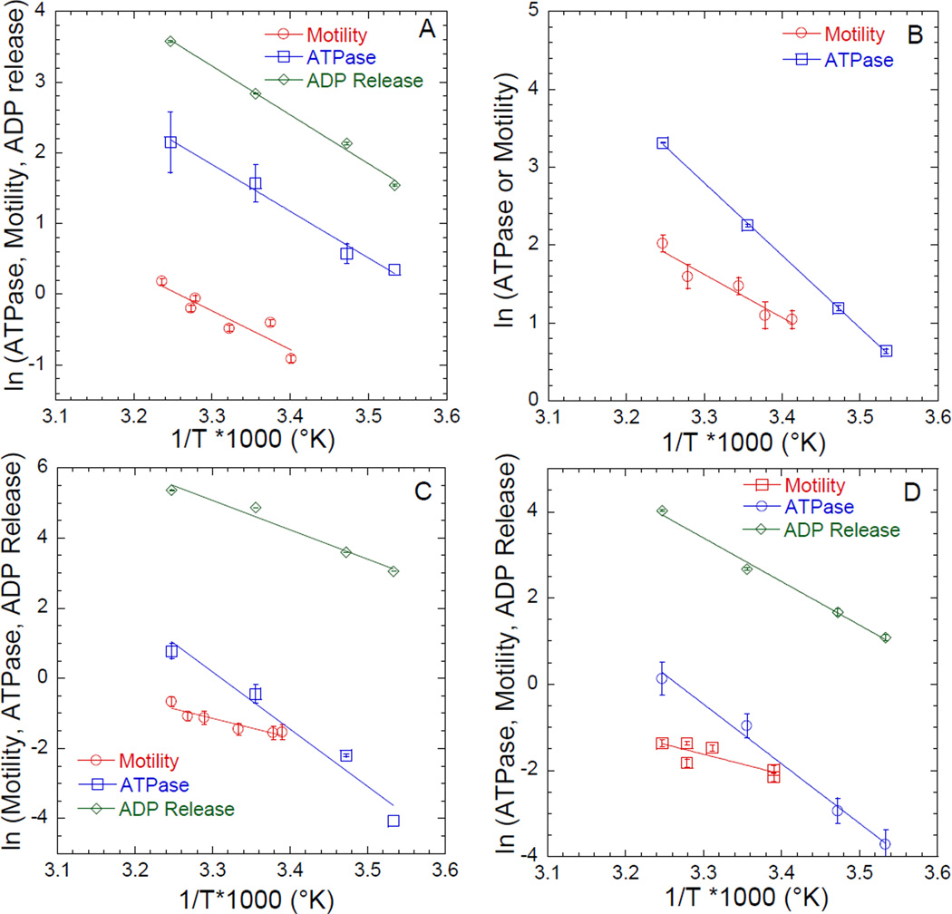
We examined the actin-activated ATPase activity of four different myosins (MV, SK, SM, and NM) as a function of temperature (10, 15, 25, 35 °C) at a fixed actin (saturating) concentration. The data for each myosin are displayed (Figure 1A–D) in Arrhenius plot format (natural log of ATPase activity plotted as a function of inverse temperature in degrees Kelvin). Previous work demonstrated that the actin-dependence of ATPase activity, which is evaluated by determining the actin concentration at which one-half maximal ATPase activity is achieved (KATPase), is temperature independent in most myosins including MV (Jacobs et al. 2011) and NM (Kovacs et al. 2004; Hu et al. 2002). Therefore, Arrhenius plots allowed for comparison of the energy of activation associated with the rate-limiting step in the ATPase reaction for each myosin. We also examined the sliding velocities in the in vitro motility assay over a range of temperatures (20–35 °C). The average sliding velocity (µm/sec) of 15–20 filaments was determined at each temperature. The data are plot in Arrhenius format on the same graph as the ATPase plots (Figure 1A–D) for each myosin to allow for comparison of the temperature dependence of solution ATPase and in vitro sliding velocity. In MV we found that the slopes of the linear-dependence of ATPase and motility are very similar (Figure 1A) which was expected because the rate-limiting step in both assays is thought to be ADP release (De La Cruz et al. 1999; Mehta et al. 1999). Interestingly, we found that the ATPase and motility assay results with SK, SM, and NM demonstrated a different dependence on temperature, as indicated by Arrhenius plot slopes that are more than two-fold different (Table 1), indicating the rate limiting steps for the assays are different in these myosins.
Figure 1. Influence of temperature on actomyosin kinetics and motility in various myosins.
Motility assays were performed at temperatures ranging from 20°C to 35°C, while ATPase assays and ADP-release measurements were performed at 10–35 °C for (A) Chicken Myosin V (MV), (B) Rabbit Skeletal Muscle Myosin II (SK), (C) Gizzard Smooth Muscle Myosin II (SM), and (D) Mouse Non-Muscle Myosin IIA (NM) (all were HMM fragments). The ATPase measurements were performed at a single actin concentration; MV (20 µM), SK (40 µM), SM (40 µM), and NM (20 µM). The ADP release rate constant was examined with dmantADP. The data is plot in Arrhenius format, and the temperature dependence of the ATPase, motility, and ADP release data fit to a linear relationship for comparison. See Table 1 for summary of ATPase activity (s−1), sliding velocity (µm•s−1), and ADP release (s−1) data.
Table 1.
Summary of temperature dependent measurements.
Temperature dependence of dmantADP release
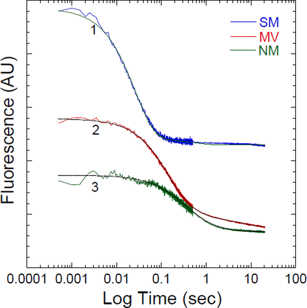
We performed direct measurements to determine the ADP release rate constant as a function of temperature using the stopped-flow apparatus in MV, SM, and NM. We did not measure the ADP release rate constant in SK because it is too fast to measure by stopped-flow at elevated temperatures (Nyitrai et al. 2006). Our results allowed us to compare the temperature dependence of ADP release to the temperature dependence of ATPase and motility. To measure the ADP release rate constant we mixed a pre-equilibrated complex of actomyosin and dmantADP with saturating ATP. The conditions were slightly different for each myosin and are indicated in the Figure 2 legend. We examined the fluorescence signal observed upon dmantADP dissociation from actomyosin (ex. 290 nm/ em. 395 nm long-pass filter). Previous studies have observed multi-exponential fluorescence transients using dmantADP or mantADP (Sweeney et al. 1998; Rosenfeld and Sweeney 2004; Kovacs et al. 2007; Forgacs et al. 2008). A complicating factor is that when both heads of the HMM dimer are bound to actin the ADP release rate constant is different in the negatively-strained (lead) head and positively-strained (trail) head (Sweeney et al. 1998; Rosenfeld and Sweeney 2004; Kovacs et al. 2007; Forgacs et al. 2008). For example, Kovacs et al. (Kovacs et al. 2007) found that for non-muscle myosin IIA HMM the rate constant for ADP release from the trail head was similar or faster than that from monomeric subfragment-1 (S1), while the rate constant for ADP release from the lead head was five to ten fold slower. In the current study we found that MV and NM contained bi- and tri-exponential ADP release transients, while SM was dominated by a single phase. We also measured with identical conditions the light scatter transient, which represents the rate constant for detachment of myosin from actin (data not shown). Because ATP binding is rapid under these conditions the detachment is limited by the ADP release rate constant. The relative amplitude of the slower components in the light scatter and dmantADP transients was more significant at lower temperatures and was completely absent at 35 °C. Therefore, we have focused on the observed fast phase of the fluorescence transients which were quite similar by following the dmantADP fluorescence and the light scatter transients. We plotted the dmantADP release rate constants along with the ATPase and motility results in Arrhenius format (Figure 1A–D). The slope of all the Arrhenius plots are similar in MV, demonstrating the ADP release rate constant is rate-limiting in both ATPase and motility assays. In SM and NM we found the slopes from the motility Arrhenius plots to be shifted toward the slopes of the ADP release plots.
Figure 2. Fluorescence transients from measuring the dmantADP release rate constant in various myosins.
A complex of actomyosin and dmantADP was mixed with saturating ATP. The fluorescence transient (trace 1) with acto-SM at 15 °C is fit a two exponential function (35.3 ± 2.1 and 2.1 ± 0.1 sec−1 with relative amplitudes of 0.95 and 0.05, respectively) (final conditions: 0.5 µM SM, 1.0 µM actin, 10 µM dmantATP, 1 mM ATP). The fluorescence transient (trace 2) with acto-MV at 15 °C is fit to a three exponential function (8.4 ± 0.1, 2.4 ± 0.1, and 0.11 ± 0.01 sec−1 with relative amplitudes of 0.64, 0.26, and 0.11, respectively) (final conditions: 0.5 µM MV, 1.0 µM actin, 10 µM dmantATP, 1 mM ATP). The fluorescence transient (trace 3) with acto-NM at 15 °C is fit to a three exponential function (5.3± 0.1, 1.5 ± 0.1, and 0.04 ± 0.01 sec−1 with relative amplitudes of 0.34, 0.59, and 0.07, respectively) (final conditions: 0.75 µM NM, 1.25 µM actin, 10 µM dmantATP, 1 mM ATP).
Correlation of ATPase activity and in vitro motility
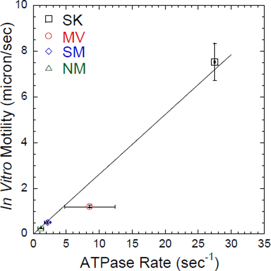
To determine if the actin-activated ATPase activity correlates with the in vitro motility in this small subset of myosins we plotted ATPase rate versus in vitro motility from our results collected at 35 (± 1 °C) in the motility assay and 35 °C in the ATPase assay (Figure 3). This analysis was inspired by the original studies performed by Barany (Barany 1967), which compared the ATPase rates and contractile velocities in a large number of different muscles types from a variety of different species. We found a relatively good correlation between ATPase rate and motility in this small subset of muscle and non-muscle myosins.
Figure 3. Correlation between ATPase activity and in vitro motility.
The sliding velocity in the in vitro motility assay (measured at 35–36 °C) is plotted as a function of actin-activated ATPase activity (measured at 35 °C). The error bars represent the standard deviation from 2–3 protein preparations. The data is fit with a linear regression (slope = 0.26 ± 0.02, R2 = 0.98) to demonstrate the correlation between ATPase activity and in vitro motility sliding velocity.
Discussion
Michael Barany’s classic paper in the Journal of General Physiology made the observation that myosin ATPase activity and muscle unloaded shortening velocity correlated reasonably well for a variety of muscles types over a large range of speeds (Barany 1967). At the time of this study, little was known about the kinetic mechanism of myosins or how some of the myosins studied are regulated. For example, we now know that molluscan myosins used by Barany require bound calcium for activation (Szent-Gyorgyi et al. 1999), whereas the vertebrate striated muscle myosins used do not. Fortunately, Barany assayed all the myosins in the presence of calcium, since the literature at the time of the study suggested that vertebrate striated muscle myosin required calcium for maximal activity (Weber and Winicur 1961). Barany was also not aware that vertebrate smooth muscle myosin (Cremo et al. 1995) and Limulus muscle myosin (Sellers 1981) must be phosporylated to obtain maximal activity since this form of regulation was not discovered until much later. His assays were conducted at a single actin concentration and under the assay conditions used this actin concentration was probably not saturating for most of the myosins studied. Finally, anyone who has performed actin-activated MgATPase assays on intact myosin knows the difficulties in obtaining reproducible results from this complicated system. Virtually all kinetic studies for the past 40 years have been carried out with the soluble fragments of myosin, S1 or HMM. Nevertheless, the data published by Barany have inspired investigators to determine the molecular step that limits contractile velocity in muscle (Siemankowski et al. 1985; Marston and Taylor 1980; Nyitrai et al. 2006; Weiss et al. 2000; Iorga et al. 2007).
Biophysical models proposed by A.F. Huxley suggested unloaded contractile velocity may be limited by the crossbridge detachment rate (Huxley 1957). This, in turn, should be determined not necessarily by the overall cycle time of the myosin, but rather by dissociation of ADP from the actin-attached myosin or possibly binding of ATP to actomyosin which would result in rapid dissociation of the two proteins. Interestingly, the Barany study also determined that the temperature dependence of shortening velocity and solution ATPase measurements differed by a factor of two (Barany 1967) at lower temperatures, suggesting the kinetic step that limits ATPase activity is different from the step that limits unloaded shortening velocity. Siemankowski et al. (Siemankowski et al. 1985) sought to test this concept more directly and compared the temperature dependence of the ADP release rate constant measured by stopped-flow to that of the unloaded shortening velocity in different vertebrate muscles (taking measurements from the literature) as well as the maximal ATPase activity of myosin isolated from these muscle. Interestingly, the rate constant for ADP release correlated well with unloaded shortening velocity. In addition, the temperature dependence of the unloaded shortening velocity was similar to that of the ADP release rate constants suggesting that ADP release from actomyosin is the kinetic step that limits unloaded shortening in muscle. They found that the temperature dependence of actin-activated ATPase rates to be steeper than that of the ADP release rate constant or shortening velocity. In summary these results provide support for the A.F. Huxley model of muscle contraction in which the rate constant for crossbridge detachment limits unloaded shortening velocity (Huxley 1957).
The relationship between sliding velocity and actomyosin kinetics has been further explored by comparing muscles of different contractile velocity. Investigators have compared the kinetic properties of pure myosin isoforms isolated from single muscle fibers of known contractile speed (Weiss et al. 2001). It was found that the ADP release kinetics of each myosin isoform (MHC1, 2A, 2X, 2B) correlated well with the unloaded shortening velocity of the muscle it was isolated from (Weiss et al. 2001). However, this group also found that the ATP binding step could be slow enough to limit the sliding velocity at low temperature, while at higher temperatures (closer to body temperature) they predicted the ADP release rate constant would be the slower step (Nyitrai et al. 2006). Single molecule optical trapping studies have the ability to measure the power stroke size (also called the displacement (d)) and attachment life-time (ton) of different myosin isoforms with vastly different contractile speeds. These studies demonstrated that the displacement was relatively invariant amongst different myosins and could not explain the differences in contractile speed (Capitanio et al. 2006; Tyska and Warshaw 2002). However, the ton for the various myosin isoforms could account for the differences in contractile velocity. Collectively, these results suggest the major determinant of unloaded shortening velocity is attachment time, which is likely mediated by ADP release, and/or ATP binding.
In the current study we have examined the actin filament sliding velocities, actin-activated ATPase rates, and rate constants for ADP release from the acto-myosin•ADP complex for two muscle myosins and two non-muscle myosins as a function of temperature, making all the measurements on the same proteins. We did not, however, measure the ADP release rate constant in rabbit skeletal muscle myosin since it is too fast to measure at higher temperatures. The myosins were chosen for their varying kinetic properties. Fast skeletal muscle and smooth muscle myosin have low duty ratios (<0.05) (Uyeda et al. 1990; Harris and Warshaw 1993) whereas that of myosin V is high (0.7) (De La Cruz et al. 1999). The duty ratio of non-muscle myosin IIA is intermediate between these two extremes (0.1) (Kovacs et al. 2003). The kinetic cycles of the myosin II family members are thought to be limited by phosphate release or other kinetic steps that affect the weak binding to strong binding transition, whereas myosin V is limited by ADP release (De La Cruz and Ostap 2004; Bloemink and Geeves 2011). We find that the temperature dependence of the solution ATPase rate has an activation energy that is almost two times greater than that of the motility assay for all the myosin II proteins studied, whereas the temperature dependence of the ADP release rate constant for non-muscle myosin IIA and smooth muscle myosin II was shifted toward the temperature dependence of in vitro motility. We used the fast phase from the ADP release transients for the temperature dependent correlations and this is likely the “unstrained” ADP release rate constant. Others have demonstrated that smooth muscle myosin is limited by the ADP release rate constant in the in vitro motility assay (Joel et al. 2003; Lauzon et al. 1998). However, the observed activation energies determined from the temperature-dependent in vitro motility and the ADP release rate constant experiments in SM and NM are similar but not exactly the same. These results suggest that other steps, such as attachment to actin or phosphate release, may play a role in limiting unloaded shortening velocity (Hooft et al. 2007). Our results show that myosin V has virtually the same activation energy for all three assays which is consistent with ADP release being the rate limiting step in both the in vitro motility and ATPase assays. Finally, we compared the calculated detachment rate constant determining using the classic velocity equation to that of the measured values of the ADP release rate constant at 35 °C and found they agree within a factor of two for SM, NM, and MV (Table 2).
Table 2.
Relationship between ADP release rate constant and in vitro sliding velocity.
| Myosin (HMM) | aMotility (nm/sec) | b1/ton(sec−1) | cADP Release (sec−1) |
|---|---|---|---|
| MV | 1199 ± 51 | 48 ± 2 | 36 ± 1 |
| SK | 7530 ± 810 | 1506 ± 162 | N.D. |
| SM | 513 ± 65 | 103 ± 13 | 213 ± 1 |
| NM | 253 ± 20 | 51 ± 4 | 56 ± 1 |
Measured in the in vitro motility assay at 35–36 °C.
The rate constant for detachment is based on the equation V = duni/ton, therefore 1/ton is the detachment rate constant. The value for (unitary displacement) duni was assumed to be 5 nm for SK, SM, and NM, and 25 nm for MV.
Measured in the stopped-flow at 35 °C. The fast phase of the fluorescence transients from dmantADP release is shown.
Our results suggest the detachment limited model of motility may be conserved in most myosin isoforms. How does ADP release become rate limiting in the motility assay in myosins that do not have rate limiting ADP release in solution? This question has been discussed in detail in many reviews (Nyitrai and Geeves 2004; Goldman 1987; Olivares and De La Cruz 2005) but is still controversial. Baker and coworkers argue that strain between neighboring myosin heads (interhead) is responsible for altering the rate constant for ADP release (Jackson and Baker 2009), while classic models propose that the strain within the myosin head (intrahead) (Huxley 1957) alters the conformation of the active site and the rate constant for ADP release. A structural change in the active site of myosin V occurs prior to the ADP release step (Jacobs et al. 2011; Rosenfeld et al. 2005; Hannemann et al. 2005), which could be a key strain sensitive conformational change in the actomyosin pathway. In addition, single molecule mechanical studies with myosin V suggest that the presence of assistive loads accelerate ADP release while resistive loads reduce ADP release (Oguchi et al. 2008; Sellers and Veigel 2010). It remains to be elucidated if the position of the lever arm can alter the conformation of the active site, providing a structural basis for strain sensitive ADP release.
Although the argument for the detachment limited model of actomyosin based motility is quite strong, it is interesting that there is a linear correlation between the ATPase activity in solution and the in vitro motility in our study when both are measured at around 35 °C. The fact that MV is a slight outlier in this relationship is likely because of its three times longer lever arm and corresponding unitary displacement compared to the myosin II motors examined (SK, SM, NM). This agrees well with the correlation that Barany observed between the ATPase rate and the unloaded shortening velocity. These results suggest that other steps in the cycle, including attachment to actin and or phosphate release may also be tuned to allow fast myosin isoforms to cycle rapidly. Indeed, mutational studies suggest that reducing actin affinity and/or phosphate release can prevent or reduce in vitro motility (Kojima et al. 2001; Onishi et al. 2006; Furch et al. 2000; Joel et al. 2001). In contrast, a recent study found that mutating a conserved loop in the actin binding region of Dictyostelium myosin severely reduced actin-activated ATPase activity, but did not alter in vitro motility rates or the ADP release rate constant (Varkuti et al. 2012). The mutated myosin was found to cause reduced force generation of body-wall muscle in a transgenic C. elegans model system. These results suggest it is possible to design a myosin with relatively fast motility but low ATPase activity. However, the consequence is a very low duty ratio and hence minimal ability to generate force even in the context of a large ensemble of myosins such as in muscle. In summary, myosins perform a diversity of biological functions, while studies continue to reveal mechanisms conserved in all forms of actomyosin-based motility.
Acknowledgements
We thank Attila Nagy for assistance with expression and purification of the non-muscle myosin IIA. We thank William Unrath, Darshan Trivedi, Anja Swenson, Pallavi Penumetcha, and Omar Quintero for assistance with collecting and analyzing the ATPase and motility results. We thank Ned Debold for help in generating Scheme 1. We thank Sean Stocker for allowing us to borrow the thermocouple for measuring the temperature in motility experiments. This work was supported by grants from NIH (EYE018141 and HL093531) to CMY, and NHLBI internal funds to JRS.
References
- Barany M. ATPase activity of myosin correlated with speed of muscle shortening. J Gen Physiol. 1967;50(6) Suppl:197–218. doi: 10.1085/jgp.50.6.197. [DOI] [PMC free article] [PubMed] [Google Scholar]
- Bloemink MJ, Geeves MA. Shaking the myosin family tree: biochemical kinetics defines four types of myosin motor. Semin Cell Dev Biol. 2011;22(9):961–967. doi: 10.1016/j.semcdb.2011.09.015. [DOI] [PMC free article] [PubMed] [Google Scholar]
- Brenner B. The stroke size of myosins: a reevaluation. J Muscle Res Cell Motil. 2006;27(2):173–187. doi: 10.1007/s10974-006-9056-7. [DOI] [PubMed] [Google Scholar]
- Capitanio M, Canepari M, Cacciafesta P, Lombardi V, Cicchi R, Maffei M, Pavone FS, Bottinelli R. Two independent mechanical events in the interaction cycle of skeletal muscle myosin with actin. Proc Natl Acad Sci U S A. 2006;103(1):87–92. doi: 10.1073/pnas.0506830102. [DOI] [PMC free article] [PubMed] [Google Scholar]
- Cremo CR, Sellers JR, Facemyer KC. Two heads are required for phosphorylation-dependent regulation of smooth muscle myosin. J Biol Chem. 1995;270(5):2171–2175. doi: 10.1074/jbc.270.5.2171. [DOI] [PubMed] [Google Scholar]
- De La Cruz EM, Ostap EM. Relating biochemistry and function in the myosin superfamily. Curr Opin Cell Biol. 2004;16(1):61–67. doi: 10.1016/j.ceb.2003.11.011. [DOI] [PubMed] [Google Scholar]
- De La Cruz EM, Sweeney HL, Ostap EM. ADP inhibition of myosin V ATPase activity. Biophys J. 2000;79(3):1524–1529. doi: 10.1016/S0006-3495(00)76403-4. [DOI] [PMC free article] [PubMed] [Google Scholar]
- De La Cruz EM, Wells AL, Rosenfeld SS, Ostap EM, Sweeney HL. The kinetic mechanism of myosin V. Proc Natl Acad Sci U S A. 1999;96(24):13726–13731. doi: 10.1073/pnas.96.24.13726. [DOI] [PMC free article] [PubMed] [Google Scholar]
- Forgacs E, Cartwright S, Sakamoto T, Sellers JR, Corrie JE, Webb MR, White HD. Kinetics of ADP dissociation from the trail and lead heads of actomyosin V following the power stroke. J Biol Chem. 2008;283(2):766–773. doi: 10.1074/jbc.M704313200. [DOI] [PubMed] [Google Scholar]
- Furch M, Remmel B, Geeves MA, Manstein DJ. Stabilization of the actomyosin complex by negative charges on myosin. Biochemistry. 2000;39(38):11602–11608. doi: 10.1021/bi000985x. [DOI] [PubMed] [Google Scholar]
- Goldman YE. Kinetics of the actomyosin ATPase in muscle fibers. Annu Rev Physiol. 1987;49:637–654. doi: 10.1146/annurev.ph.49.030187.003225. [DOI] [PubMed] [Google Scholar]
- Hannemann DE, Cao W, Olivares AO, Robblee JP, De La Cruz EM. Magnesium, ADP, and actin binding linkage of myosin V: evidence for multiple myosin V-ADP and actomyosin V-ADP states. Biochemistry. 2005;44(24):8826–8840. doi: 10.1021/bi0473509. [DOI] [PubMed] [Google Scholar]
- Harris DE, Warshaw DM. Smooth and skeletal muscle myosin both exhibit low duty cycles at zero load in vitro. J Biol Chem. 1993;268(20):14764–14768. [PubMed] [Google Scholar]
- Hooft AM, Maki EJ, Cox KK, Baker JE. An accelerated state of myosin-based actin motility. Biochemistry. 2007;46(11):3513–3520. doi: 10.1021/bi0614840. [DOI] [PubMed] [Google Scholar]
- Hu A, Wang F, Sellers JR. Mutations in human nonmuscle myosin IIA found in patients with May-Hegglin anomaly and Fechtner syndrome result in impaired enzymatic function. J Biol Chem. 2002;277(48):46512–46517. doi: 10.1074/jbc.M208506200. [DOI] [PubMed] [Google Scholar]
- Huxley AF. Muscle structure and theories of contraction. Prog Biophys Biophys Chem. 1957;7:255–318. [PubMed] [Google Scholar]
- Iorga B, Adamek N, Geeves MA. The slow skeletal muscle isoform of myosin shows kinetic features common to smooth and non-muscle myosins. J Biol Chem. 2007;282(6):3559–3570. doi: 10.1074/jbc.M608191200. [DOI] [PubMed] [Google Scholar]
- Jackson DR, Jr, Baker JE. The energetics of allosteric regulation of ADP release from myosin heads. Phys Chem Chem Phys. 2009;11(24):4808–4814. doi: 10.1039/b900998a. [DOI] [PMC free article] [PubMed] [Google Scholar]
- Jacobs DJ, Trivedi D, David C, Yengo CM. Kinetics and thermodynamics of the rate-limiting conformational change in the actomyosin v mechanochemical cycle. J Mol Biol. 2011;407(5):716–730. doi: 10.1016/j.jmb.2011.02.001. [DOI] [PMC free article] [PubMed] [Google Scholar]
- Joel PB, Sweeney HL, Trybus KM. Addition of lysines to the 50/20 kDa junction of myosin strengthens weak binding to actin without affecting the maximum ATPase activity. Biochemistry. 2003;42(30):9160–9166. doi: 10.1021/bi034415j. [DOI] [PubMed] [Google Scholar]
- Joel PB, Trybus KM, Sweeney HL. Two conserved lysines at the 50/20-kDa junction of myosin are necessary for triggering actin activation. J Biol Chem. 2001;276(5):2998–3003. doi: 10.1074/jbc.M006930200. [DOI] [PubMed] [Google Scholar]
- Kojima S, Konishi K, Katoh K, Fujiwara K, Martinez HM, Morales MF, Onishi H. Functional roles of ionic and hydrophobic surface loops in smooth muscle myosin: their interactions with actin. Biochemistry. 2001;40(3):657–664. doi: 10.1021/bi0011328. [DOI] [PubMed] [Google Scholar]
- Kovacs M, Thirumurugan K, Knight PJ, Sellers JR. Load-dependent mechanism of nonmuscle myosin 2. Proc Natl Acad Sci U S A. 2007;104(24):9994–9999. doi: 10.1073/pnas.0701181104. [DOI] [PMC free article] [PubMed] [Google Scholar]
- Kovacs M, Toth J, Nyitray L, Sellers JR. Two-headed binding of the unphosphorylated nonmuscle heavy meromyosin.ADP complex to actin. Biochemistry. 2004;43(14):4219–4226. doi: 10.1021/bi036007l. [DOI] [PubMed] [Google Scholar]
- Kovacs M, Wang F, Hu A, Zhang Y, Sellers JR. Functional divergence of human cytoplasmic myosin II: kinetic characterization of the non-muscle IIA isoform. J Biol Chem. 2003;278(40):38132–38140. doi: 10.1074/jbc.M305453200. [DOI] [PubMed] [Google Scholar]
- Kron SJ, Toyoshima YY, Uyeda TQ, Spudich JA. Assays for actin sliding movement over myosin-coated surfaces. Methods Enzymol. 1991;196:399–416. doi: 10.1016/0076-6879(91)96035-p. [DOI] [PubMed] [Google Scholar]
- Lauzon AM, Tyska MJ, Rovner AS, Freyzon Y, Warshaw DM, Trybus KM. A 7-amino-acid insert in the heavy chain nucleotide binding loop alters the kinetics of smooth muscle myosin in the laser trap. J Muscle Res Cell Motil. 1998;19(8):825–837. doi: 10.1023/a:1005489501357. [DOI] [PubMed] [Google Scholar]
- Lymn RW, Taylor EW. Mechanism of adenosine triphosphate hydrolysis by actomyosin. Biochemistry. 1971;10(25):4617–4624. doi: 10.1021/bi00801a004. [DOI] [PubMed] [Google Scholar]
- Malnasi-Csizmadia A, Kovacs M. Emerging complex pathways of the actomyosin powerstroke. Trends Biochem Sci. 2010 doi: 10.1016/j.tibs.2010.07.012. [DOI] [PMC free article] [PubMed] [Google Scholar]
- Malnasi-Csizmadia A, Pearson DS, Kovacs M, Woolley RJ, Geeves MA, Bagshaw CR. Kinetic resolution of a conformational transition and the ATP hydrolysis step using relaxation methods with a Dictyostelium myosin II mutant containing a single tryptophan residue. Biochemistry. 2001;40(42):12727–12737. doi: 10.1021/bi010963q. [DOI] [PubMed] [Google Scholar]
- Margossian SS, Lowey S. Preparation of myosin and its subfragments from rabbit skeletal muscle. Methods Enzymol. 1982;85(Pt B):55–71. doi: 10.1016/0076-6879(82)85009-x. [DOI] [PubMed] [Google Scholar]
- Marston SB, Taylor EW. Comparison of the myosin and actomyosin ATPase mechanisms of the four types of vertebrate muscles. J Mol Biol. 1980;139(4):573–600. doi: 10.1016/0022-2836(80)90050-9. [DOI] [PubMed] [Google Scholar]
- Mehta A. Myosin learns to walk. J Cell Sci. 2001;114(Pt 11):1981–1998. doi: 10.1242/jcs.114.11.1981. [DOI] [PubMed] [Google Scholar]
- Mehta AD, Rock RS, Rief M, Spudich JA, Mooseker MS, Cheney RE. Myosin-V is a processive actin-based motor. Nature. 1999;400(6744):590–593. doi: 10.1038/23072. [DOI] [PubMed] [Google Scholar]
- Meijering E, Dzyubachyk O, Smal I. Methods for cell and particle tracking. Methods Enzymol. 2012;504:183–200. doi: 10.1016/B978-0-12-391857-4.00009-4. [DOI] [PubMed] [Google Scholar]
- Moore JR, Krementsova EB, Trybus KM, Warshaw DM. Myosin V exhibits a high duty cycle and large unitary displacement. J Cell Biol. 2001;155(4):625–635. doi: 10.1083/jcb.200103128. [DOI] [PMC free article] [PubMed] [Google Scholar]
- Nyitrai M, Geeves MA. Adenosine diphosphate and strain sensitivity in myosin motors. Philos Trans R Soc Lond B Biol Sci. 2004;359(1452):1867–1877. doi: 10.1098/rstb.2004.1560. [DOI] [PMC free article] [PubMed] [Google Scholar]
- Nyitrai M, Rossi R, Adamek N, Pellegrino MA, Bottinelli R, Geeves MA. What limits the velocity of fast-skeletal muscle contraction in mammals? J Mol Biol. 2006;355(3):432–442. doi: 10.1016/j.jmb.2005.10.063. [DOI] [PubMed] [Google Scholar]
- Oguchi Y, Mikhailenko SV, Ohki T, Olivares AO, De La Cruz EM, Ishiwata S. Load-dependent ADP binding to myosins V and VI: implications for subunit coordination and function. Proc Natl Acad Sci U S A. 2008;105(22):7714–7719. doi: 10.1073/pnas.0800564105. [DOI] [PMC free article] [PubMed] [Google Scholar]
- Olivares AO, De La Cruz EM. Holding the reins on myosin V. Proc Natl Acad Sci U S A. 2005;102(39):13719–13720. doi: 10.1073/pnas.0507068102. [DOI] [PMC free article] [PubMed] [Google Scholar]
- Onishi H, Mikhailenko SV, Morales MF. Toward understanding actin activation of myosin ATPase: the role of myosin surface loops. Proc Natl Acad Sci U S A. 2006;103(16):6136–6141. doi: 10.1073/pnas.0601595103. [DOI] [PMC free article] [PubMed] [Google Scholar]
- Pardee JD, Spudich JA. Purification of muscle actin. Methods Enzymol. 1982;85(Pt B):164–181. doi: 10.1016/0076-6879(82)85020-9. [DOI] [PubMed] [Google Scholar]
- Putkey JA, Donnelly PV, Means AR. Bacterial expression vectors for calmodulin. Methods Enzymol. 1987;139:303–317. doi: 10.1016/0076-6879(87)39094-9. [DOI] [PubMed] [Google Scholar]
- Rock RS, Rice SE, Wells AL, Purcell TJ, Spudich JA, Sweeney HL. Myosin VI is a processive motor with a large step size. Proc Natl Acad Sci U S A. 2001;98(24):13655–13659. doi: 10.1073/pnas.191512398. [DOI] [PMC free article] [PubMed] [Google Scholar]
- Rosenfeld SS, Houdusse A, Sweeney HL. Magnesium regulates ADP dissociation from myosin V. J Biol Chem. 2005;280(7):6072–6079. doi: 10.1074/jbc.M412717200. [DOI] [PubMed] [Google Scholar]
- Rosenfeld SS, Sweeney HL. A model of myosin V processivity. J Biol Chem. 2004;279(38):40100–40111. doi: 10.1074/jbc.M402583200. [DOI] [PubMed] [Google Scholar]
- Sakamoto T, Wang F, Schmitz S, Xu Y, Xu Q, Molloy JE, Veigel C, Sellers JR. Neck length and processivity of myosin V. J Biol Chem. 2003;278(31):29201–29207. doi: 10.1074/jbc.M303662200. [DOI] [PubMed] [Google Scholar]
- Sakamoto T, Yildez A, Selvin PR, Sellers JR. Step-size is determined by neck length in myosin V. Biochemistry. 2005;44(49):16203–16210. doi: 10.1021/bi0512086. [DOI] [PubMed] [Google Scholar]
- Sellers JR. Phosphorylation-dependent regulation of Limulus myosin. J Biol Chem. 1981;256(17):9274–9278. [PubMed] [Google Scholar]
- Sellers JR. Myosins. 2nd edn. Oxford University Press; Oxford, UK: 1999. [Google Scholar]
- Sellers JR. Myosins: a diverse superfamily. Biochim Biophys Acta. 2000;1496(1):3–22. doi: 10.1016/s0167-4889(00)00005-7. [DOI] [PubMed] [Google Scholar]
- Sellers JR, Veigel C. Direct observation of the myosin-Va power stroke and its reversal. Nat Struct Mol Biol. 2010;17(5):590–595. doi: 10.1038/nsmb.1820. [DOI] [PMC free article] [PubMed] [Google Scholar]
- Siemankowski RF, Wiseman MO, White HD. ADP dissociation from actomyosin subfragment 1 is sufficiently slow to limit the unloaded shortening velocity in vertebrate muscle. Proc Natl Acad Sci U S A. 1985;82(3):658–662. doi: 10.1073/pnas.82.3.658. [DOI] [PMC free article] [PubMed] [Google Scholar]
- Sun M, Oakes JL, Ananthanarayanan SK, Hawley KH, Tsien RY, Adams SR, Yengo CM. Dynamics of the upper 50-kDa domain of myosin V examined with fluorescence resonance energy transfer. J Biol Chem. 2006;281(9):5711–5717. doi: 10.1074/jbc.M508103200. [DOI] [PubMed] [Google Scholar]
- Sun M, Rose MB, Ananthanarayanan SK, Jacobs DJ, Yengo CM. Characterization of the pre-force-generation state in the actomyosin cross-bridge cycle. Proc Natl Acad Sci U S A. 2008;105(25):8631–8636. doi: 10.1073/pnas.0710793105. [DOI] [PMC free article] [PubMed] [Google Scholar]
- Sweeney HL, Houdusse A. Structural and functional insights into the Myosin motor mechanism. Annu Rev Biophys. 2010;39:539–557. doi: 10.1146/annurev.biophys.050708.133751. [DOI] [PubMed] [Google Scholar]
- Sweeney HL, Rosenfeld SS, Brown F, Faust L, Smith J, Xing J, Stein LA, Sellers JR. Kinetic tuning of myosin via a flexible loop adjacent to the nucleotide binding pocket. J Biol Chem. 1998;273(11):6262–6270. doi: 10.1074/jbc.273.11.6262. [DOI] [PubMed] [Google Scholar]
- Szent-Gyorgyi AG, Kalabokis VN, Perreault-Micale CL. Regulation by molluscan myosins. Mol Cell Biochem. 1999;190(1–2):55–62. [PubMed] [Google Scholar]
- Tyska MJ, Mooseker MS. Myosin-V motility: these levers were made for walking. Trends Cell Biol. 2003;13(9):447–451. doi: 10.1016/s0962-8924(03)00172-7. [DOI] [PubMed] [Google Scholar]
- Tyska MJ, Warshaw DM. The myosin power stroke. Cell Motil Cytoskeleton. 2002;51(1):1–15. doi: 10.1002/cm.10014. [DOI] [PubMed] [Google Scholar]
- Urbanke C, Wray J. A fluorescence temperature-jump study of conformational transitions in myosin subfragment 1. Biochem J. 2001;358(Pt 1):165–173. doi: 10.1042/0264-6021:3580165. [DOI] [PMC free article] [PubMed] [Google Scholar]
- Uyeda TQ, Kron SJ, Spudich JA. Myosin step size. Estimation from slow sliding movement of actin over low densities of heavy meromyosin. J Mol Biol. 1990;214(3):699–710. doi: 10.1016/0022-2836(90)90287-V. [DOI] [PubMed] [Google Scholar]
- Varkuti BH, Yang Z, Kintses B, Erdelyi P, Bardos-Nagy I, Kovacs AL, Hari P, Kellermayer M, Vellai T, Malnasi-Csizmadia A. A novel actin binding site of myosin required for effective muscle contraction. Nat Struct Mol Biol. 2012;19(3):299–306. doi: 10.1038/nsmb.2216. [DOI] [PubMed] [Google Scholar]
- Veigel C, Wang F, Bartoo ML, Sellers JR, Molloy JE. The gated gait of the processive molecular motor, myosin V. Nat Cell Biol. 2002;4(1):59–65. doi: 10.1038/ncb732. [DOI] [PubMed] [Google Scholar]
- Weber A, Winicur S. The role of calcium in the superprecipitation of actomyosin. J Biol Chem. 1961;236:3198–3202. [PubMed] [Google Scholar]
- Weiss S, Chizhov I, Geeves MA. A flash photolysis fluorescence/light scattering apparatus for use with sub microgram quantities of muscle proteins. J Muscle Res Cell Motil. 2000;21(5):423–432. doi: 10.1023/a:1005690106951. [DOI] [PubMed] [Google Scholar]
- Weiss S, Rossi R, Pellegrino MA, Bottinelli R, Geeves MA. Differing ADP release rates from myosin heavy chain isoforms define the shortening velocity of skeletal muscle fibers. J Biol Chem. 2001;276(49):45902–45908. doi: 10.1074/jbc.M107434200. [DOI] [PubMed] [Google Scholar]


