Summary
The anterior cruciate ligament (ACL) consists of two bundles, the anteromedial (AM) and posterolateral bundle (PM). Double bundle reconstructions appear to give better rotational stability. The usual technique is to make two tunnels in the femur and two in the tibia. This is difficult and in small knees may not even be possible. We have developed a foreign material free press fit fixation for double bundle ACL reconstruction using a single femoral tunnel (R). This is based on the ALL PRESS FIT ACL reconstruction. It is suitable for the most common medium and, otherwise difficult, small sizes of knees.
Method:
Using diamond edged wet grinding hollow reamers, bone cylinders in different diameters are harvested from the implantation tunnels of the tibia and femur and used for the press fit fixation. Using the press fit technique the graft is first fixed in tibia. It is then similarly fixed under tension in the femoral side with the knee in 120 degree flexion. This is called Bottom To Top Fixation (BTT). On extending the knee the graft tension is self adapting. Depending on the size of the individual knee, the diameter of the femoral bone plug is varied from 8 to 13 mm to achieve an anatomic spread with a double bundle-like insertion. The tibia tunnel can be applied with two 7 or 8 mm diameter tunnels overlapping to a semi oval tunnel between 10 to 13 mm.
Results:
Since May 2003 we have carried out ACL-reconstructions with Hamstring grafts without foreign material using the ALL PRESS FIT technique. Initially, an 8 mm press fit fixation was used proximally with good results. Since April 2008, the range of diameters was increased up to 13 mm. The results of the Lachman tests have been good to excellent. Results of the Pivot shift test suggested more stability with femoral broader diameters of 9,5 to 13 mm.
Conclusions:
The foreign material free fixation of ham-string in the ALL PRESS FIT Bottom To Top Fixation is a successful method for ACL Reconstruction. The Diamond Instruments and tubed guiding devices are precise, reliable and easy to manage. On this basis a double bundle reconstruction is achieved using a single tunnel. A broad anatomic femoral insertion with autogenous bone plugs inserted near the cortex seems to improve rotational stability.
Keywords: ACL, press fit, double bundle, single tunnel, foreign material free, bottom to top
Introduction
The ACL is made up of two functional bundles of tissue, the anteromedial (AM) and posterolateral (PL) bundle (2, 3, 9, 16, 17). These bundles are first seen during fetal development (10, 14). The AM bundle primarily controls anterior movement of the tibia relative to the femur. The PL bundle controls rotational stability of the knee from the last 30° to complete extension (12, 25). For better rotational stability, ACL reconstruction ought to be anatomic with a two bundle reconstruction. Two tunnels in the tibia and in the femur are necessary to reconstruct the two bundles. In bigger knees this is easy to realize, but in small knees it is difficult or impossible. Individual anatomy makes it necessary to reconstruct individually. Technically, it is easier to prepare one tunnel in the tibia and one in the femur. With this in mind, we developed a single tunnel fixation in the femur with a spread insertion near the origin from 8 mm up to 13 mm diameter with a foreign material free press fit fixation. This was done using the ALL PRESS FIT technique with diamond edged wet grinding instruments (7).
Surgical technique
The use of diamond wet grinding instruments (Surgical Diamond Instruments, SDI) gives a reproducible precision of 0,2 mm for the bone dowels and press-fit fixation in different diameters. Based on this method we developed a technique for hamstring in 2003 (5, 7).
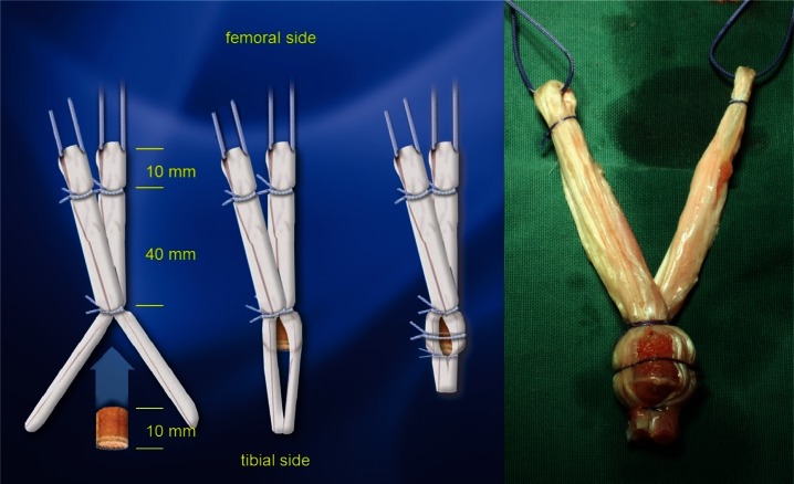
We developed tubed guiding devices for the tibial and the femoral tunnel for the use with diamond instruments (7). Disposable diamond edged bone core harvesters with diameters from 6 to 13 mm were introduced. These guarantee sharpness and sterility. Semitendinosus and gracilis tendon are harvested at the pes anserinus. The tendons are doubled or quadrupled into two bundles. The graft length is about 70 mm and with a diameter of 8 to 9 mm. Sutures mark the 10 mm minimum depth in the femoral tunnel. At a distance of 3 to 4 cm an 11 mm cortical- cancellous bone cylinder from the medial tibia condyle is sutured into the graft (Fig. 1). The cancellous surface of this cylinder is placed pointing proximaly into the hamstring graft and is beveled manually using bone cutters to facilitate insertion.
Figure 1.
Semitendinosus- and Gracilis tendon are prepared in two bundles. A suture indicates the minimum length in the femoral tunnel. In the tibia side sutures keep the 11 mm bone cylinder from the tibia head. The bone-window is necessary for bone-bone-contact healing.
The femoral tunnel is placed at 2,30/9,30 o’clock through the anteromedial port. The minimum depth is 30 mm with a diameter of 8 to 13 mm depending on the size of the knee and the original footprint. The intercondylar bifurcate ridge as the border between AM and PL bundles has to be in the centre of the tunnel.
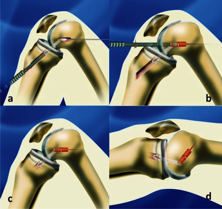
The tibial tunnel is cored using the tibia tubed guiding device. The diameter of the tibial tunnel is 8 to 9 mm depending on the graft size and is placed in the centre of the footprint. In big knees we used two 7 or 8 mm tunnel overlapping to a semi oval tunnel in the tibia up to 13 mm in sagittal length. The bone cylinders from the tunnels were harvested. The distal graft is positioned anatomically with the bundles and pulled in from distal to proximal and fixed press-fit with the 11mm cortico-cancellous bone cylinder directly under the tibial plateau. The proximal portion of the graft (first PM and second AM with different loops in the anatomical position) is pulled into the femoral tunnel and fixed with the femoral bone cylinder in 120° knee flexion (Fig. 2).
Figure 2 a,b,c,d.
The graft is implanted bottom to top (BTT). The distal bone cylinder is impacted press fit under the tibia plateau (a). The proximal graft is tensioned and fixed press fit in 120 degree knee flexion (b). The tension of the graft is self adapting in extension (c,d).
The crescent femoral attachment of the graft is taking on the appearance of two bundles (Fig. 3). The optimum tensioning of the graft is self adapting and achieved by BTT implantation and (Fig. 4). The tibial bone defect is filled completely with the rest of the bone cylinder.
Figure 3.
The width of a 11 mm diamond instrument overlaps the insertion of AM and PL bundle. The bone cylinder harvested from this tunnel is given back press fit and forms the two bundles.
Figure 4.
In extension the bundles self adapt the graft tension. After a year the bundles show regular signal and anatomic position.
What is Bottom to Top (BTT) fixation?
Usually the graft is fixed top down first in the femoral tunnel and then tensioned and fixed in 30° knee flexion in the tibia. We inverted this common procedure to achieve the “bottom to top” tensioning. First the ligament is fixed press fit with the tibial bone cylinder near the joint beyond the tibial spine. The graft is pulled into the femoral tunnel with the knee in 120 degrees flexion. The cortico-cancellous bone dowel harvested from the femoral tunnel is pushed parallel to the graft into the femoral tunnel and fixes the ligament near the joint (Fig. 1). The ligament is now tightened under flexion. On extending the knee, the graft becomes tighter and achieves the necessary and optimum tension for its mechanical function. The graft takes a right angle turn as it goes from the joint space into the femoral tunnel. The fixation in the femur is so strong that the graft can glide slightly out of the tibia tunnel (Figs. 2, 4).
Rehabilitation
The postoperative treatment is based on early functional rehabilitation for both BTB and hamstring grafts. The bone dowels are incorporated over the next 4–6 weeks.
Free extension and flexion is allowed for patients with normal bone quality. For those with osteoporotic bone we restrict the range of motion to between 30 degrees flexion to 90 degrees flexion in a brace for 3–4 weeks. In our hands this is enough for stable fixation. A brace is worn for six to eight weeks if the muscles are not strong enough.
Complete weight-bearing is usually reached after one week. Muscle training with electro-muscular stimulation (EMS) starts during the first days. We have shown the positive effects of Aquasprint (6) and proprioceptive vibration training on the quadriceps muscle (8). At the earliest, proprioceptive vibration training is used after two to three weeks postop two times a week.
Preliminary Results
The primary goal of this paper is to present the surgical technique.
Since April 2008, we increased the diameter of the femoral fixation initially from 11,5 to 13 mm. 46 patients (17W, 29M; age 28,3 y) were assessed 3 and 6 months post op. No complications were found. All patients followed their prescribed training according to the rehabilitation protocol. 8 of them then started special training for soccer and 5 for team handball. In 24 patients the femoral fixation was done with a 9,5 mm cylinder, 18 with 11,5 mm cylinder and 4 with 13 mm cylinder, all using single tunnel fixation. 8 patients of the 11,5 mm femoral diameter and al 4 with 13 mm femoral diameter had a semi oval tibia tunnel between 11 and 13 mm length. The subjective results of the patients were good and excellent. The Lachman Tests confirmed stability with a mean difference of 0,9 mm compared with the uninjured knee.
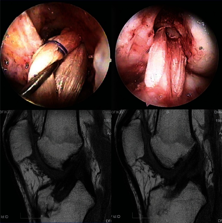
Compared to the 8 mm proximal insertion of the standard fixation with 1,2 mm difference we saw the same good and excellent clinical results in Lachman position and pivot shift. Pivot shift seemed to be more stable in the first 6 months in the broader insertions from 9,5, 11,5, 13 mm. No patients had a positive pivot shift or glide. In a case of a 40 years old male a rerupture happened 7 months after reconstruction with a 13 mm diameter press fit fixation on the femoral side. The insertion of the AM and PL bundle could be observed separated with a ridge as evidence of a double bundle en-grafting on the bonecylinder and reorganization of the origin insertion (Fig. 5).
Figure 5.
40 y, M, femoral single tunnel with 13 mm diameter and original ridge (left). ACL rerupture with adequate trauma after 7 months. The broad insertion of the AM and PL bundle of the graft with a reorganized bifurcate ridge can be observed as evidence of a two bundle engrafting on the bonecylinder and reorganization of the origin insertion (central, right).
Discussion
The ACL is made out of two functional bundles, the anteromedial (AM) and posterolateral (PL) bundle (2, 3, 10, 16, 17). The AM bundle of the ACL primarily controls anterior movement of the tibia underneath the femur. The PL bundle controls rotational stability of the knee during the last 30° to complete extension in movements such as pivoting, twisting, running, and jumping (12, 25). In the author’s opinion the native ACL is not only a double bundle it is a multi bundle construction. Every fiber of the ligament counts and stabilizes the knee. Single bundle ACL reconstruction does not adequately restore knee stability, particularly tibial rotation (1, 2, 13, 19, 20, 22). Anatomic double bundle ACL reconstruction restores knee stability better compared with single bundle reconstruction (2, 11, 18, 21–23). Usually two tunnels in the tibia and in the femur are necessary to reconstruct both bundles (2, 11, 18, 21–23). In bigger knees this is easy to achieve but in small knees difficult or impossible. Individual anatomy makes it necessary to reconstruct individually. Active sportsmen occasionally report some laxity after ACL reconstruction. In these cases in our patients we sometimes found a glide of the pivot shift while Lachman test was stable. In anatomic studies Siebold et al. could measure a femoral insertion from 50 to 156 mm2 and on tibia 70 to 260 mm2 (15, 16). Kopf et al. made measurements by arthroscopy and found for both bundles on the tibial side a 7–11 mm width and sagittal length of 11–20 mm and on the femoral side a width of 7–10 mm and a sagittal length of 12–20 mm (15). The standard single tunnel reconstruction has diameters of the tunnels between 6 and 8 mm. Replacement through the anteromedial portal at 2,30/9,30 o’clock allows a partial reconstruction with the AM bundle in small knees. ACL reconstruction should be anatomic with a broad femoral insertion similar to the two bundles in every knee size. In our former press fit fixation with patella BTB and hamstring in one 8 mm tunnel we observed different tensioning of the fibers and described this as an “imitation” of the bundles (5, 7). From this observations we developed a single tunnel fixation in the femur with a spread insertion near the origin from 9,5 mm up to actually 13 mm diameter with foreign material free press fit fixation. The tibia side was reconstructed in these cases with one 8 or 9 mm tunnel. 8 patients of the 11,5 mm femoral diameter and al 4 with 13 mm femoral diameter had a semi oval tibia tunnel between 11 and 13 mm length. We found a good rotational stability with a single tunnel reconstruction from 9.5 to 13 mm diameter. Differences to the semi oval tibia tunnel could not be obtained. It seems as the broad insertion on the femur is a leading factor for rotational stability. This also defines the better results by application of the femoral tunnel through the anteromedial portal compared to the high noon fixation. More studies are necessary to prove these findings and make ACL reconstruction more perfect.
Conclusion
Double bundle reconstruction has been done with a single tunnel fixed with the technique of the ALL PRESS FIT method. Broad anatomic femoral insertions with fixation near the cortex adjacent to the joint would appear to be better for rotational stability. The foreign material free fixation for hamstring in the ALL PRESS FIT Bottom To Top Fixation is successful easily reproducible. The Diamond Instruments, tubed guiding devices and applicators are precise, reliable and easy to manage.
Advantages of ALL PRESS FIT ACL Reconstruction are:
- Double bundle reconstruction with a single tunnel
- Fixation close to the anatomical insertion
- No bone defects
- Economical, no screws ore other foreign material
- Arthroscopic implantation
References
- 1.Anderson AF, Snyder RB, Lipscomb AB., Jr Anterior cruciate ligament reconstruction. A prospective randomized study of three surgical methods. Am J Sports Med. 2001;29:272–279. doi: 10.1177/03635465010290030201. [DOI] [PubMed] [Google Scholar]
- 2.Colombet P, Robinson J, Christel P, et al. Using navigation to measure rotation kinematics during ACL reconstruction. Clin Orthop Rel Res. 2007;454:59–65. doi: 10.1097/BLO.0b013e31802baf56. [DOI] [PubMed] [Google Scholar]
- 3.Edwards A, Bull AM, Amis AA. The attachments of the anteromedial and posterolateral fibre bundles of the anterior cruciate ligament. Part 2: femoral attachment. Knee Surg Sports Traumatol Arthrosc. 2008;16:29–36. doi: 10.1007/s00167-007-0410-0. [DOI] [PubMed] [Google Scholar]
- 4.Edwards A, Bull AM, Amis AA. The attachments of the anteromedial and posterolateral fibre bundles of the anterior cruciate ligament: Part 1: tibial attachment. Knee Surg Sports Traumatol Arthrosc. 2007;15:1414–1421. doi: 10.1007/s00167-007-0417-6. [DOI] [PubMed] [Google Scholar]
- 5.Felmet G. All-Press-FIT, eine neue Operationsmethode zum vorderen Kreuzbandersatz mit gleichzeitiger femoraler und tibialer Press-Fit-Verankerung. Arthroskopie. 1999;12:299–304. [Google Scholar]
- 6.Felmet G. Rehabilitation nach Kreuzbandersatz, der Stellenwert des „Aquasprint“ im frühfunktionellen Nachbehandlungsprogramm. Kongress CompactVerlag; Belin: 2002. pp. 188–196. (Arthroskopische Gelenkchirurgie, Gestern-heute-morgen, Standortbestimmung; G. Frenzel, H. Wuschech. [Google Scholar]
- 7.Felmet G. ALL PRESS FIT, gelenknah fixierter vorderer Kreuzbandersatz mit Semitendinosus-& Gracilissehne, eine neue OP-Technik, 21. Kongress der deutschsprachigen Arbeitsgemeinschaft für Arthroskopie AGA, 1. und 2. Luzern, Switzerland: Oktober. 2004. [Google Scholar]
- 8.Felmet G. Der Stellenwert des propriozeptiven Vibration-strainings im Nachbehandlungsprogramm nach Kreuzbandersatz, DGOOC. Berlin: Oktober. 2004. Abstr. E10-1404. [Google Scholar]
- 9.Felmet G. Implant-free press-fit fixation for bone-patellar tendon-bone ACL reconstruction: 10-year results. Arch Orthop Trauma Surg. 2010;130:985–992. doi: 10.1007/s00402-010-1050-2. [DOI] [PubMed] [Google Scholar]
- 10.Ferretti M, Levicoff EA, Macpherson TA, Moreland MS, Cohen M, Fu FH. The fetal anterior cruciate ligament: An anatomic and histologic study. Arthroscopy. 2007;23:278–283. doi: 10.1016/j.arthro.2006.11.006. [DOI] [PubMed] [Google Scholar]
- 11.Fu FH, Shen W, Starman JS, Okeke N, Irrgang JJ. Primary Anatomic Double-Bundle Anterior Cruciate Ligament Reconstruction: A Preliminary 2-Year Prospective Study. Am J Sports Med. 2008;36:1263–1274. doi: 10.1177/0363546508314428. [DOI] [PubMed] [Google Scholar]
- 12.Gabriel MT, Wong EK, Wool SL, et al. Distribution of in situ forces in the anterior cruciate ligament in response to rotatory loads. J Orthop Res. 2004;22:85–89. doi: 10.1016/S0736-0266(03)00133-5. [DOI] [PubMed] [Google Scholar]
- 13.Georgoulis AD, Ristanis S, Chouliaras V, et al. Tibial rotation is not restored after ACL reconstruction with a ham-string graft. Clin Ortho Rel Res. 2007;454:89–94. doi: 10.1097/BLO.0b013e31802b4a0a. [DOI] [PubMed] [Google Scholar]
- 14.Girgis FG, Marshall JL, Monajem A. The cruciate ligaments of the knee joint. Anatomical, functional and experimental analysis. Clin Orthop. 1975;106:216–231. doi: 10.1097/00003086-197501000-00033. [DOI] [PubMed] [Google Scholar]
- 15.Kopf S, Pombo M, Szczodry M, Shen W, Irrgang J, Fu FH. Pittsburgh Interoperative Größenbestimmung der femoralen und tibialen Insertion des vorderen Kreuzbandes“. AGA Interlaken; Sep 25–27, 2008. [Google Scholar]
- 16.Siebold R, Ellert T, Metz S, Metz J. Tibial insertions of the anteromedial and posterolateral bundles of the anterior cruciate ligament: morphometry, arthroscopic landmarks, and orientation model for bone tunnel placement. Arthroscopy. 2008;24:154–161. doi: 10.1016/j.arthro.2007.08.006. [DOI] [PubMed] [Google Scholar]
- 17.Siebold R, Ellert T, Metz S, Metz J. Femoral insertions of the anteromedial and posterolateral bundles of the anterior cruciate ligament: morphometry, arthroscopic landmarks, and orientation model for bone tunnel placement. Arthroscopy. 2008;24:585–592. doi: 10.1016/j.arthro.2007.08.006. [DOI] [PubMed] [Google Scholar]
- 18.Siebold R, Dehler C, Ellert T. Prospective randomized comparison of double-bundle versus single-bundle anterior cruciate ligament reconstruction. Arthroscopy. 2008;24:137–145. doi: 10.1016/j.arthro.2007.11.013. [DOI] [PubMed] [Google Scholar]
- 19.Tashman S, Collon D, Anderson K, et al. Abnormal rotational knee motion during running after anterior cruciate ligament reconstruction. Am J Sports Med. 2004;32:975–983. doi: 10.1177/0363546503261709. [DOI] [PubMed] [Google Scholar]
- 20.Woo SL, Kanamori A, Zeminski J, Yagi M, Papageorgiou C, Fu FH. The effectiveness of reconstruction of the anterior cruciate ligament with hamstrings and patellar tendon. A Cadaveric study comparing anterior tibial and rotational loads. J Bone Joint Surg Am. 2002;84:907–914. doi: 10.2106/00004623-200206000-00003. [DOI] [PubMed] [Google Scholar]
- 21.Yagi M, Kuroda R, Nagamune K, Yoshiya S, Kurosaka M. Doublebundle ACL reconstruction can improve rotational stability. Clin Orthop Relat Res. 2007;454:100–107. doi: 10.1097/BLO.0b013e31802ba45c. [DOI] [PubMed] [Google Scholar]
- 22.Yagi M, Wong EK, Kanamori A, et al. Biomechanical Analysis of an Anatomic Anterior Cruciate Ligament Reconstruction. Am J Sports Med. 2002;30:660–666. doi: 10.1177/03635465020300050501. [DOI] [PubMed] [Google Scholar]
- 23.Yasuda K, Kondo E, Ichiyama H, et al. Clinical evaluation of anatomic Double-Bundle anterior cruciate ligament reconstruction procedure using hamstring tendon grafts: comparisons among 3 different procedures. Arthroscopy. 2006;22:240–251. doi: 10.1016/j.arthro.2005.12.017. [DOI] [PubMed] [Google Scholar]
- 24.Zantop T, Herbort M, Raschke MJ, Fu FH, Petersen W. The role of the anteromedial and posterolateral bundles of the anterior cruciate ligament in anterior tibial translation and internal rotation. Am J Sports Med. 2007;35:223–227. doi: 10.1177/0363546506294571. [DOI] [PubMed] [Google Scholar]


