Summary
The purpose of this study was to evaluate the neuromuscular efficiency of the vastus medialis obliquus and postural balance in high-performance soccer athletes after anterior cruciate ligament (ACL) reconstruction, compared to the uninvolved leg. A cross-sectional study was conducted with 22 male professional soccer players after ACL reconstruction (4–12 months postoperatively). The athletes were submitted to functional rehabilitation with an accelerated protocol on the soccer team. They were evaluated using isokinetic dynamometer, surface electromyography and electronic baropodometer. There was no decrease or difference between neuromuscular efficiency of the VMO when comparing both the limbs after ACL reconstruction in the professional soccer athletes under treatment. The same result was found in postural balance. It can be concluded that the NME of the VMO in the involved member and postural balance were successfully re-established after the reconstruction procedure of the ACL in the sample group studied.
Keywords: soccer, anterior cruciate ligament, electromyography, torque, postural balance
Introduction
Rupture of the anterior cruciate ligament (ACL) is a recurrent injury in soccer athletes, with a high prevalence in high-performance athletes. On average, one ACL is injured every 2000 hours of sport practice, 14% of knee sprains injure this structure and each club disputes 12.8 games before losing a team member because of the rupture of this ligament1.
After partial or total rupture of the ACL the individual may have a deficit in conscious joint positioning2, deficiency in the perception of change in position during passive movement3 and a decrease in latency of reflex hamstring contraction4.
These proprioceptive alterations inhibit the action of the motor units in the knee extensors, decreasing the strength and the muscle power and tending to decrease the amplitude of the active movement in this articulation5.
One way to evaluate neuromuscular performance is quantifying the responsiveness of the contractile elements to an electric stimulus of the sarcolemma and, consequently, establishing a relationship with force production. Thereby, the individual is considered more efficient that requires lower activation to produce a given force. This ratio of myoelectrical activation, given by the average of the positive EMG signal (Root Mean Square - RMS), divided by the peak torque value generated by the muscle is called Neuromuscular Efficiency (NME)6, 7.
Another physical valence which can be affected by ACL injury is postural balance, which can be harmed by the loss of information supplied by mechanoreceptors in the knee joint8. These sensory receptors are responsible for providing information about knee position and the movements performed by this joint9. When there is rupture of this ligament the neural feedback mechanism is stopped and the motor control of the knee is damaged10. In addition to the consequences mentioned there is also a mechanical restraint to excessive movement in this joint11. These factors may harm the postural balance of the individual12.
Reconstruction of the ACL is indicated to restore the mechanical stability of the knee and allow a return to high-intensity functional activities, as required by soccer athletes13. The literature does not offer consensus on somatosensory activity restoration after this ligament reconstruction, with questions remaining about the restoration of postural balance after surgery14.
The purpose of this study was to evaluate the neuromuscular efficiency of the vastus medialis obliquus (VMO) and postural balance in high-performance soccer athletes after anterior cruciate ligament (ACL) reconstruction, between 4 and 12 month post-operation, comparing to the unaffected limb.
Materials and methods
Sample
This study was approved by the committee of ethics in research of the Universidade Federal of Ceará (COMEPUFC), protocol number 230/2011, and participants signed a free and informed consent form, confirming their voluntary participation on research. The collection happened at the Human Movement Analysis Lab, in 2011. A sample of twenty-two men were selected, all high-performance soccer players from three professional soccer teams in Brazil and within four to twelve months postoperative for ACL reconstruction. All individuals were undergoing functional rehabilitation on an accelerated protocol. The athletes were referred spontaneously by the rehabilitation department of the teams to which they belonged. Athletes were allowed to participate in the research who did not present changes in their cardiovascular system, such as uncontrolled hypertension, angina pectoris or arrhythmia and athletes were excluded who had acute musculoskeletal pain before or during testing (Analog Pain Scale less than 70 mm), untreated injuries or any other factor that affected the athlete’s performance during the evaluation.
Procedures
Evaluation protocol
Stabilometry
The first stage of evaluation was stabilometry, which aims to evaluate postural balance. The procedure was performed on an electronic baropodometer, trademark Diagnostic Support Italy, in a 3.2 m-long platform and composed of 4800 pressure sensors arranged continuously at a distance of 1.6 m in the center platform. This equipment, composed of electronic sensors which recognize oscillations in the center of gravity, permits the analysis of static equilibrium through a stabilometric test, which was carried out in both bipodalic and monopodalic forms. For the bipodalic evaluation, the athlete was instructed to remain in a standing position with feet put into a triangular shape, which accompanies the appliance, at an external rotation of 15º, along with arms at their sides, gaze directed to the horizon, and keep their temporomadibular joint relaxed (open mouth) for 51 seconds. For the monopodalic test, the individuals were supported by their left foot, keeping the right foot elevated with the knee flexed and then reversed, holding each position for 5 seconds. All tests were performed twice, first with open eyes and the second with closed eyes.
Isokinetic evaluation and neuromuscular efficiency
The next procedure was to evaluate the NME. For this, a Biodex® (System 4 Pro) isokinetic dynamometer was used, which was capable of evaluating the peak torque in the analyzed segments. In addition, surface electromyography (Miotool 400, trademark Miotec®, system composed of 4 channels, with a sampling rate frequency of 2000Hz per channel) was used, which was capable of recording total myoelectric activity from active muscle fibers.
The athlete warmed up freely for 5 minutes15. The isokinetic chair of the dynamometer was positioned so that the subject’s hip stayed at 85° of flexion and the movement axis of the equipment was aligned with the knee joint line. Next, the subject sat in the dynamometer chair and their position was stabilized with the use of seat belts on the trunk, abdomen and the unevaluated thigh, to prevent accessory movements. The lever arm of the equipment was fixed 2 centimeters above the medial malleolus. Preparation of the skin to collect the electromyography signal involved shaving, cleaning and abrasion of the skin surface of the individual with paper towel moistened with 70% alcohol, following standardization of the SENIAM. The electrodes were placed in the region of the vastus medialis obliquus muscle, with 20mm distance between the centers, and the reference electrode was placed at the lateral epicondyle of the humerus. The analog signal was converted to digital signal by sensor SDS500. Meditrace conducting electrodes for adults were used for the electrocardiogram in this research. The collection date was transmitted to the dynamometer computer by USB connection. The software used was Miograph, which accompanied the equipment.
The test always started with the dominant member. The isokinetic protocol established involved the following steps: concentric contractions to agonist and antagonist muscles, two velocities: 60° and 300°/s, 5 and 15 repetitions respectively and 30 seconds of rest between the series of exercises. The amplitude of movement was calibrated to start from maximal flexion to maximal extension, where the reference point was 90° of flexion. The member was weighed by the isokinetic dynamometer to exclude the gravity action bias. To normalize the electromyographic signal, 3 series of 3 maximal voluntary isometric contractions for 5 seconds were accomplished, wherein the rest between the series were 1 minute and between the repetitions were 5 seconds. Both evaluations happened at the same time.
After the procedures for positioning, alignment and normalization of the EMG signal, the test subjects were asked to perform five movements of knee flexion and extension at submaximal intensity in order to complete the period of muscle activation and also for familiarization with the equipment and test procedure. The upper limbs were fixed laterally to the chair in an appropriate place. Subsequently, the test was started, where a voice command was given by the same evaluator during all tests.
Electromyographic signal processing
In the post-collection phase, for analysis of EMG data, an interval of 1 second before the onset of contraction and 1 second after the end of contractions was duplicated and filtered. A digital filter was applied to the signal: Butterworth-type 4th-order band pass and cutoff frequencies between 20 and 450 Hz.
Statistical Analysis
Descriptive measurements were used to describe the sample characteristics, such as the measure of central tendency (mean) and dispersion (standard deviation). To compare the mean variables between the various treatments we used the Student-t test for independent sample analysis to compare the involved limb group with the uninvolved limb group. Data were analyzed with the SPSS software application (Statistical Package for Social Sciences). We accepted 5% as the significance level.
Results
The sample had a mean age of 21.77±4.45 years, mean weight 76.41±7.99 kg, mean height 1.79±0.06 and mean body mass index 23.70±1.54 Kg/m2. Amongst the athletes assessed, 54.5% were right-handed and 31.8% had the dominant limb involved in the procedure (Tab. 1).
Table 1.
Descriptive sample data
| Sample data | Values |
|---|---|
| Age | 21.77±4.45 years* |
| Weight | 76.41±7.99 kg* |
| Height | 1.79±0.06 m* |
| Body mass index | 23.70±1.54 Kg/m²* |
| Right-handed | 54.5% |
| Dominant limb involved in the procedure | 31.8% |
Mean ± Standard deviation
Neuromuscular Efficiency
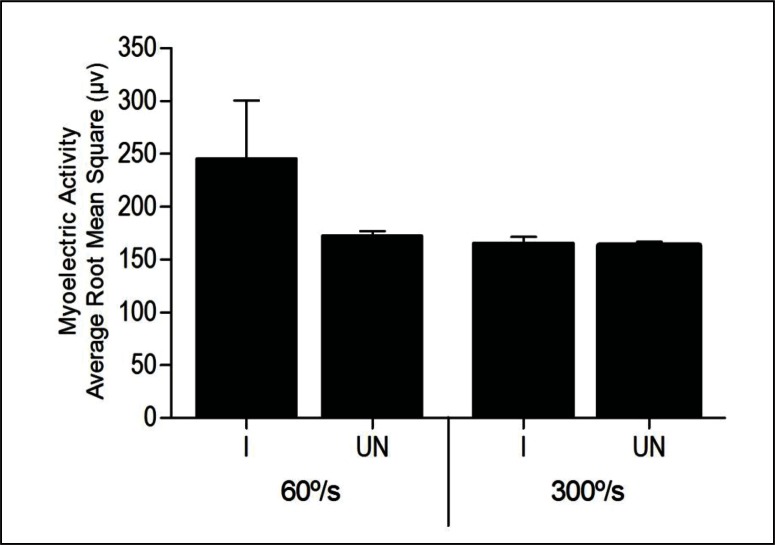
The myoelectric activity speed of 60°/s of VMO in the involved member (166.3±30.2 μV) did not present a statistic difference when compared to the uninvolved member (172.6±18.6 μV) p=0.44, while at a speed of 300°/s, where the mean of involved member was 165.6±25.9 μV and the uninvolved member 163.3±14.4 μV p=0.74 (Fig. 1).
Figure 1.
Myoelectric activity at speeds of 60°/s and 300°/s of the VMO in the involved limb (I) and uninvolved limb (UN). The values are expressed as means and standard deviation. * - p<0.05 Student-t Test
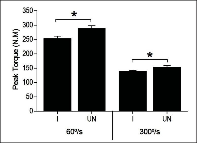
However, the quadriceps peak torque at speed 60°/s of the involved knee (253.2±40.7 N.m) was statistically lower when compared to the peak torque of the uninvolved knee (287.7±49.2 N.m), p=0.01. In the same way, when evaluating the peak torque at a speed of 300°/s, the involved limb (138.2±18.3 N.m) had an average lower than the average values of peak torque of the uninvolved limb (153.6±24.9 N.m) p= 0.03 (Fig. 2).
Figure 2.
Peak torque at speeds of 60°/s and 300°/s of the quadriceps in the involved limb (I) and uninvolved limb (UN). The values are expressed as means and standard deviation. * - p<0.05 Student-t Test.
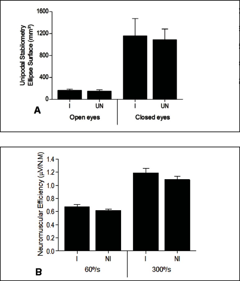
The NME showed no statistical difference at the different speeds assessed. At 60°/s, the involved limb showed a mean of 0.7±0.1 μV/N.m and the uninvolved member of 0.6±0.1 μV/N.m p=0.14, while at 300°/s the involved limb had a mean of 1.2±0.3 μV/N.m and the uninvolved limb, 1.0±0.2 μV/N.m, p=0.27 (Fig. 3).
Figure 3.
Neuromuscular efficiency at speeds of 60°/s and 300°/s for the VMO in the involved limb (I) and the uninvolved limb (UN). The values are expressed as means and standard deviation. * - p<0.05 Student-t Test.
Postural balance
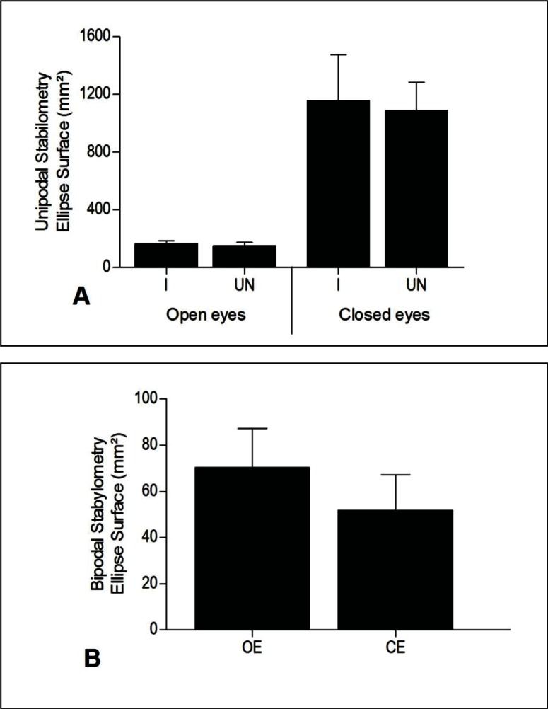
Comparing the two members in unilateral support for the oscillation surface with eyes open (165.8 ± 21.80 mm² for the involved member and 151.3 ± 23.67 mm² for the uninvolved member, p = 0.66) and oscillation surface with closed eyes (1157.8 ± 1314,3 mm² for the involved member and 1090.4 ± 801.6 mm² for the uninvolved member, p = 0.86), it can be observed that there was no statistical difference (Fig. 4A). The same happens in the anteroposterior variables mean displacement velocity with open eyes (15.8 ± 4.6 mm/s for the involved member and 16.9 ± 5.6 mm/s for the uninvolved member, p = 0.55) and with closed eyes (36.3 ± 4.5 mm/s involved member and 35.5 ± 2.6 mm/s for the uninvolved member, p = 0.87) and lateral-lateral mean displacement velocity with eyes open (10.4 ± 3.9 mm/s involved member and 10.6 ± 3.3 mm/s for the uninvolved member, p = 0.89) and eyes closed (15.6 ± 29.4 mm/s for the involved member and 28.7 ± 11.2 mm/s for the uninvolved member, p = 0.88). When comparing the variables in bipodal support, surface oscillation with eyes open (70.3 ± 7.,2 mm2) and eyes closed (51.79 ± 63.6 mm2) p = 0.42 (Fig. 4B), mean anteroposterior displacement with eyes open (4.14 ± 1.5 mm/s) and eyes closed (3.7 ± 0.9 mm/s) p = 0.31 and average lateral-lateral speed with eyes open (4.9 ± 1.8 mm/s) and eyes closed (4.1 ± 1.3 mm/s) p = 0.17, it can also be noted that these data showed no statistical difference.
Figure 4.
Ellipse surface with unipodal stabilometry (A) and bipodal stabilometry (B) comparing the involved limb (I) and un-involved limb (UN). The values are expressed as means and standard deviation. * - p<0.05 Student-t Test
Using the Romberg Index, no significant difference was found between the involved limb (832.1 ± 1074.2) and the uninvolved one (929.7 ± 813.7) for unipodal support (p = 0.77).
Discussion
This study had the purpose of evaluating peak torque of the knee extensor muscles, myoelectric activity and neuromuscular efficiency of VMO, and postural balance of soccer players who underwent ACL reconstruction. To compare the variables, the averages of the athletes’ own uninvolved members were used. This procedure has been used in several studies2, 11, 15.
Male soccer players were chosen to compose the sample owing to the high prevalence of ACL injuries in this sport. The reason for choosing the VMO was the fact that this is one of more susceptible muscles to atrophy after ACL rupture5. The NME is a rare topic in literature. The evaluation of this valence is a strategy for evaluating neuromuscular performance and success of the rehabilitation process of injured athletes. The restoration of postural balance and muscle strength after anterior cruciate ligament reconstruction are issues much discussed in the literature, and therefore, are evaluated in this study.
In terms of myoelectrical activation, no statistical difference was observed when comparing the two members of the individual. This result differs from that found in a study in which thirteen individuals in a post-surgical period after ACL ligamentoplasty showed a significantly higher rate of myoelectrical activation of the vastus medialis and vastus lateralis in the affected limb when compared to the unaffected one15.
The peak torque in the involved limb performed better than the uninvolved one. Another study, which accompanied the rehabilitation of twenty patients for twelve weeks after surgery for ACL reconstruction showed that the difference in peak torque at a speed of 180°/s between the two members was 38%. After isokinetic exercises for 20 minutes for 12 weeks, at a speed of 240°/s in the first six weeks and 180°/s in the last six weeks, there was an average 23.8% reduction in the difference between the peak torque of the two members16. This result confirms what was found in another study that assessed the same variable in eighteen soccer players, where the authors evaluated the peak torque before surgery and four months after rehabilitation, concluding that this deficit, especially in knee extensors muscles, was still great four months after surgery17. Other authors have demonstrated in their study that the deficit of the peak torque was maintained only at a slower speed, indicating that power was restored, but not force18.
By comparing the difference in percentage of the peak torque of the two members, it was found that the member subjected to the procedure showed 88% of strength and 90% of power of the control member, and the difference recommended for patient discharged is 80%. This result does not corroborate with findings in a study of 120 patients undergoing the same procedure for 6 months, showing that only half of competitive athletes had achieved this result. This difference must be a functional target reached 4–6 months after surgery19.
In postural balance, there were no significant differences in any analyzed variable, at any posture adopted. One study compared 19 healthy subjects between 18 and 30 years of age and 19 patients with unilateral ACL injury on the postural control with unilateral support, alternating the sides and the opening and closing of the eyes. The results showed that balance is changed in unilateral support on both sides after unilateral ACL rupture, which is more evident in the affected limb20.
According to the results presented and discussed, the accelerated rehabilitation protocol appears effective at restoring myoelectric activity, neuromuscular efficiency and postural control in subjects who underwent anterior cruciate ligamentoplasty.
For this research, it was not possible to monitor neuromuscular performance and fitness of athletes who comprised the sample, because this was done in the rehabilitation department of each team. Due to the difficulty to isolate the activity of a single quadriceps muscle, it was necessary to evaluate the myoelectric activity of the vastus medialis obliquus and peak torque of the quadriceps, to calculate the values of neuromuscular efficiency. Ideally, however, data would be collected related to the muscle that was studied, in this case, the vastus medialis obliquus.
This study used laboratory equipment of high technology in data collection, such as isokinetic dynamometer. Although this is an important tool to evaluate dynamic muscle work21, the isokinetic test is not sensitive and specific to analyze functional movements and sports skills as one leg hop test, co-contraction test, shuttle run test, carioca test22. Thus is important to combine information from laboratory tests with functional performance tests as a criterion for return to sport. For future studies it is suggested that the rehabilitation of the sample be monitored and there be periodic assessments to compare the results at different cutoff points in the rehabilitation phases.
Conclusion
According to the results we can conclude that the NME of the VMO of the limb involved in the procedure of ACL ligamentoplasty was reestablished with the protocol used to rehabilitate the patients studied. For postural balance the result was the same, no significant difference between unilateral support for the two members, suggesting that the protocol used was adequate to restore the equilibrium variables analyzed.
References
- 1.Walden M, Hagglund M, Magnusson H, Ekstrand J. Anterior cruciate ligament injury in elite football: a prospective three-cohort study. Knee Surg Sports Traumatol Arthrosc. 2011;19(1):11–19. doi: 10.1007/s00167-010-1170-9. [DOI] [PubMed] [Google Scholar]
- 2.Katayama M, Higuchi H, Kimura M, Kobayashi A, Hatayama K, Terauchi M, et al. Proprioception and performance after anterior cruciate ligament rupture. Int Orthop. 2004;28(5):278–281. doi: 10.1007/s00264-004-0583-9. [DOI] [PMC free article] [PubMed] [Google Scholar]
- 3.Dhillon MS, Bali K, Prabhakar S. Proprioception in anterior cruciate ligament deficient knees and its relevance in anterior cruciate ligament reconstruction. Indian J Orthop. 2011;45(4):294–300. doi: 10.4103/0019-5413.80320. [DOI] [PMC free article] [PubMed] [Google Scholar]
- 4.Beard DJ, Kyberd PJ, Fergusson CM, Dodd CA. Proprioception after rupture of the anterior cruciate ligament. An objective indication of the need for surgery? J Bone Joint Surg Br. 1993;75(2):311–315. doi: 10.1302/0301-620X.75B2.8444956. [DOI] [PubMed] [Google Scholar]
- 5.Alves PHdM, Silva DCdO, Lima FC, Pereira ML, Silva Z. Lesão do ligamento cruzado anterior e atrofia do músculo quadríceps femoral. Biosci J. 2009;25(1):146–156. [Google Scholar]
- 6.Muraro ARC. Efeito de um protocolo de fadiga concêntrico x excêntrico na resposta eletromiográfica e no torque dos extensores e flexores do joelho de jogadores de futebol [Monografia] Porto Alegre: Universidade Federal do Rio Grande do Sul; 2010. [Google Scholar]
- 7.Deschenes MR, Giles JA, McCoy RW, Volek JS, Gomez AL, Kraemer WJ. Neural factors account for strength decrements observed after short-term muscle unloading. Am J Physiol Regul Integr Comp Physiol. 2002;282(2):R578–583. doi: 10.1152/ajpregu.00386.2001. [DOI] [PubMed] [Google Scholar]
- 8.Alonso AC, Brech GC, Greve JMDA. Técnicas de avaliação proprioceptiva do ligamento cruzado anterior do joelho. Acta Fisioatr. 2010;17(3):134–140. [Google Scholar]
- 9.Johansson H, Sjolander P, Sojka P. A sensory role for the cruciate ligaments. Clin Orthop Relat Res. 1991;(268):161–178. [PubMed] [Google Scholar]
- 10.Bonfim TR, Jansen Paccola CA, Barela JA. Proprioceptive and behavior impairments in individuals with anterior cruciate ligament reconstructed knees. Arch Phys Med Rehabil. 2003;84(8):1217–1223. doi: 10.1016/s0003-9993(03)00147-3. [DOI] [PubMed] [Google Scholar]
- 11.Mattacola CG, Perrin DH, Gansneder BM, Gieck JH, Saliba EN, McCue FC., 3rd Strength, Functional Outcome, and Postural Stability After Anterior Cruciate Ligament Reconstruction. J Athl Train. 2002;37(3):262–268. [PMC free article] [PubMed] [Google Scholar]
- 12.Trulsson A, Roos EM, Ageberg E, Garwicz M. Relationships between postural orientation and self reported function, hop performance and muscle power in subjects with anterior cruciate ligament injury. BMC Musculoskelet Disord. 2010;11:143. doi: 10.1186/1471-2474-11-143. [DOI] [PMC free article] [PubMed] [Google Scholar]
- 13.Ardern CL, Webster KE, Taylor NF, Feller JA. Return to sport following anterior cruciate ligament reconstruction surgery: a systematic review and meta-analysis of the state of play. Br J Sports Med. 2011;45(7):596–606. doi: 10.1136/bjsm.2010.076364. [DOI] [PubMed] [Google Scholar]
- 14.Howells BE, Ardern CL, Webster KE. Is postural control restored following anterior cruciate ligament reconstruction? A systematic review. Knee Surg Sports Traumatol Arthrosc. 2011;19(7):1168–1177. doi: 10.1007/s00167-011-1444-x. [DOI] [PubMed] [Google Scholar]
- 15.Briant AL, Kelly J, Hohmann E. Neuromuscular adaptations and correlates of knee functionality following ACL reconstruction. Journal of orthopedics research. 2008;26(1):126–135. doi: 10.1002/jor.20472. [DOI] [PubMed] [Google Scholar]
- 16.Fabis J. The impact of a isokinetic training program on the peak torque of the quadriceps and knee flexors after anterior cruciate ligament reconstruction with hamstrings. Ortop Traumatol Rehabil. 2007;9(5):527–531. [PubMed] [Google Scholar]
- 17.Olivier N, Rogez J, Masquelier B. Benefit of isokinetic evaluations of knee before and after anterior cruciate ligament reconstruction in soccer players. Ann Readapt Med Phys. 2007;50(7):564–569. doi: 10.1016/j.annrmp.2007.03.004. [DOI] [PubMed] [Google Scholar]
- 18.Karasel S, Akpinar B, Gulbahar S, Baydar M, El O, Pinar H, et al. Clinical and functional outcomes and proprioception after a modified accelerated rehabilitation program following anterior cruciate ligament reconstruction with patellar tendon autograft. Acta Orthop Traumatol Turc. 2010;44(3):220–228. doi: 10.3944/AOTT.2010.2293. [DOI] [PubMed] [Google Scholar]
- 19.Carter TR, Edinger S. Isokinetic evaluation of anterior cruciate ligament reconstruction: hamstring versus patellar tendon. Arthroscopy. 1999;15(2):169–172. doi: 10.1053/ar.1999.v15.0150161. [DOI] [PubMed] [Google Scholar]
- 20.Tookuni KS, Neto RB, Pereira CAM, Souza DRd, Greve JMDA, Ayala ADA. Análise comparativa do controle postural de indivíduos com e sem lesão do ligamento cruzado anterior do joelho. Acta Ortop Bras. 2005;13(3):115–119. [Google Scholar]
- 21.Bosco C, Belli A, Astrua M, Tihanyi J, Pozzo R, Kellis S, et al. A dynamometer for evaluation of dynamic muscle work. Eur J Appl Physiol Occup Physiol. 1995;70(5):379–386. doi: 10.1007/BF00618487. [DOI] [PubMed] [Google Scholar]
- 22.Kong DH, Yang SJ, Ha JK, Jang SH, Seo JG, Kim JG. Validation of functional performance tests after anterior cruciate ligament reconstruction. Knee Surg Relat Res. 2012;24(1):40–45. doi: 10.5792/ksrr.2012.24.1.40. [DOI] [PMC free article] [PubMed] [Google Scholar]


