Abstract
An increase in cardiac troponin I (cTnI) occurs often after aneurysmal subarachnoid hemorrhage (SAH), but its significance is not well understood. One hundred three patients with SAH were prospectively evaluated in the SAHMII Study to determine the relations of cTnI to clinical severity, systolic and diastolic cardiac function, pulmonary congestion, and length of intensive care unit stay. Echocardiographic ejection fraction, wall motion score, mitral inflow early diastolic (E) and mitral annular early (E′) velocities were assessed. Thirty patients (29%) had mildly positive cTnI (0.1 to 1.0 ng/ml), 24 (23%) had highly positive cTnI (>1.0 ng/ml), and 49 (48%) had negative cTnI (<0.1 ng/ml). Highly positive cTnI was associated with worse neurologic disease, longer intensive care unit stay, and slight depression of ejection fraction (51 ± 11% [p <0.05] vs 59 ± 8% and 63 ± 6% in mildly positive or negative cTnI groups, respectively). Highly positive cTnI was also associated with abnormal wall motion acutely (>1.31 ng/ml; 76% sensitivity, 91% specificity), which typically resolved within 5 to 10 days. Both mildly or highly positive cTnI were associated with acute diastolic dysfunction, with E/E′ of 17 ± 6 and 16 ± 6 (both p <0.05) vs 13 ± 4 in patients with negative cTnI. Prevalences of pulmonary congestion were 79% (p <0.05) in patients with highly positive cTnI, 53% (p <0.05) in patients with mildly positive cTnI, and 29% in cTnI-negative patients. In conclusion, highly positive cTnI with SAH was associated with clinical neurologic severity, systolic and diastolic cardiac dysfunction, pulmonary congestion, and longer intensive care unit stay. Even mild increases in cTnI were associated with diastolic dysfunction and pulmonary congestion.
Aneurysmal subarachnoid hemorrhage (SAH) is a devastating neurologic disease associated with variable degrees of acute cardiac dysfunction.1,2 Although not clearly understood, a catecholamine-surge hypothesis was the most widely accepted theory for SAH-induced cardiac injury.3 Patients with aneurysmal SAH had an increase in systematic and local norepinephrine release4–7 that may have been related to myocardial injury deflected by increased serum cardiac troponin I (cTnI).2,8,9 Cardiac troponin is the current standard method of serologic detection of myocardial injury in patients with acute coronary syndromes.10 In addition, increases in cTnI were associated with increased mortality in critically ill patients,11 and even slight increases in cTnI (≥0.04 ng/ml) were associated with increased mortality rates in patients with advanced chronic heart failure.12 Although the cardiac injury associated with SAH was usually temporary and normalized over time, the degree of cardiac dysfunction was highly variable,2,7,13–16 and the clinical significance of acutely increased cTnI after SAH, especially when mildly positive, was not well understood. Accordingly, our objective was to test the hypothesis that increases in cTnI in patients with aneurysmal SAH were related to severity of the clinical neurologic condition, cardiac systolic and diastolic function, and occurrence of acute pulmonary congestion.
Methods
From May 2004 to January 2007, we prospectively enrolled consecutive patients admitted to the University of Pittsburgh Medical Center NeuroVascular Intensive Care Unit (Pitts-burgh, Pennsylvania) with a diagnosis of aneurysmal SAH documented using angiography and computed tomography as part of the Subarachnoid Hemorrhage and Myocardial Infarction and Ischemia (SAHMII) study. All patients were adults (age 18 to 75 years) with Fisher score ≥2 and/or Hunt-Hess grade ≥3. Patients were excluded if they had preexisting neurologic disease or traumatic or mycotic aneurysms. Patients were also excluded from this analysis if they had severe renal insufficiency or disease. The study protocol was approved by the Institutional Review Board at the University of Pittsburgh, and informed consent was obtained from each patient or an appropriate designee before study entry.
Severity of neurologic injury on admission was graded using the Hunt-Hess score to classify SAH severity using clinical symptoms, defined as 1 = asymptomatic or mild headache and slight nuchal rigidity; 2 = cranial nerve palsy, moderate to severe headache, or nuchal rigidity; 3 = mild focal deficit, lethargy, or confusion; 4 = stupor, moderate to severe hemiparesis, or early decerebrate rigidity; and 5 = deep coma, decerebrate rigidity, or moribund appearance.17
Clinical diagnosis, aneurysm site, and Hunt-Hess criteria were evaluated and graded by the admitting physician. Aneurysms were secured in the operating room using surgical clip placement or in the interventional radiology suite using endovascular coil and embolization. All patients were managed according to the standard practice guidelines for patients with SAH. Early routine care included prophylactic nimodipine (generally 60 mg every 4 hours) and magnesium replacement. Blood pressure management consisted of antihypertensives (labetolol, nicardipine, or nitroprusside) or vasopressor or inotropic therapy (phenylephrine, norepinephrine, dopamine, or dobutamine). Normovolemia was maintained using crystalloids and albumin guided by central venous pressure monitoring. cTnI was measured using a standard chemiluminescence immunoassay (Bayer Heath Care, Tarrytown, New York) on admission and daily for the first 5 days after SAH, and maximum daily cTnI was used as a marker of myocardial injury.
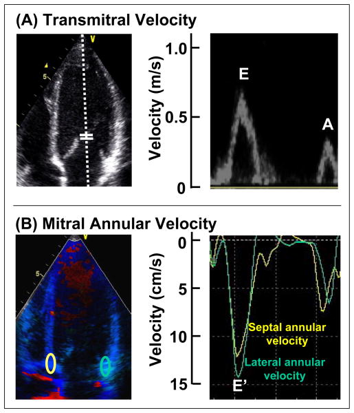
Transthoracic echocardiography was performed <72 hours after SAH and repeated 5 to 10 days after SAH onset in a subset of patients (Vivid; GE-Vingmed, Horton, Norway). Left ventricular ejection fraction was assessed using biplane Simpson’s rule with manual tracing of apical 4- and 2-chamber views in a blinded manner.18 Wall motion score was calculated using a standard 16-segment model. Segment scores were 1 = normal, 2 = hypokinesis, 3 = akinesis, and 4 = dyskinesis. Wall motion score index was calculated as the sum of all segment scores divided by the number of segments visualized.18 Pulse-wave Doppler–derived transmitral velocity and digital color tissue Doppler images were also obtained from apical views in a subset of patients.19 Early diastolic (E) and atrial (A) wave velocities and E wave deceleration time were measured using pulse-wave Doppler recording. Septal and lateral mitral annular early diastolic velocities (E′) were measured from an apical 4-chamber view using off-line color tissue Doppler images.20 E′ velocities from 2 sites (lateral and septal annulus) were averaged (E′), and E/E′ was calculated for assessment of left ventricular filling pressure (Figure 1).21
Figure 1.
Examples of echocardiographic data obtained. (A) Transmitral velocities with pulse-wave Doppler in an apical 4-chamber view at the mitral leaflet tips for E and A wave velocity and E wave deceleration time. (B) Mitral annular velocities of septal and lateral mitral annulus (E′) using color tissue Doppler images. Septal and lateral annular velocities were averaged, with E/E′ used as an estimate of left ventricular filling pressure.
Chest radiographs were completed on admission and through day 5 (or as long as clinically indicated). Interpretation of chest radiographs by a radiologist was used for assessment of pulmonary congestion. A positive finding of pulmonary congestion was radiographically defined as the presence of any of hilar edema, hilar fullness, interstitial edema, pulmonary congestion, or pulmonary edema on the radiology report. The presence of any of these findings on any of the first 5 days was defined as a positive chest radiograph for pulmonary congestion.
All group data were presented as mean ± SD and compared using 2-tailed Student’s t test for unpaired data. Analysis of variance and Bonferroni’s correction for post hoc tests were used for 3-group comparison. Proportional differences were evaluated using Fisher’s exact test or chi-square test as appropriate. The 95% confidence intervals (CIs) were calculated using Fisher’s r to z transformation. Receiver-operating characteristic curves were analyzed to determine the optimal cut-off values for cTnI to predict regional wall motion abnormality and pulmonary congestion. Statistical significance was p <0.05.
Results
One hundred three patients who were enrolled had complete baseline data sets, including complete clinical evaluation, cTnI, echocardiogram, and chest radiographs. Ninety-five (92%) also had complete Doppler measurements performed at baseline, and 88 (85%) had follow-up echocardiograms performed at 5 to 10 days. The group was predominately women (73%) with a mean age of 53 ± 10 years and mean Hunt-Hess grade of 2.6 ± 1.1 (Table 1). Only 7 patients (7%) had a history of coronary artery disease. Serum cTnI was undetectable (<0.1 ng/ml) in 49 patients (48%). In 54 patients (52%) with positive cTnI, mean levels were 2.7 ± 6.6 ng/ml, with a wide range of 0.1 to 42.7. Thirty patients (29%) were classified as having mildly positive cTnI (0.1 to 1.0 ng/ml), and 24 patients (23%), highly positive cTnI (>1.0 ng/ml). Highly positive cTnI was associated with worse SAH clinical severity (higher mean Hunt-Hess grade) than negative cTnI (p <0.05). Specifically, cTnI >1.31 ng/ml was associated with a significantly higher mean Hunt-Hess grade (3.4 ± 1.1) compared with patients with lower cTnI (2.5 ± 1.0; p <0.05; Figures 2 and 3).
Table 1.
Patient characteristics in groups classified by peak cardiac troponin I (cTnI)
| Peak cTnI (ng/ml)
|
p Value | |||
|---|---|---|---|---|
| Negative <0.1 (n = 49) | Mildly Positive 0.1–1.0 (n = 30) | Highly Positive >1.0 (n = 24) | ||
| Age (yrs) | 51 ± 10 | 55 ± 9 | 56 ± 11 | NS |
| Women | 35 (71%) | 23 (77%) | 17 (71%) | NS |
| Hunt-Hess grade | ||||
| Mean | 2.4 ± 1.0 | 2.6 ± 0.9 | 3.2 ± 1.2* | <0.01 |
| 1 | 12 (24%) | 4 (13%) | 2 (8%)† | <0.05 |
| 2 | 13 (27%) | 8 (27%) | 4 (17%) | NS |
| 3 | 18 (37%) | 13 (43%) | 9 (38%) | NS |
| 4 | 6 (12%) | 5 (17%) | 5 (21%) | NS |
| 5 | 0 (0%) | 0 (0%) | 4 (17%) | NS |
Data expressed as mean ± SD or number (percent).
p <0.01 versus negative and mildly positive cTnI group.
p <0.01 versus negative cTnI group.
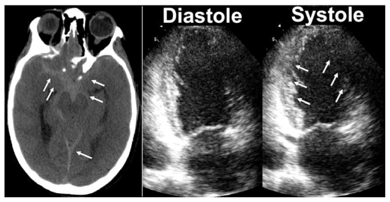
Figure 2.
An illustrative case example of a patient with highly increased cTnI (10 ng/ml) and left ventricular wall motion abnormalities after SAH. Hunt-Hess score on admission was 3, and brain computed tomography showed Fisher score of 3 (left). SAH shown by arrows. Echocardiogram on admission (right) showed midventricular wall motion abnormalities (arrows) in the apical 2-chamber view. Wall motion score was 1.9, and ejection fraction was 30%.
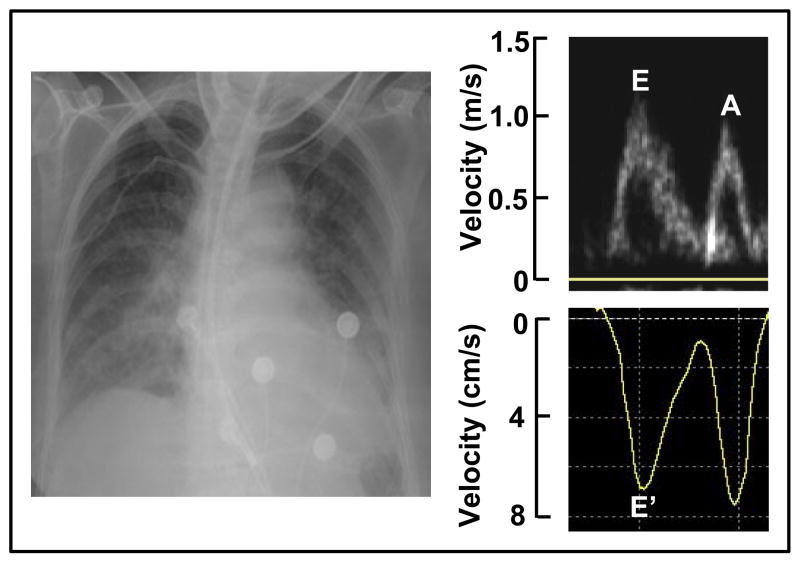
Figure 3.
A illustrative case example of a patient with mildly increased cTnI (0.2 ng/ml) and pulmonary congestion using chest X-ray after SAH (left). E/A ratio was 1.2, and E deceleration time was 160 ms using transmitral velocity (right upper). Mitral annular velocity was 7 cm/s, and E/E′ was 16 (right lower). Wall motion score was normal (1.0), and ejection fraction was 58%.
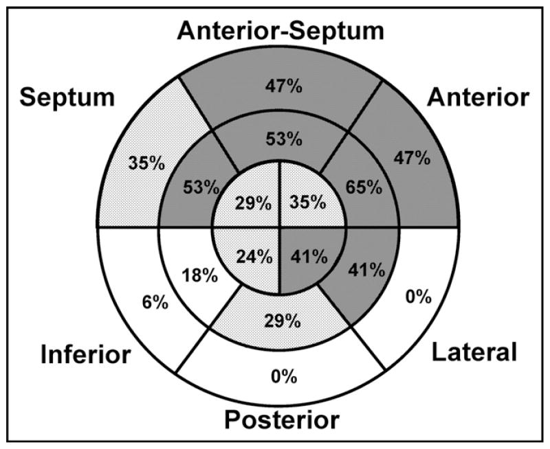
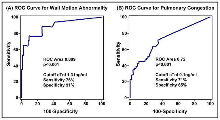
Overall, positive cTnI was not associated with abnormal ejection fraction in patients with SAH (Table 2). Only highly positive cTnI was associated with relatively depressed ejection fraction at 51 ± 11% (p <0.05 vs other groups) compared with 59 ± 8% and 63 ± 6% in the mildly positive or negative cTnI groups, respectively. Specifically, no patient with negative cTnI had an abnormal ejection fraction, and only 1 patient with mildly positive cTnI had an abnormal ejection fraction of 40%. Of patients with highly positive cTnI, only 4 had abnormal ejection fractions of 27%, 30%, 40%, and 41%. The overall degree of left ventricular systolic dysfunction was modest, and inotropic or vasopressor support was infrequent and administered only briefly. No patient had cardiogenic shock. Only 17 patients (17%) had acute wall motion abnormalities, and they tended to be modest. They were observed in several left ventricular anatomic regions, but appeared to be more frequent in the basal anterior and anteroseptal segments (Figure 4). All patients with an abnormal wall motion score at baseline had a follow-up study at 5 to 10 days after SAH onset; 8 patients (47%) improved and 4 patients (24%) normalized their wall motion scores, whereas 5 (29%) had persistence or worsening of their baseline wall motion scores (including 1 patient with a history of coronary artery disease). No patient with a normal wall motion score at baseline showed abnormal wall motion at follow-up. There was a higher prevalence of left ventricular wall motion abnormalities in patients with highly positive cTnI (54%; p <0.05 vs other groups) compared with the mildly positive (10%) or negative cTnI groups (2%), respectively. Using receiver-operating characteristic curve analysis, cTnI >1.31 ng/ml predicted wall motion score abnormalities with 76% sensitivity and 91% specificity (Figure 5).
Table 2.
Association of cardiac troponin I (cTnI) with systolic function
| Peak cTnI (ng/ml)
|
p Value | |||
|---|---|---|---|---|
| Negative <0.1 (n = 49) | Mildly Positive 0.1–1.0 (n = 30) | Highly Positive >1.0 (n = 24) | ||
| Wall motion score | 1.0 ± 0.0 | 1.0 ± 0.1 | 1.3 ± 0.3* | <0.001 |
| Abnormal wall motion | 1 (2%) | 3 (10%) | 13 (54%)* | <0.001 |
| Ejection fraction (%) | 63 ± 6 | 59 ± 8 | 51 ± 11* | 0.001 |
Data presented as mean ± SD or number (percent).
p <0.01 versus negative and mildly positive cTnI groups.
Figure 4.
Distribution of abnormal segments in patients with wall motion abnormalities after SAH using a 16-segment model showing outer, middle, and inner represent basal, midventricular, and apical segments of the left ventricle. Numbers in the segment show percentages of patients with segmental wall motion (n = 17).
Figure 5.
Receiver-operating characteristic (ROC) curves to determine cut-off values for cTnI to predict (A) regional wall motion abnormalities and (B) pulmonary congestion. ROC areas, cut-off cTnI values, sensitivities, and specificities are shown.
Left ventricular diastolic function evaluation is listed in Table 3. In the 3 groups classified using cTnI, E/A ratio was not statistically significant, but the group mean E wave deceleration time in the highly positive cTnI group was significantly shorter than that in the mildly positive group (170 vs 194 ms; p <0.05), indicating a relative decrease in diastolic function. Significant differences in mitral annular velocity were observed between groups, and patients in both the mildly positive and highly positive cTnI groups had lower E′ (7.0 ± 1.9 and 6.7 ± 1.4 [both p <0.05] vs 8.8 ± 2.2 cm/s in the cTnI negative group), consistent with modest diastolic dysfunction. E/E′ as an indicator of left ventricular filling pressure was significantly increased at 17 ± 6 (p<0.05) in the mildly cTnI positive group to 16 ± 6 (p<0.05) in the highly cTnI positive group compared with 13 ± 4 in the cTnI-negative group. Patients with positive cTnI (≥0.1 ng/ml) showed significantly lower E′ than patients with negative cTnI (6.9 ± 1.7 vs 8.8 ± 1.7 cm/s; p <0.05). Similarly, patients with positive cTnI (≥0.1 ng/ml) showed significantly higher E/E′ than patients with negative cTnI (17 ± 0.6 vs 13 ± 4; p <0.05).
Table 3.
Association of cardiac troponin I (cTnI) with diastolic function and pulmonary congestion
| Peak cTnI (ng/ml)
|
p Value | |||
|---|---|---|---|---|
| Negative <0.1 (n = 49) | Mildly Positive 0.1–1.0 (n = 30) | Highly Positive >1.0 (n = 24) | ||
| Transmitral velocity (n = 97) | ||||
| E/A ratio | 1.4 ± 0.4 | 1.3 ± 0.5 | 1.4 ± 0.5 | NS |
| E deceleration time | 181 ± 25 | 194 ± 46 | 170 ± 35* | <0.05 |
| Annular velocity (n = 95) | ||||
| E′ velocity (cm/s) | 8.8 ± 2.2 | 7.0 ± 1.9† | 6.7 ± 1.4† | <0.001 |
| Left ventricular filling pressure (n = 95) | ||||
| E/E′ ratio | 13 ± 4 | 17 ± 6† | 16 ± 6† | 0.001 |
| Chest X-ray (n = 103) | ||||
| Pulmonary congestion (%) | 14 (29%) | 16 (53%)† | 19 (79%)* | <0.001 |
Data presented as mean ± SD or number (percent).
p <0.01 versus negative and mildly positive cTnI groups.
p <0.05 versus negative cTnI group.
Patients in the highly positive cTnI group had a high prevalence of pulmonary congestion using chest radiographs at 79% (95% CI 60 to 91). Furthermore, 53% (95% CI 36 to 70) of patients with mildly positive cTnI had pulmonary congestion, which was significantly more than observed in the cTnI-negative group at 29% (95% CI 17 to 43, p <0.05; Figure 5). A cut-off value for positive cTnI (≥0.1 ng/ml) predicted the occurrence of pulmonary congestion with 71% sensitivity and 65% specificity using receiver-operating characteristic curve analysis (Figure 5). Intensive care unit stay was significantly longer in the highly positive cTnI group at 14.0 ± 7.3 days compared with 11.6 ± 5.8 in patients with negative or mildly positive cTnI (p <0.05).
Discussion
This study was a prospective evaluation of the clinical significance of cTnI in a large series of consecutive patients with acute aneurysmal SAH. It showed the relation of cTnI to clinical severity of neurologic condition, systolic and diastolic cardiac dysfunction, occurrence of pulmonary congestion, and length of intensive care unit stay. It added to existing reports by showing associations of increases in cTnI with diastolic function assessed using E/E′ and cut-off values predictive of acute cardiac dysfunction and pulmonary congestion. A highly positive cTnI was associated with severity of neurologic damage and longer intensive care unit stay, as well as evidence of global and regional left ventricular systolic dysfunction, although degrees of ejection fraction depression and wall motion abnormalities were typically modest and transient. Mild increases in cTnI were associated with diastolic dysfunction, and importantly, the occurrence of pulmonary congestion increased with cTnI. Even mild increases in cTnI were associated with an increased incidence of pulmonary congestion. These data support the relation between severity of SAH and clinical manifestations of acute cardiac injury reflected by cTnI.
Myocardial injury and increased troponin after aneurysmal SAH have been previously reported.1,3,14 Hypotheses regarding the cause of myocardial injury have included supply-demand mismatch, epicardial coronary vasospasm, and coronary thrombosis.7 However, growing evidence supported a catecholamine hypothesis in which SAH led to an acute surge in catecholamine release at the time of rupture and subsequent myocardial injury and dysfunction. The proposed mechanism included increased autonomic output in response to SAH, increased systemic catecholamine release, increased blood pressure, vascular wall stress, and coronary vasospasm.5,6 A central component of the catecholamine hypothesis of myocardial injury included increased local norepinephrine release resulting in a cascade of cellular events ultimately leading to myocardial necrosis.4,7 In this study, patients presenting with severe neurologic damage and systolic dysfunction were more often seen in the highly positive cTnI group, consistent with previous observations.2,22 These observations suggested that severe neurologic damage may cause an increase in systematic and local norepinephrine release that induces myocardial injury, represented as systolic dysfunction and increased cTnI.
The catecholamine hypothesis has been used to describe the unusual patterns and modest distribution of wall motion abnormalities observed in patients with SAH, which were not consistent with epicardial coronary atherosclerosis.1,7,23 Khush et al23 found wall motion abnormalities in patients with SAH more frequently in anterior-anteroseptal walls of mid to basal segments with relative sparing of apical segments. Apical ballooning syndrome, also known as Takotsubo cardiomyopathy, shared the same hypothesized mechanism of massive catecholamine release, although this appeared to be a different disease than myocardial dysfunction associated with SAH.3,24 In the present study, 71% of patients with SAH with abnormal wall motion improved 5 to 10 days after SAH. Only 5 patients did not recover acutely, and 1 of these patients had a possible preexisting wall motion abnormality because of a history of coronary artery disease. Ejection fraction depression was an uncommon observation, and the potential long-term clinical consequence of persistence of depressed ejection fraction was not known in these patients.
Although left ventricular systolic dysfunction was either absent or only modest in the group with highly positive cTnI, diastolic function was detected in many patients with SAH, even when only mildly positive. The finding of positive cTnI was often associated with pulmonary congestion. Heart failure often occurred with preserved systolic function, termed diastolic heart failure or heart failure with preserved ejection fraction.25 Recent results from the Candesartan in Heart failure: Assessment of Reduction in Mortality and morbidity (CHARM)-Preserved study showed diastolic dysfunction in ⅔of patients with heart failure with preserved systolic function.26 Although the acute diastolic dysfunction observed in patients with SAH may likely result from a different mechanism, its association with pulmonary congestion indicated that these echocardiographic Doppler measures may be more sensitive markers of myocardial injury than global ejection fraction or wall motion scores. These observations suggest that patients with even mild cTnI increases had a degree of myocardial injury important enough to result in diastolic dysfunction and pulmonary congestion.
There were several limitations to this study. Diastolic function assessed using Doppler echocardiography had its potential limitations.27 Left ventricular diastolic velocities using tissue Doppler of the mitral annulus were less dependent on preload than transmitral flow velocities, but not completely load independent.28 Another limitation was that E′ used directly and for E/E′ was measured using color tissue Doppler in the present study, which was technically different from that measured using pulse tissue Doppler.29,30 McCulloch et al21 directly compared tissue velocities measured using color tissue Doppler with pulse tissue Doppler and reported significant correlations, but that myocardial velocities measured using color tissue Doppler significantly underestimated velocities using pulse tissue Doppler. Consequently, E/E′ using color tissue Doppler may have overestimated E/E′. E′ and E/E′ values in this study cannot be compared with previous reports using pulse tissue Doppler. However, comparisons between groups remain valid as a surrogate marker of left ventricular filling pressure. Another limitation was the use of chest radiographs to evaluate pulmonary congestion. However, a positive chest radiograph did not denote clinical heart failure conclusively. Many clinical trials of patients with heart failure previously used acute pulmonary congestion on chest radiographs as a marker for heart failure, although we acknowledge that patients with SAH represent a different patient population. Finally, we did not have long-term outcome data in the present study, and the clinical significance of our observations was limited to the acute in-hospital course.
Acknowledgments
We thank Leslie Hoffman, RN, PhD; the research staff at the University of Pittsburgh School of Nursing; and the physicians and staff in the Neurovascular Intensive Care Unit at the University of Pittsburgh Medical Center-Presbyterian for assistance and cooperation.
This work was supported by Grants RO1HL074316 and K24 HL04503-02 from the National Institutes of Health, National Heart, Lung, and Blood Institute, Bethesda, Maryland.
References
- 1.Zaroff JG, Rordorf GA, Ogilvy CS, Picard MH. Regional patterns of left ventricular systolic dysfunction after subarachnoid hemorrhage: evidence for neurally mediated cardiac injury. J Am Soc Echocardiogr. 2000;13:774–779. doi: 10.1067/mje.2000.105763. [DOI] [PubMed] [Google Scholar]
- 2.Kothavale A, Banki NM, Kopelnik A, Yarlagadda S, Lawton MT, Ko N, Smith WS, Drew B, Foster E, Zaroff JG. Predictors of left ventricular regional wall motion abnormalities after subarachnoid hemorrhage. Neurocrit Care. 2006;4:199–205. doi: 10.1385/NCC:4:3:199. [DOI] [PubMed] [Google Scholar]
- 3.Lee VH, Oh JK, Mulvagh SL, Wijdicks EF. Mechanisms in neurogenic stress cardiomyopathy after aneurysmal subarachnoid hemorrhage. Neurocrit Care. 2006;5:243–249. doi: 10.1385/NCC:5:3:243. [DOI] [PubMed] [Google Scholar]
- 4.Mertes PM, Carteaux JP, Jaboin Y, Pinelli G, el Abassi K, Dopff C, Atkinson J, Villemot JP, Burlet C, Boulange M. Estimation of myocardial interstitial norepinephrine release after brain death using cardiac microdialysis. Transplantation. 1994;57:371–377. doi: 10.1097/00007890-199402150-00010. [DOI] [PubMed] [Google Scholar]
- 5.Naredi S, Lambert G, Eden E, Zall S, Runnerstam M, Rydenhag B, Friberg P. Increased sympathetic nervous activity in patients with nontraumatic subarachnoid hemorrhage. Stroke. 2000;31:901–906. doi: 10.1161/01.str.31.4.901. [DOI] [PubMed] [Google Scholar]
- 6.Masuda T, Sato K, Yamamoto S, Matsuyama N, Shimohama T, Matsunaga A, Obuchi S, Shiba Y, Shimizu S, Izumi T. Sympathetic nervous activity and myocardial damage immediately after subarachnoid hemorrhage in a unique animal model. Stroke. 2002;33:1671–1676. doi: 10.1161/01.str.0000016327.74392.02. [DOI] [PubMed] [Google Scholar]
- 7.Banki NM, Kopelnik A, Dae MW, Miss J, Tung P, Lawton MT, Drew BJ, Foster E, Smith W, Parmley WW, Zaroff JG. Acute neurocardiogenic injury after subarachnoid hemorrhage. Circulation. 2005;112:3314–3319. doi: 10.1161/CIRCULATIONAHA.105.558239. [DOI] [PubMed] [Google Scholar]
- 8.Miss JC, Kopelnik A, Fisher LA, Tung PP, Banki NM, Lawton MT, Smith WS, Dowd CF, Zaroff JG. Cardiac injury after subarachnoid hemorrhage is independent of the type of aneurysm therapy. Neurosurgery. 2004;55:1244–1251. doi: 10.1227/01.neu.0000143165.50444.7f. [DOI] [PubMed] [Google Scholar]
- 9.Horowitz MB, Willet D, Keffer J. The use of cardiac troponin-I (cTnI) to determine the incidence of myocardial ischemia and injury in patients with aneurysmal and presumed aneurysmal subarachnoid hemorrhage. Acta Neurochir (Wien) 1998;140:87–93. doi: 10.1007/s007010050063. [DOI] [PubMed] [Google Scholar]
- 10.Braunwald E, Antman EM, Beasley JW, Califf RM, Cheitlin MD, Hochman JS, Jones RH, Kereiakes D, Kupersmith J, Levin TN, et al. ACC/AHA 2002 guideline update for the management of patients with unstable angina and non-ST-segment elevation myocardial infarction—summary article: a report of the American College of Cardiology/American Heart Association task force on practice guidelines (Committee on the Management of Patients With Unstable Angina) J Am Coll Cardiol. 2002;40:1366–1374. doi: 10.1016/s0735-1097(02)02336-7. [DOI] [PubMed] [Google Scholar]
- 11.Lim W, Cook DJ, Griffith LE, Crowther MA, Devereaux PJ. Elevated cardiac troponin levels in critically ill patients: prevalence, incidence, and outcomes. Am J Crit Care. 2006;15:280–288. [PubMed] [Google Scholar]
- 12.Horwich TB, Patel J, MacLellan WR, Fonarow GC. Cardiac troponin I is associated with impaired hemodynamics, progressive left ventricular dysfunction, and increased mortality rates in advanced heart failure. Circulation. 2003;108:833–838. doi: 10.1161/01.CIR.0000084543.79097.34. [DOI] [PubMed] [Google Scholar]
- 13.Banki NM, Zaroff JG. Neurogenic cardiac injury. Curr Treat Options Cardiovasc Med. 2003;5:451–458. doi: 10.1007/s11936-003-0034-8. [DOI] [PubMed] [Google Scholar]
- 14.Naidech AM, Kreiter KT, Janjua N, Ostapkovich ND, Parra A, Commichau C, Fitzsimmons BF, Connolly ES, Mayer SA. Cardiac troponin elevation, cardiovascular morbidity, and outcome after subarachnoid hemorrhage. Circulation. 2005;112:2851–2856. doi: 10.1161/CIRCULATIONAHA.105.533620. [DOI] [PubMed] [Google Scholar]
- 15.Banki N, Kopelnik A, Tung P, Lawton MT, Gress D, Drew B, Dae M, Foster E, Parmley W, Zaroff J. Prospective analysis of prevalence, distribution, and rate of recovery of left ventricular systolic dysfunction in patients with subarachnoid hemorrhage. J Neurosurg. 2006;105:15–20. doi: 10.3171/jns.2006.105.1.15. [DOI] [PubMed] [Google Scholar]
- 16.Kopelnik A, Fisher L, Miss JC, Banki N, Tung P, Lawton MT, Ko N, Smith WS, Drew B, Foster E, Zaroff J. Prevalence and implications of diastolic dysfunction after subarachnoid hemorrhage. Neurocrit Care. 2005;3:132–138. doi: 10.1385/NCC:3:2:132. [DOI] [PubMed] [Google Scholar]
- 17.Hunt WE, Hess RM. Surgical risk as related to time of intervention in the repair of intracranial aneurysms. J Neurosurg. 1968;28:14–20. doi: 10.3171/jns.1968.28.1.0014. [DOI] [PubMed] [Google Scholar]
- 18.Lang RM, Bierig M, Devereux RB, Flachskampf FA, Foster E, Pellikka PA, Picard MH, Roman MJ, Seward J, Shanewise JS, et al. Recommendations for chamber quantification: a report from the American Society of Echocardiography’s Guidelines and Standards Committee and the Chamber Quantification Writing Group, developed in conjunction with the European Association of Echocardiography, a branch of the European Society of Cardiology. J Am Soc Echocardiogr. 2005;18:1440–1463. doi: 10.1016/j.echo.2005.10.005. [DOI] [PubMed] [Google Scholar]
- 19.Gulati VK, Katz WE, Follansbee WP, Gorcsan J., III Mitral annular descent velocity by tissue Doppler echocardiography as an index of global left ventricular function. Am J Cardiol. 1996;77:979–984. doi: 10.1016/s0002-9149(96)00033-1. [DOI] [PubMed] [Google Scholar]
- 20.Nagueh SF, Mikati I, Kopelen HA, Middleton KJ, Quinones MA, Zoghbi WA. Doppler estimation of left ventricular filling pressure in sinus tachycardia. A new application of tissue Doppler imaging. Circulation. 1998;98:1644–1650. doi: 10.1161/01.cir.98.16.1644. [DOI] [PubMed] [Google Scholar]
- 21.McCulloch M, Zoghbi WA, Davis R, Thomas C, Dokainish H. Color tissue Doppler myocardial velocities consistently underestimate spectral tissue Doppler velocities: impact on calculation peak transmitral pulsed Doppler velocity/early diastolic tissue Doppler velocity (E/Ea) J Am Soc Echocardiogr. 2006;19:744–748. doi: 10.1016/j.echo.2006.01.020. [DOI] [PubMed] [Google Scholar]
- 22.Tung P, Kopelnik A, Banki N, Ong K, Ko N, Lawton MT, Gress D, Drew B, Foster E, Parmley W, Zaroff J. Predictors of neurocardiogenic injury after subarachnoid hemorrhage. Stroke. 2004;35:548–551. doi: 10.1161/01.STR.0000114874.96688.54. [DOI] [PubMed] [Google Scholar]
- 23.Khush K, Kopelnik A, Tung P, Banki N, Dae M, Lawton M, Smith W, Drew B, Foster E, Zaroff J. Age and aneurysm position predict patterns of left ventricular dysfunction after subarachnoid hemorrhage. J Am Soc Echocardiogr. 2005;18:168–174. doi: 10.1016/j.echo.2004.08.045. [DOI] [PubMed] [Google Scholar]
- 24.Bybee KA, Kara T, Prasad A, Lerman A, Barsness GW, Wright RS, Rihal CS. Systematic review: transient left ventricular apical ballooning: a syndrome that mimics ST-segment elevation myocardial infarction. Ann Intern Med. 2004;141:858–865. doi: 10.7326/0003-4819-141-11-200412070-00010. [DOI] [PubMed] [Google Scholar]
- 25.Senni M, Redfield MM. Heart failure with preserved systolic function. A different natural history? J Am Coll Cardiol. 2001;38:1277–1282. doi: 10.1016/s0735-1097(01)01567-4. [DOI] [PubMed] [Google Scholar]
- 26.Persson H, Lonn E, Edner M, Baruch L, Lang CC, Morton JJ, Ostergren J, McKelvie RS. Diastolic dysfunction in heart failure with preserved systolic function: need for objective evidence: results from the CHARM Echocardiographic Substudy-CHARMES. J Am Coll Cardiol. 2007;49:687–694. doi: 10.1016/j.jacc.2006.08.062. [DOI] [PubMed] [Google Scholar]
- 27.Redfield MM, Jacobsen SJ, Burnett JC, Jr, Mahoney DW, Bailey KR, Rodeheffer RJ. Burden of systolic and diastolic ventricular dysfunction in the community: appreciating the scope of the heart failure epidemic. JAMA. 2003;289:194–202. doi: 10.1001/jama.289.2.194. [DOI] [PubMed] [Google Scholar]
- 28.Jacques DC, Pinsky MR, Severyn D, Gorcsan J., III Influence of alterations in loading on mitral annular velocity by tissue Doppler echocardiography and its associated ability to predict filling pressures. Chest. 2004;126:1910–1918. doi: 10.1378/chest.126.6.1910. [DOI] [PubMed] [Google Scholar]
- 29.Sohn DW, Chai IH, Lee DJ, Kim HC, Kim HS, Oh BH, Lee MM, Park YB, Choi YS, Seo JD, Lee YW. Assessment of mitral annulus velocity by Doppler tissue imaging in the evaluation of left ventricular diastolic function. J Am Coll Cardiol. 1997;30:474–480. doi: 10.1016/s0735-1097(97)88335-0. [DOI] [PubMed] [Google Scholar]
- 30.Ommen SR, Nishimura RA, Appleton CP, Miller FA, Oh JK, Redfield MM, Tajik AJ. Clinical utility of Doppler echocardiography and tissue Doppler imaging in the estimation of left ventricular filling pressures: a comparative simultaneous Doppler-catheterization study. Circulation. 2000;102:1788–1794. doi: 10.1161/01.cir.102.15.1788. [DOI] [PubMed] [Google Scholar]


